| PMC full text: | Cornea. Author manuscript; available in PMC 2024 Apr 1. Published in final edited form as: |
Fig. 3.

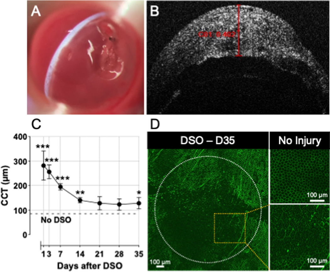
(A) Stripped corneal endothelium and increased corneal thickness can be visualized on slit-lamp exam using a narrow slit-lamp light beam. (B) AS-OCT examination shows corneal thickness greater than 400 μm at post-operative day 1. (C) Serial corneal thickness measurements via OCT examination show a decrease in corneal thickness through 35 days of follow-up. Data is presented as mean ± SEM; comparison was made by multiple paired t-test at each time point (DSO vs. No DSO [Pre-injury]) N=6/group; *, P<0.05; **, P<0.01, ***, P<0.001. CCT, Central Corneal Thickness. (D) Staining of corneal endothelium with ZO-1 35 days (7wks) after DSO showed impaired corneal wound healing, evidenced by enlarged and dysmorphic corneal endothelial cells as compared to uninjured corneas.


