| PMC full text: |
|
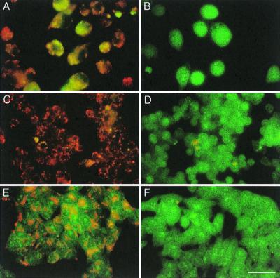
FIG. 3

Effect of BAF on vacuolar acidification after staining with acridine orange. BAF-treated (B, D, and F) or control untreated cells (A, C, and E) were incubated for 5 min with acridine orange (5 μg/ml) in PBS and then visualized using a Zeiss Axiophot microscope with an ×65 oil immersion lens. (A and B) J774 cells. (C and D) RAW cells. (E and F) Henle cells. Bar, 20 μm.


