| PMC full text: | Published online 2023 Dec 7. doi: 10.7759/cureus.50139
|
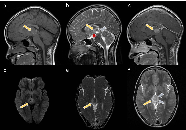
Figure 3

Selected MRIs of the patient after the recurrence of the mass
The images show the pineal mass larger than the initial lesion (yellow arrows). The mass shows low to intermediate T1 signal intensity with the bright surrounding rim (a) and heterogeneous low T2 signal intensity (b, f)) with intralesional cysts (red arrow). The mass is enhanced in the post-contrast T1WI sequence (c) and restricted on the DWI (d,e). Noted is the surrounding edema (blue arrows) of the bilateral thalami and tectum on the T2WI.