| PMC full text: | Published online 2024 Jul 11. doi: 10.1172/JCI169994
|
Figure 1

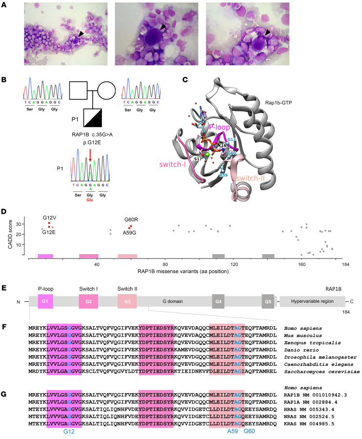
(A) May-Grünwald-Giemsa staining of P1 BM smear at age of M18 showing reduced richness for the patient’s age, elements at all stages of maturation, predominance of the granular lineage, absence of atypical cells, and presence of rare hypolobed megakaryocytes (black arrowheads). Original magnification, ×500 (left); ×1,000 (center and right). (B) P1 pedigree and familial segregation. Sanger sequencing of RAP1B in whole peripheral blood from P1 and his parents shows the heterozygous RAP1B c.35G>A (p.G12E) variant in P1 (red arrow), but not in P1’s parents, confirming its de novo nature. (C) Ribbon representation of the 3D structure of rat Rap1B bound to a nonhydrolyzable GTP analog (GppNHp, pdb 3X1X) (42). The sequences of rat Rap1B and human RAP1B are identical, except for C139 in the human sequence, which is replaced by serine in the rat sequence. This surface residue is far from the nucleotide-binding site. P-loop, switch I, and switch II regions are shown in pink. Magnesium ion is shown in green, water molecules in red, and the residues G12, A59, and G60, which have been found mutated in patients (Table 3), are in blue. (D) CADD score and amino acid position of all human RAP1B missense variants listed in gnomAD (11) as of February 6, 2023. RAP1B variants reported in patients are shown in red: G12E (P1) and G12V, A59G, and G60R (P2, P3, and P4) (5, 6). (E) Schematic representation of the secondary structure of human RAP1B with G domain–containing P-loop (G1), switch I and switch II (G2 and G3), G4, and G5 functional domains (33, 61), and hypervariable region. (F) Multiple sequence alignment of RAP1B G1–G3 functional domains from different species (62). (G) Multiple sequence alignment of G1–G3 functional domains of human small GTPases: RAP1B, RAP1A, HRAS, NRAS, and KRAS (62).


