| PMC full text: | Published online 2024 Oct 7. doi: 10.18632/aging.206121
|
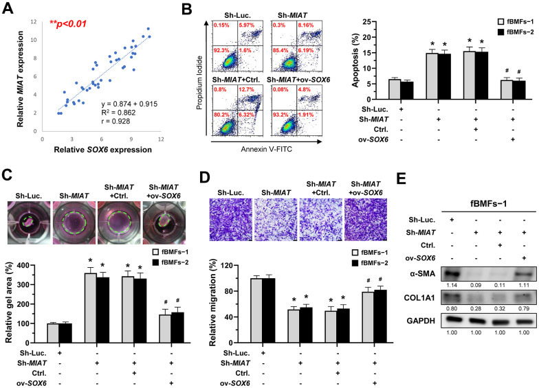
Figure 7

MIAT increases the myofibroblastic properties by positively regulating SOX6. (A) A significant positive correlation between the expression of MIAT and SOX6 in fibrotic tissue sample (OSF; n=43). (B–E) Fibrotic BMFs (−1 and −2) were co-transfected with lentiviruses expressing the following constructs in the indicated combinations: non-targeting ShRNA (Sh-Luc.), Sh-MIAT, control vector (Ctrl.), and SOX6 (ov-SOX6). Cell apoptosis (annexin V+ or annexin V+/PI+) was determined using flow cytometry (B). Cells (fBMFs−1 and −2) were cultured in collagen gel for an additional 48 hours. The resulting gel area after cell contraction was measured (C). Cells were cultured in Transwell system for an additional 24 hours, and their migration ability was assessed. Scale bar, 50 μm (D). Data are presented as mean ± SD (n=3); *p < 0.05 vs. Sh-Luc.; #p < 0.05 vs. Sh-MIAT with Ctrl. (B–D). The protein expression of α-SMA and COL1A1 in fBMFs−1 was analyzed using Western blotting (E).


