Abstract
Free full text

Enhanced Expression of an α2,6-Linked Sialic Acid on MDCK Cells Improves Isolation of Human Influenza Viruses and Evaluation of Their Sensitivity to a Neuraminidase Inhibitor
Abstract
The extensive use of neuraminidase (NA) inhibitors to treat influenza virus infections mandates close monitoring for resistant variants. Cultured cells do not provide a reliable means of evaluating the susceptibility of human influenza virus isolates to NA inhibitors. That is, the growth of such viruses in cell lines (e.g., Madin-Darby canine kidney [MDCK] cells) is not inhibited by these drugs, even though their sialidase activity is drug-sensitive. Matrosovich et al. (J. Virol. 77:8418-8425, 2003) showed that an MDCK cell line overexpressing the human β-galactoside α2,6-sialyltransferase I (ST6Gal I) gene has the potential to assess the sensitivity of human influenza virus isolates to NA inhibitors, based on studies with a limited number of viruses. Here, we asked whether clinical isolates of influenza virus are universally sensitive to an NA inhibitor (oseltamivir) in an MDCK cell line expressing the ST6Gal I gene. The sensitivity of viruses to oseltamivir correlated with the sensitivity of viral sialidase to the compound, demonstrating the potential utility of this modified cell line for detecting NA inhibitor-resistant viruses. Moreover, in ST6Gal I-overexpressing cells, the growth of human influenza viruses was up to 2 logs higher than in MDCK cells. We conclude that the human ST6Gal I-expressing MDCK cell line is useful not only for evaluating their sensitivity to NA inhibitors, but also for isolation of influenza viruses from clinical samples.
Influenza epidemics continue to impose a significant toll on the world's population in terms of hospitalizations and deaths (3, 26). Since the use of influenza virus neuraminidase (NA)-specific inhibitors, especially oseltamivir, is rapidly increasing, the emergence of drug-resistant variants has become a major concern. In fact, one recent study showed that the frequency of NA inhibitor resistance among young children was considerably higher than previously thought (15). Thus, careful monitoring for the emergence of NA inhibitor-resistant variants is essential if we intend to derive maximum therapeutic benefit from this class of antiviral agents.
Influenza A and B viruses bind to sialyloligosaccharides on host cell surface glycolipids or glycoproteins via the hemagglutinin (HA) protein, a surface spike protein on virions. Human influenza viruses preferentially bind to sialyloligosaccharides containing terminal N-acetyl sialic acid linked to galactose by an α2,6-linkage (NeuAcα2,6Gal), while avian influenza viruses mainly bind to those containing an α2,3-linkage (NeuAcα2,3Gal) (4, 25).Human airway epithelial cells mainly contain NeuAcα2,6Gal (5), although NeuAcα2,3Gal can be found in cultured ciliated epithelial cells derived from humans (19). By contrast, epithelial cells in duck intestine possess mainly NeuAcα2,3Gal (14). Thus, viral receptor specificity and the predominant type of sialic acid-galactose linkage in sialyloligosaccharides in host epithelial cells are critical determinants of the extent of viral replication in different hosts.
Although both NeuAcα2,6Gal and NeuAcα2,3Gal are present in Madin-Darby canine kidney (MDCK) cells (13), the amount of NeuAcα2,6Gal may not be as high as that of epithelial cells in human airway, leading to suboptimal growth in these cells. A plaque reduction assay with MDCK cells was used to evaluate the susceptibility of laboratory strains of human influenza viruses to NA inhibitors (9). However, this MDCK cell assay cannot be used with clinical human isolates, especially recent strains, as the growth of many recent human influenza isolates is not inhibited by NA inhibitors in these cells, and the viruses are apparently judged to be resistant to the drugs even though their enzymatic activities are drug sensitive (1, 7, 27, 30, 32). In lieu of a reliable cell culture assay for determining the susceptibility of influenza viruses to NA inhibitors, a sialidase enzyme assay has been adapted for this purpose (22, 27), but it is not suitable for large-scale screening.
Matrosovich et al. (18) recently addressed this problem by generating MDCK cells overexpressing the β-galactoside α2,6-sialyltransferase I (ST6Gal I) gene, whose product catalyzes α2,6-sialylation of galactose on glycoproteins (28, 29). The growth of clinical isolates of human influenza viruses was shown to be inhibited by oseltamivir carboxylate in these mutant cells, but only four clinical isolates were tested, leaving in question whether such cells can be used universally to evaluate the sensitivity of human clinical isolates to NA inhibitors. Another problem stems from the inability of some recent human isolates to form clear plaques in MDCK cells, limiting the use of this assay in drug susceptibility assays. Thus, as reported here, we established a cell line expressing a larger amount of NeuAcα2,6Gal that would mimic the receptor environment in human airway cells, and then tested these cells for their utility in monitoring the sensitivity of influenza viruses to NA inhibitors.
MATERIALS AND METHODS
Cells.
An MDCK cell line originally obtained from the American Type Culture Collection was passaged using standard laboratory techniques, prior to being used for these experiments. MDCK cells were maintained in minimal essential medium containing 5% newborn calf serum and antibiotics at 37°C in 5% CO2.
Clinical specimens and viruses.
Clinical specimens (throat swabs, nasal swabs, or nasal washes) shown to be influenza virus-positive by a rapid diagnostic kit were obtained from Keiyu Hospital, Kawasaki Municipal Hospital, Nippon Kokan Hospital, and Yokohama City Institute of Health between 2002 and 2004 and stored at −80°C until use. Rapid diagnostic kits used for diagnosis in these institutes were Capilia FluA,B (Nippon Becton Dickinson and Company, Japan), Directigen FluA+B (Nippon Becton Dickinson and Company, Japan) and Espline Influenza A&B-N (Fujirebio Inc., Japan). Clinical specimens prior to 2001 were all obtained from Yokohama City Institute of Health.
Influenza viruses were isolated in MDCK cells and passaged less than three times in these cells. Viral subtypes were determined by conventional hemagglutinin and neuraminidase inhibition assays (31). The oseltamivir-resistant variants of clinical isolates (H3N2) bearing an R292K or an E119V mutation in their NA were isolated from patients treated with oseltamivir as described previously (15). The R292K and E119V mutants were shown to be 104- to 105-fold and 500-fold, respectively, more resistant to oseltamivir than their parent viruses in an enzymatic assay.
Construction of plasmid expressing the ST6Gal I gene.
Human ST6Gal I cDNA in the pGEM-T vector (Promega) was prepared as described elsewhere (12). The ST6Gal I gene was amplified by PCR with 5′-CAAGGCGGCCGCGATGATTCACACCAACCTGAAGAAAAAGTTCAGCTGCT-3′ and 5′-AGATCTGCTAGCTCGAGTAAGCAGTGAATGGTCCGGAAGCCAGGCAGTGTGGC-3′, containing NotI and BglII restriction sites, respectively. The PCR product was cloned into a NotI- and BglII-cut pCAGGS-FLAG expression vector, which was constructed by adding the FLAG epitope sequence downstream of the BglII site of eukaryotic expression vector pCAGGS/MCS (16, 23), resulting in pCAGGS-FLAG-ST6Gal I.
Escherichia coli DH5α competent cells (Toyobo) were transformed by the ligated product and the plasmid was purified with the Plasmid Maxiprep kit (Marligen). pCAGGS-FLAG-ST6Gal I was digested with ApaI and XhoI. The ApaI- and XhoI-digested fragment containing ST6Gal I tagged with the FLAG epitope was subcloned into the pCAGGS-PUR plasmid, carrying the puromycin N-acetyltransferase gene, between the ApaI and XhoI restriction sites, resulting in pCAGGS-FLAG-PUR-ST6Gal I.
Establishment of a stable cell line expressing ST6Gal I.
The pCAGGS-FLAG-PUR-ST6Gal I plasmid was transfected into MDCK cells with the Trans IT-293 transfection reagent (Mirus) according to the manufacturer's instructions. Briefly, on the day before transfection, MDCK cells were plated at 5 × 10⁵ cells/100-mm dish. On day 1, 10 μg of plasmid DNA was mixed with 20 μg of Trans IT-293 in 0.3 ml of OptiMEM (Gibco) and was incubated with these cells at 37°C in 5% CO2 overnight. On day 2, the transfection mixture was replaced with a complete medium (modified Eagle's medium [MEM] supplemented with 5% newborn calf serum containing 5 μg/ml of puromycin dihydrochloride [Nacalai Tesque]). Eleven days later, the medium was replaced with a complete medium containing 7.5 μg/ml of puromycin. The next day, puromycin-resistant clones were isolated and transferred to 24-well plates. Each clone was passaged an additional three to five times in the presence of 7.5 μg/ml of puromycin. ST6Gal I-expressing cells were maintained in the presence of 7.5 μg/ml of puromycin except when they were used for viral infection.
Immunostaining.
Cells were fixed in 4% paraformaldehyde for 30 min at room temperature and permeated with 0.5% Triton X-100 in phosphate-buffered saline (PBS) for 30 min. The cells were then washed with PBS and incubated for 1 h at room temperature with the anti-FLAG monoclonal antibody M2 (Sigma), diluted 1:1,000 in PBS, and then washed four times with PBS. This was followed by a 30-min incubation with biotinylated anti-mouse immunoglobulin G, and the reaction was then detected by incubating with the Vectastain ABC reagent (Vectastain ABC kit, Vector Laboratories) using diaminobenzidine as a substrate (SIGMA FAST, Sigma).
Flow cytometric analysis of sialic acid expression on cells.
To examine the levels of sialic acid linked to galactose on the cell surface by the α2,3 linkage (SAα2,3Gal) or the α2,6 linkage (SAα2,6Gal), we used digoxigenin-labeled lectins, Sambucus nigra agglutinin specific for SAα2,6Gal and Maackia amurensis agglutinin specific for SAα2,3Gal (digoxigenin-glycan differentiation kit, Roche). The anti-digoxigenin fluorescein-conjugated antibody (Roche) was used as a secondary antibody. Fluorescence was determined on a FACSCalibur flow cytometer (Becton Dickinson) by measuring the fluorescence of a minimum of 10,000 cells. Approximately 106 parental or plasmid-transfected MDCK cells were washed twice with PBS containing 10 mM glycine and then washed once with buffer 1 (50 mM Tris-HCl, 0.15 M NaCl, 1 mM MgCl2, 1 mM MnCl2, 1 mM CaCl2, pH 7.5). The cells were blocked with a blocking solution from the digoxigenin kit for 1 h on ice, and then washed in the same manner as described above. After centrifugation, the cell pellet was incubated with digoxigenin-labeled lectins (either 1 μl of S. nigra agglutinin or 1 μl of M. amurensis agglutinin) in 30 μl of buffer 1 for 1 h on ice. After two washes with PBS containing glycine and one with buffer 1, the cells were incubated with 1 μl of anti-digoxigenin-fluorescein conjugated antibody in 30 μl of buffer 1 for 1 h on ice. After another three washes with PBS, the fluorescence intensities were quantified by fluorescence-activated cell sorter (FACS) analysis.
Viral infection.
To determine virus titers, we performed plaque assays with either MDCK cells or ST6Gal I-expressing MDCK cells. In a preliminary study, we titrated clinical isolates at 33°C and 37°C using both MDCK and ST6Gal I-expressing cells. We did not see a difference in titers of the viruses tested at 33°C or 37°C in either cell line, with one exception. Also, plaques were larger at 33°C than at 37°C in MDCK cells, while they were larger at 37°C than at 33°C in ST6Gal I-expressing cells. Therefore, virus in MEM containing 7.5% bovine serum albumin (BSA-MEM) was incubated with cells for 60 min at 37°C. The inoculum was then removed and the cells were washed once with BSA-MEM. They were then cultured in infection medium (7.5% bovine serum albumin, 0.5 μg/ml of trypsin, 1% agarose in MEM). Two to three days later, the overlays were removed, and the cell monolayers were stained with 0.1% crystal violet in 20% methanol.
Determination of viral sensitivity to oseltamivir carboxylate in cell culture.
Confluent monolayers of MDCK cells or ST6Gal I-expressing MDCK cells in six-well tissue culture plates were inoculated with 50 to 100 PFU per well of virus in BSA-MEM. After 60 min at 37°C, the inoculum was removed and the cells were washed once. They were then cultured in 3 ml of infection medium with or without serial 10-fold dilutions of oseltamivir carboxylate (GS4071, Roche Products), the active compound of the ethyl ester prodrug oseltamivir phosphate. Two to three days later, the cell monolayers were stained with 0.1% crystal violet as described above.
Sialidase sensitivity to oseltamivir carboxylate.
The sensitivity of viral sialidase to oseltamivir was determined as described by Gubareva et al. (8). Briefly, viral sialidase activity was determined with 2′-(4-methylumbelliferyl)-α-d-N-acetylneuraminic acid (Sigma) used as a substrate. To 20 μl of diluted virus (serially diluted twofold) in V-bottomed microtiter plates, we added 30 μl of a 0.1 mM solution of 2′-(4-methylumbelliferyl)-α-d-N-acetylneuraminic acid in calcium-MES buffer (33 mM 2-[N-morpholino]ethanesulfonic acid [MES], 4 mM CaCl2; pH 6.0). The mixtures were incubated at 37°C for 60 min, and the reaction was stopped by the addition of 150 μl of stop solution (0.1 M NaOH in 80% ethanol; pH 10.0).
Fluorescence was quantified at an excitation wavelength of 360 nm and an emission wavelength of 465 nm with a SPECTRA max GEMINI XS spectrophotometer (Molecular Devices). We then used dilutions of virus with 800 to 1,200 fluorescence units in the sialidase inhibition assay. Ten μl of the dilutions and 10 μl of the drug (0.01 nM to 1 mM) in calcium-MES buffer were mixed and incubated at 37°C for 30 min, followed by the addition of 30 μl of the substrate. After 1 h of incubation at 37°C, the reaction was stopped by adding 150 μl of the stop solution. Fluorescence was quantified in the manner described above. The 50% inhibitory concentrations (IC50s) were determined by extrapolation of the relation between the concentration of inhibitor and the proportion of fluorescence inhibition.
RESULTS
ST6Gal I-expressing MDCK cells overexpressing α2,6-linked sialic acids on the cell surface.
To obtain cells constitutively expressing a high level of α2,6-linked sialic acids, we transfected MDCK cells with pCAGGS-FLAG-PUR-ST6Gal I and selected cell clones resistant to puromycin. Among the randomly selected puromycin-resistant clones, four were shown by immunostaining to express the FLAG epitope (Fig. (Fig.1).1). The effect of ST6Gal I expression in these clones was studied by testing the cells' reactivity with sialic acid linkage-specific lectins (S. nigra agglutinin, specific for α2,6-linked sialic acids, and M. amurensis agglutinin, specific for α2,3-linked sialic acids) by FACS. In two of the four anti-FLAG-positive clones, the reactivity with S. nigra agglutinin increased by approximately 50% compared with the parent MDCK cells (Fig. (Fig.2;2; data for only one clone are shown). By contrast, the reactivity with M. amurensis agglutinin was similar between the parent and FLAG tag-positive clones. One of these two ST6Gal I-expressing cell clones was used in subsequent studies.
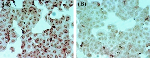
Generation of a cell line constitutively expressing ST6Gal I. MDCK cells were transfected with a plasmid containing the ST6Gal I and puromycin N-acetyltransferase genes and selected with puromycin. Expression of ST6Gal I was confirmed by monoclonal antibody to the FLAG epitope used for tagging ST6Gal I, as described in Materials and Methods. (A) ST6Gal I-transfected MDCK cells. (B) Parent MDCK cells.
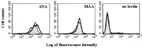
Reactivity of sialic acid linkage-specific lectins with ST6Gal I-expressing or parent MDCK cells. Cells were incubated with digoxigenin-labeled S. nigra agglutinin, specific for α2,6-linked sialic acids, or M. amurensis agglutinin, specific for α2,3-linked sialic acids, lectins, followed by the anti-digoxigenin-fluorescein conjugated antibody, and then analyzed by FACS. Open profiles, ST6Gal I-expressing cells; shaded profiles, parent MDCK cells. Far left panel, binding of the S. nigra agglutinin lectin; middle panel, binding of the M. amurensis agglutinin lectin; far right panel, negative control (no lectin added). The ratio of the mean fluorescence intensity (ST6Gal I-expressing cells/MDCK cells) was 1.45 with S. nigra agglutinin lectin and 1.06 with M. amurensis agglutinin lectin.
Clinical isolates of human influenza viruses grow better in ST6Gal I-expressing cells than in the parent MDCK cells.
To test whether a higher expression level of α2,6-linked sialic acid on the cell surface affects their susceptibility to human influenza viruses, we performed a plaque assay using clinical isolates of influenza viruses. Among 20 clinical specimens (eight H3N2, seven H1N1, and five type B viruses), titers were appreciably higher in the ST6Gal I-expressing cells compared with MDCK cells (Table (Table1).1). With multiple samples that were positive by a rapid diagnostic kit, it was not possible to isolate any viruses in MDCK cells, whereas more than 102 PFU/ml of viruses were detected with ST6Gal I-expressing cells. Our inability to isolate virus using MDCK cells from samples that were positive with a rapid diagnostic kit was likely due to the subsequent declines in virus viability in these clinical specimens, which had undergone two freezing-thawing cycles after being tested with the rapid diagnostic kit.
TABLE 1.
ST6Gal I-expressing cells are superior to MDCK cells in the isolation of human influenza viruses from clinical samplesa
| Virus type | Virus isolate | Yr of isolation | Antigenic property | Virus titer (PFU/ml) | |
|---|---|---|---|---|---|
| MDCK cells | ST6Gal I cells | ||||
| H3N2 | 1 | 2004 | A/Fujian/411/2002-like | 3.4 × 10⁴ | 5.5 × 10⁵ |
| 2 | 2004 | A/Fujian/411/2002-like | <6.3 | 2.8 × 10² | |
| 3 | 2004 | A/Fujian/411/2002-like | <6.3 | 1.0 × 10² | |
| 4 | 2004 | A/Fujian/411/2002-like | <6.3 | 1.9 × 10² | |
| 5 | 2003 | A/Fujian/411/2002-like | <6.3 | 6.9 × 10 | |
| 6 | 2001 | A/Panama/2007/99-like | 9.2 × 10⁴ | 1.0 × 10⁶ | |
| 7 | 1999 | A/Sydney/5/97-like | <6.3 | 1.0 × 10² | |
| 8 | 1999 | A/Sydney/5/97-like | 2.0 × 10² | 2.2 × 10³ | |
| H1N1 | 9 | 2002 | A/New Calledonia/20/99-like | 1.1 × 10² | 2.6 × 10⁴ |
| 10 | 2002 | A/New Calledonia/20/99-like | 4.3 × 10⁴ | 1.8 × 10⁵ | |
| 11 | 2002 | A/New Calledonia/20/99-like | 1.3 × 10² | 6.3 × 10² | |
| 12 | 2002 | A/New Calledonia/20/99-like | 2.5 × 10³ | 1.1 × 10⁴ | |
| 13 | 2002 | A/New Calledonia/20/99-like | 6.3 | 1.3 × 10 | |
| 14 | 2000 | A/Beijing/262/95-like | 2.0 × 10² | 3.3 × 10³ | |
| 15 | 2000 | A/Beijing/262/95-like | 4.0 × 10 | 4.5 × 10² | |
| B | 16 | 2003 | B/Shandong/7/97-like | <6.3 | 6.3 |
| 17 | 2003 | B/Shandong/7/97-like | <6.3 | 5.6 × 10² | |
| 18 | 2003 | B/Shandong/7/97-like | <6.3 | 3.1 × 10 | |
| 19 | 2003 | B/Shandong/7/97-like | <6.3 | 1.9 × 10² | |
| 20 | 2002 | B/Shandong/7/97-like | 2.4 × 10⁶ | 1.5 × 10⁷ | |
In the second series of experiments, 23 viruses (eight H3N2, eight H1N1, and seven type B viruses) that had been isolated from clinical samples and propagated in MDCK cells were tested. All of these viruses grew to higher titers (20 times higher in some instances) in ST6Gal I-expressing cells than in MDCK cells (Table (Table2).2). By contrast, the A/Aichi/1/68 (H3N2) virus grown in eggs replicated with similar efficiencies in ST6Gal I-expressing cells and MDCK cells (data not shown).
TABLE 2.
Comparison of replicative efficiencies of clinical isolates of human influenza viruses in ST6Gal I-expressing cells versus MDCK cells
| Virus type | Virusa | Yr of isolation | Antigenic property | Virus titer (PFU/ml)b | Ratio (ST6Gal I titer/MDCK titer) | |
|---|---|---|---|---|---|---|
| MDCK cells | ST6Gal I-expressing cells | |||||
| H3N2 | 1 | 2004 | A/Fujian/411/2002-like | 3.9 × 10⁵ | 1.1 × 10⁶ | 2.8 |
| 2 | 2002 | A/Panama/2007/99-like | 6.5 × 10⁶ | 1.7 × 10⁷ | 2.6 | |
| 3 | 2002 | A/Panama/2007/99-like | 1.5 × 10⁶ | 2.4 × 10⁷ | 16.0 | |
| 4 | 2002 | A/Panama/2007/99-like | 3.4 × 10⁵ | 5.9 × 10⁶ | 17.4 | |
| 5 | 2002 | A/Panama/2007/99-like | 1.9 × 10⁷ | 2.5 × 10⁷ | 1.3 | |
| 6 | 2002 | A/Panama/2007/99-like | 3.5 × 10⁶ | 4.0 × 10⁶ | 1.1 | |
| 7 | 1995 | A/Akita/1/94-like | 6.1 × 10⁶ | 4.9 × 10⁷ | 8.0 | |
| 8 | 1995 | A/Akita/1/94-like | 1.2 × 10⁵ | 2.0 × 10⁶ | 16.7 | |
| H1N1 | 9 | 2002 | A/New Calledonia/20/99-like | 3.0 × 10⁵ | 1.7 × 10⁶ | 5.7 |
| 10 | 2002 | A/New Calledonia/20/99-like | 1.7 × 10⁷ | 4.8 × 10⁷ | 2.8 | |
| 11 | 2002 | A/New Calledonia/20/99-like | 8.5 × 10⁶ | 1.3 × 10⁷ | 1.5 | |
| 12 | 2002 | A/New Calledonia/20/99-like | 3.4 × 10⁶ | 7.8 × 10⁷ | 22.9 | |
| 13 | 2002 | A/New Calledonia/20/99-like | 4.8 × 10⁶ | 1.8 × 10⁷ | 3.8 | |
| 14 | 2002 | A/New Calledonia/20/99-like | 1.5 × 10⁷ | 4.6 × 10⁷ | 3.1 | |
| 15 | 1995 | A/Yamagata/32/89-like | 5.7 × 10⁶ | 1.7 × 10⁷ | 3.0 | |
| 16 | 1995 | A/Yamagata/32/89-like | 4.8 × 10⁴ | 2.0 × 10⁵ | 4.2 | |
| B | 17 | 2003 | B/Shandong/7/97-like (Victoria) | 1.3 × 10⁷ | 3.2 × 10⁷ | 2.5 |
| 18 | 2003 | B/Shandong/7/97-like (Victoria) | 1.2 × 10⁷ | 3.7 × 10⁷ | 3.1 | |
| 19 | 2003 | B/Shandong/7/97-like (Victoria) | 1.9 × 10⁸ | 3.8 × 10⁸ | 2.0 | |
| 20 | 2003 | B/Shandong/7/97-like (Victoria) | 5.3 × 10⁶ | 1.4 × 10⁷ | 2.6 | |
| 21 | 2003 | B/Shandong/7/97-like (Victoria) | 1.0 × 10⁸ | 2.7 × 10⁸ | 2.7 | |
| 22 | 1995 | B/Mie/1/93-like (Yamagata) | 1.3 × 10⁸ | 2.2 × 10⁸ | 1.7 | |
| 23 | 1995 | B/Mie/1/93-like (Yamagata) | 6.4 × 10⁷ | 2.4 × 10⁸ | 3.8 | |
Clinical isolates of human influenza viruses, especially recent ones, often produce small, fuzzy plaques in MDCK cells, and some appear incapable of forming any plaques in these cells (27, 32). In ST6Gal I-expressing cells, however, such isolates consistently generated large, clear plaques, in sharp contrast to the results in MDCK cells (Fig. (Fig.3).3). This finding was noted with both subtypes of type A as well as type B influenza viruses from clinical samples. Considered together, these observations indicate that ST6Gal I-expressing cells are more suitable than MDCK cells for isolating human influenza viruses from clinical samples.
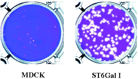
Plaque formation by a clinical isolate of human influenza virus in ST6Gal I-expressing compared with MDCK cells. Confluent monolayers of ST6Gal I-expressing cells (ST6Gal I) and MDCK cells (MDCK) in six-well plates were inoculated with the same amount of an H1N1 influenza virus isolated from a clinical sample.
ST6Gal I-expression in MDCK cells universally increases the sensitivity of influenza A and B viruses to oseltamivir.
To determine whether ST6Gal I expression universally increases the sensitivity of human influenza viruses to oseltamivir, we performed plaque assays in the presence of this compound using cells modified to express ST6Gal I, and then compared the IC50 values with those obtained from the sialidase inhibition assay. With most viruses (14 of 17 samples tested [six H3N2, six H1N1, and five type B isolates]), the NA inhibitor reduced plaque size rather than plaque number in ST6Gal I-expressing cells. We therefore defined the IC50 as the inhibitory concentration at which the plaque size was reduced by more than 50% in a majority (>90%) of the plaques, compared with the no-drug negative control, a standard that has been used in previous reports (8, 20).
In MDCK cells, all but one of these viruses produced plaques of equal size and number, whether or not oseltamivir carboxylate was present, even though the viral sialidases were highly sensitive to oseltamivir in the enzyme inhibition assay. By contrast, in tests with ST6Gal I-expressing cells, the drug inhibited the growth of all viruses. As shown in Fig. Fig.4,4, plaque formation was inhibited by drug concentrations exceeding 10 nM (for H1N1 and H3N2 isolates) and 100 nM (for type B virus). The IC50 values of clinical isolates grown in ST6Gal I-expressing cells correlated well with viral sialidase sensitivity to oseltamivir, although values obtained with ST6Gal I-expressing cells were uniformly about 10-fold higher than those from the sialidase enzyme inhibition assay (Table (Table3).3). Thus, ST6Gal I-expressing cells appear generally suited to assessing the sensitivity of clinical isolates of human influenza viruses to oseltamivir and likely other neuraminidase inhibitors as well.
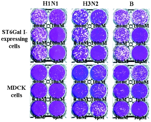
Oseltamivir sensitivity of clinical isolates of influenza viruses in ST6Gal I-expressing cells and MDCK cells. Plaque reduction assays were performed as described in Materials and Methods using clinical influenza virus isolates. The results shown represent experiments with a virus of each subtype and type (left panel, H1N1 virus; middle panel, H3N2 virus; right panel, type B virus). The same amount of virus was incubated with cells in each well in the presence of serial 10-fold dilutions of oseltamivir or in its absence (none).
TABLE 3.
Comparison of oseltamivir IC50 values in cell culture assays and in the sialidase enzyme assay for clinical isolates of influenza viruses
| Virus type | Virusa | IC50 in plaque reduction assayb (nM) | IC50 in sialidase enzyme assay (nM) | |
|---|---|---|---|---|
| MDCK cells | ST6Gal I- expressing cells | |||
| H3N2 | 1 | >1,000 | 10 | 0.5 |
| 2 | >1,000 | 10 | 0.5 | |
| 3 | >1,000 | 10 | 0.3 | |
| 4 | >1,000 | 10 | 0.2 | |
| 5 | >1,000 | 10 | 0.3 | |
| 6 | >1,000 | 10 | 1.2 | |
| H1N1 | 7 | >1,000 | 10 | 6.8 |
| 8 | >1,000 | 10 | 8.4 | |
| 9 | 1,000 | 100 | 72.9 | |
| 10 | >1,000 | 100 | 12.5 | |
| 11 | >1,000 | 10 | 5.6 | |
| 12 | >1,000 | 10 | 8.4 | |
| B | 13 | >1,000 | 1,000 | 109.3 |
| 14 | >1,000 | 1,000 | 131.3 | |
| 15 | >1,000 | 1,000 | 214.9 | |
| 16 | >1,000 | 1,000 | 59.6 | |
| 17 | >1,000 | 1,000 | 89.5 | |
We were still uncertain whether ST6Gal I-expressing cells would be useful for detecting variants resistant to the NA inhibitor, leading us to test clinical isolates of oseltamivir-resistant viruses (H3N2) bearing an R292K or an E119V mutation in the NA. Plaque formation by the resistant strains in ST6Gal I-expressing cells was inhibited by oseltamivir concentrations that were approximately 10,000-fold (four R292K variants) or 1,000-fold (E119V variant) higher than those required to inhibit the growth of the respective parent viruses. In ST6Gal I-expressing cells, plaques of the drug-resistant R292K viruses were much smaller than those of the parent drug-sensitive viruses (Fig. (Fig.5)5) as reported previously (18), although the E119V mutant produced plaques that were similar in size to those of its parent virus. Overall, the plaque morphology of drug-resistant and parent viruses in MDCK cells appeared similar. These results show that ST6Gal I-expressing MDCK cells are not only suitable for isolating viruses from clinical specimens, but can also be used to identify human clinical isolates that are resistant to NA inhibitors.
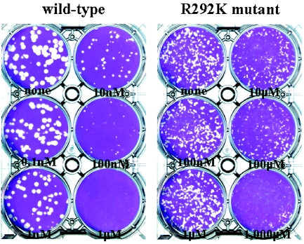
Effect of oseltamivir on plaque formation by a wild-type human H3N2 influenza virus and its NA R292K mutant in ST6Gal I-expressing cells. A plaque reduction assay in the presence of oseltamivir (serially diluted as indicated in the figure) with ST6Gal I-expressing cells was performed as described in Materials and Methods. Left panel, wild-type human H3N2 influenza virus. Right panel, NA R292K mutant recovered from a patient treated with oseltamivir. The top left well of each plate contains no inhibitor. Note that a 1,000-fold higher concentration of the compound is used for the R292K mutant than for its wild-type parent.
DISCUSSION
Here we show that human influenza viruses in clinical samples grow better and produce clearer plaques in cells expressing an increased level of SAα2,6Gal than in MDCK cells. These results can be attributed to the relatively low level of SAα2,6Gal on the MDCK cell surfaces compared with those on human airway cells. That some influenza viruses in clinical specimens grew more than 100-fold better in ST6Gal I-expressing cells than in the parent MDCK cells suggests that viruses isolated in MDCK cells may be variants resulting from growth in a suboptimal receptor environment. This likelihood warrants further analysis of receptor specificity of clinical isolates grown in MDCK versus ST6Gal I-expressing cells. In practical terms, the substitution of ST6Gal I-expressing cells for MDCK cells in the isolation of human influenza viruses from clinical samples would be expected to increase the rate of virus recovery.
Our results also show that ST6Gal I-expressing cells can be universally used to monitor the sensitivity of clinical isolates of human influenza viruses to NA inhibitors. The apparent lack of sensitivity of such isolates to NA inhibitors in MDCK cells relates directly to differences between the properties of the receptor environment on human cells and that on MDCK cells (5, 13). The essential role of NA's sialidase activity in influenza virus replication is to remove terminal sialic acid residues bound on the viral glycoproteins and cell surface glycoconjugates, allowing the release of progeny virions from the virus-infected cells and preventing self-aggregation of progeny virions (17, 24). It is likely that in MDCK cells, clinical isolates of human viruses bind to MDCK cells with relatively low affinity, making the viruses less dependent on sialidase activity for release. By contrast, ST6Gal I-expressing cells have an increased level of SAα2,6Gal, enabling these viruses to bind to the mutant cells with a higher affinity, in turn making them more dependent on sialidase activity for replication.
The reduced growth of drug-resistant variants with an NA mutation in ST6Gal I-expressing cells is interesting and may be due to the low sialidase activities of the variant viruses; the sialidase activities of the oseltamivir-resistant viruses tested were 6- to 20-fold lower than those of their parent viruses (S. Hatakeyama, M. Kiso, and Y. Kawaoka, unpublished data). The NA mutations that confer resistance to NA inhibitors reduce enzyme function and stability (6, 21). Thus, their growth may be limited in an SAα2,6Gal-abundant environment. In support of this hypothesis, the sialidase activity of a variant virus with an E119V mutation in the NA, which grew well in ST6Gal I-expressing cells as well as in MDCK cells, was not reduced by comparison with that of the parent virus. These in vitro findings with ST6Gal I-expressing cells are consistent with those of in vivo studies; that is, the infectivity and transmissibility of clinical isolates carrying an R292K (2, 10), but not E119V (11), mutation in the NA were compromised in the mouse and/or ferret models. Although a hemagglutinin mutation was not detected in the R292K variants in at least one of the two in vivo studies mentioned above, further studies are needed to evaluate whether resistant variants possessing NA mutations would be attenuated in humans.
Acknowledgments
We thank Larisa Gubareva (Department of Internal Medicine, University of Virginia Health Sciences Center) for providing us with a protocol for the neuraminidase inhibition assay, John Gilbert for editing the manuscript, and Krisna Wells for technical assistance. Oseltamivir was provided by Roche Products.
This work was supported by grants of Core Research for Evolutional Science and Technology (CREST) from Japan Science and Technology Agency (JST), Japan, by grants-in-aid from the Ministry of Education, Culture, Sports, Science and Technology and the Ministry of Health, Labor and Welfare, Japan, and by grants from the National Institutes of Health, National Institute of Allergy and Infectious Diseases. S.H.'s salary was supported in part by grants from the 21st Century Center of Excellence Programme of the Ministry of Education, Culture, Sports, Science and Technology, Japan
REFERENCES
Articles from Journal of Clinical Microbiology are provided here courtesy of American Society for Microbiology (ASM)
Full text links
Read article at publisher's site: https://doi.org/10.1128/jcm.43.8.4139-4146.2005
Read article for free, from open access legal sources, via Unpaywall:
https://europepmc.org/articles/pmc1233980?pdf=render
Citations & impact
Impact metrics
Article citations
Viral interference between severe acute respiratory syndrome coronavirus 2 and influenza A viruses.
PLoS Pathog, 20(7):e1012017, 22 Jul 2024
Cited by: 2 articles | PMID: 39038029 | PMCID: PMC11293641
Intranasal mask for protecting the respiratory tract against viral aerosols.
Nat Commun, 14(1):8398, 18 Dec 2023
Cited by: 3 articles | PMID: 38110357 | PMCID: PMC10728126
Stability of the Virucidal Activity of Commercial Disinfectants against Avian Influenza Viruses under Different Environmental Conditions.
Pathogens, 12(12):1382, 24 Nov 2023
Cited by: 0 articles | PMID: 38133267 | PMCID: PMC10745779
Contemporary human H3N2 influenza A viruses require a low threshold of suitable glycan receptors for efficient infection.
Glycobiology, 33(10):784-800, 01 Oct 2023
Cited by: 7 articles | PMID: 37471650 | PMCID: PMC10629718
Glyco-engineered MDCK cells display preferred receptors of H3N2 influenza absent in eggs used for vaccines.
Nat Commun, 14(1):6178, 04 Oct 2023
Cited by: 3 articles | PMID: 37794004 | PMCID: PMC10551000
Go to all (104) article citations
Similar Articles
To arrive at the top five similar articles we use a word-weighted algorithm to compare words from the Title and Abstract of each citation.
Susceptibility of human influenza viruses from Australasia and South East Asia to the neuraminidase inhibitors zanamivir and oseltamivir.
Antiviral Res, 62(1):37-45, 01 Apr 2004
Cited by: 75 articles | PMID: 15026200
Selection of influenza virus mutants in experimentally infected volunteers treated with oseltamivir.
J Infect Dis, 183(4):523-531, 11 Jan 2001
Cited by: 196 articles | PMID: 11170976
Overexpression of the alpha-2,6-sialyltransferase in MDCK cells increases influenza virus sensitivity to neuraminidase inhibitors.
J Virol, 77(15):8418-8425, 01 Aug 2003
Cited by: 227 articles | PMID: 12857911 | PMCID: PMC165236
Neuraminidase inhibitors as antiviral agents.
Curr Drug Targets Infect Disord, 5(4):401-409, 01 Dec 2005
Cited by: 20 articles | PMID: 16535861
Review


