| PMC full text: | Published online 2006 Aug 17. doi: 10.1038/sj.emboj.7601283
|
Figure 8
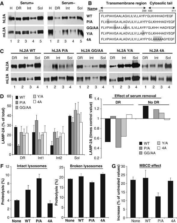
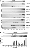
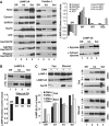
Lysosomes containing a mutant LAMP-2A unable to move into discrete lysosomal membrane regions show enhanced CMA activity. (A) Lysosomes from mouse fibroblasts transfected with wild-type hLAMP-2A and maintained in the presence (Serum+) or absence (Serum−) of serum were collected by centrifugation and subjected to Triton X-114 extraction, sucrose density gradient centrifugation and immunobloting for human (hL2A) or mouse (mL2A) LAMP-2A. Lane 1: homogenate. (B) Amino-acid sequence of the transmembrane and cytosolic region of hLAMP-2A and the mutations performed. Replaced amino acids are shadowed. (C) Lysosomes from mouse fibroblasts transfected with the indicated forms of hLAMP-2A and maintained in the presence of serum were processed as in (A). (D) The percentage of total lysosomal hLAMP-2A present in each fraction was determined by densitometric quantification of immunoblots as the ones shown here. Values, corrected for the amount of mLAMP-2A in each fraction, are mean+s.e. of 3–4 experiments. (E) Changes in hLAMP-2A distribution induced by serum removal. Values are expressed as times the values in cells maintained in the presence of serum and are the mean of two experiments. Changes in the three nonDR regions are shown together (no DR). (F) Proteolysis of a pool of 3[H]labeled cytosolic proteins by intact (left) or disrupted (right) lysosomes isolated from untransfected mouse fibroblasts (none) or fibroblasts transfected with wild-type (WT) or the indicated mutant forms of hLAMP-2A. Values are the mean+s.e. of two experiments with triplicate samples. (G) Part of the lysosomes from (F) was treated with MBCD before the incubation with the radiolabeled proteins. Values are expressed as percentage of degradation by untreated lysosomes and are the mean+s.e. of two experiments with triplicate samples.
