| PMC full text: |
|
Figure 1

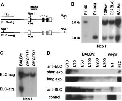
Genomic analysis of the murine ELC locus shows the presence of one ELC-atg gene and several ELC-acg genes and deletion of the ELC-atg gene in plt/plt mice. (A) Genomic organization of ELC-atg and ELC-acg genes. White boxes show 5′- and 3′-untranslated sequences, black boxes the coding sequence for ELC-atg, and gray boxes the corresponding regions of ELC-acg. The positions of NcoI restriction sites are indicated, with the location of sites in italics based upon Southern blot analysis only. (B) Southern blot of NcoI-digested P1 DNA and mouse genomic DNA to identify ELC-atg (≈2.8 kb) and ELC-acg genes (≈5.0 kb). (C) plt/plt mice lack the ELC-atg gene. Southern blot of NcoI-digested DNA from one BALB/c and two plt/plt mice. The probe used in B and C is indicated in A. (D) Western blot of extracts from pooled LNs and spleen from wild-type (BALB/c) or plt/plt mice, probed with antibodies to ELC or SLC. Extracts from BALB/c mice were titrated from 8 of 10 (80%) to 1 of 500 (0.2%). Short and long exposures of the anti-ELC probed blot are shown. A nonspecific high molecular weight band detected with the anti-ELC serum is shown as a loading control.


