Abstract
Free full text

Apoptosis: A Basic Biological Phenomenon with Wide-ranging Implications in Tissue Kinetics
Abstract
The term apoptosis is proposed for a hitherto little recognized mechanism of controlled cell deletion, which appears to play a complementary but opposite role to mitosis in the regulation of animal cell populations. Its morphological features suggest that it is an active, inherently programmed phenomenon, and it has been shown that it can be initiated or inhibited by a variety of environmental stimuli, both physiological and pathological.
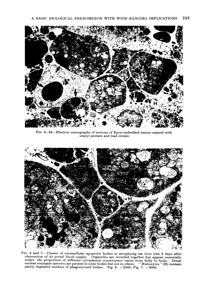
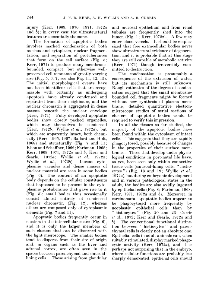
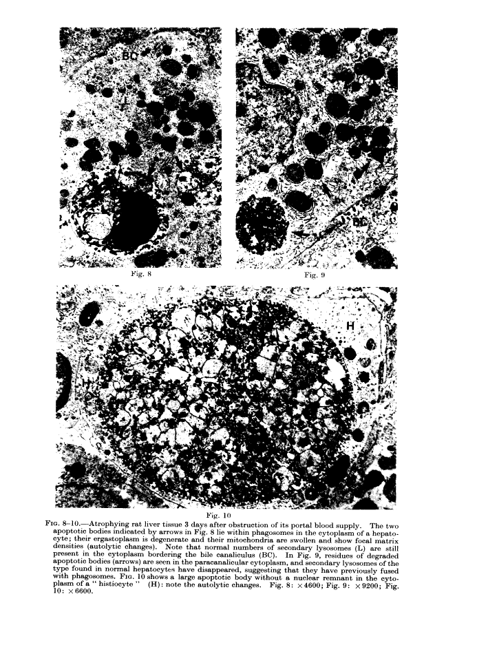
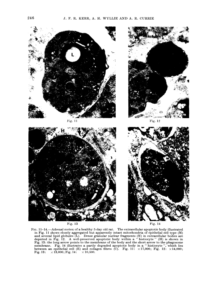
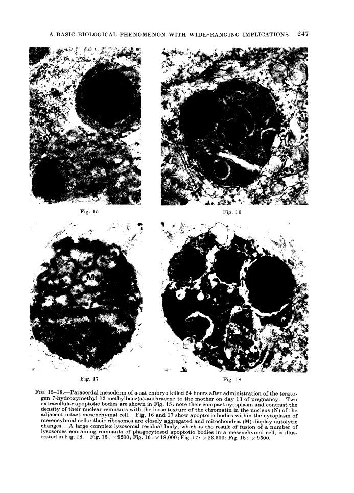
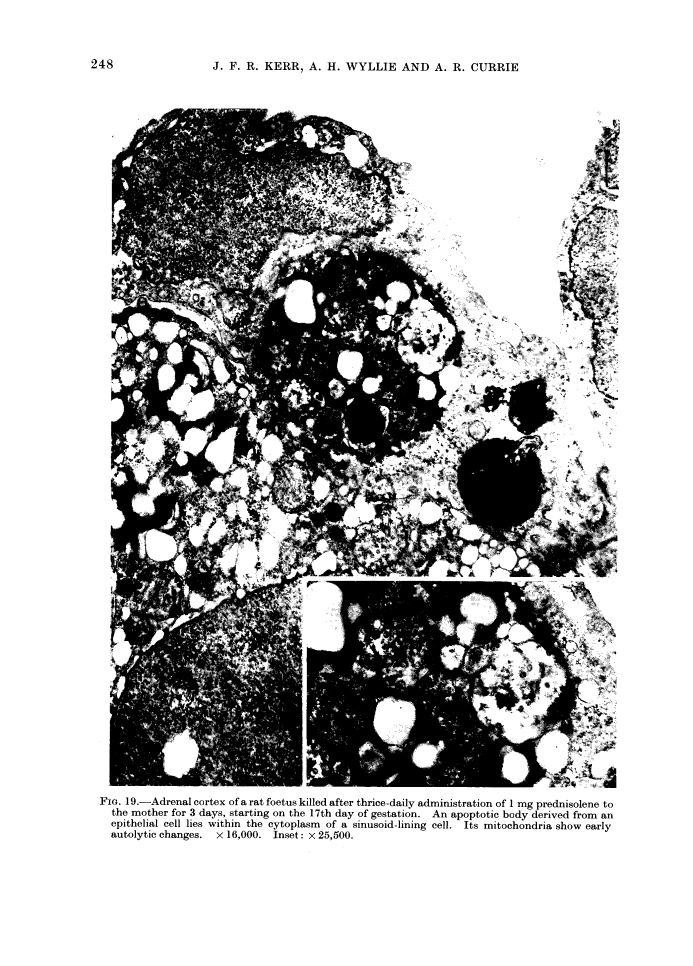
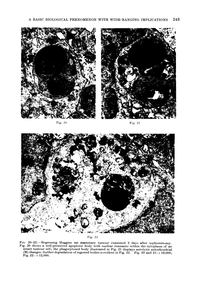
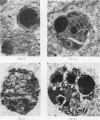
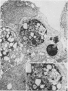
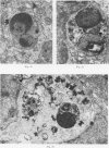
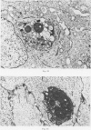
The structural changes take place in two discrete stages. The first comprises nuclear and cytoplasmic condensation and breaking up of the cell into a number of membrane-bound, ultrastructurally well-preserved fragments. In the second stage these apoptotic bodies are shed from epithelial-lined surfaces or are taken up by other cells, where they undergo a series of changes resembling in vitro autolysis within phagosomes, and are rapidly degraded by lysosomal enzymes derived from the ingesting cells.
Apoptosis seems to be involved in cell turnover in many healthy adult tissues and is responsible for focal elimination of cells during normal embryonic development. It occurs spontaneously in untreated malignant neoplasms, and participates in at least some types of therapeutically induced tumour regression. It is implicated in both physiological involution and atrophy of various tissues and organs. It can also be triggered by noxious agents, both in the embryo and adult animal.
Full text
Full text is available as a scanned copy of the original print version. Get a printable copy (PDF file) of the complete article (8.2M), or click on a page image below to browse page by page. Links to PubMed are also available for Selected References.
Images in this article
Selected References
These references are in PubMed. This may not be the complete list of references from this article.
- Abraham R, Morris M, Hendy R. Lysosomal changes in epithelial cells of the mouse thymus after hydrocortisone treatment. Histochemie. 1969;17(4):295–311. [Abstract] [Google Scholar]
- Anton E, Brandes D. Lysosomes in mice mammary tumors treated with cyclophosphamide. Distribution related to course of disease. Cancer. 1968 Mar;21(3):483–500. [Abstract] [Google Scholar]
- Ballard KJ, Holt SJ. Cytological and cytochemical studies on cell death and digestion in the foetal rat foot: the role of macrophages and hydrolytic enzymes. J Cell Sci. 1968 Jun;3(2):245–262. [Abstract] [Google Scholar]
- Biava C, Mukhlova-Montiel M. Electron Microscopic Observations on Councilman-Like Acidophilic Bodies and Other Forms of Acidophilic Changes in Human Liver Cells. Am J Pathol. 1965 May;46(5):775–802. [Europe PMC free article] [Abstract] [Google Scholar]
- Clifton KH, Yatvin MB. Cell population growth and cell loss in the MTG-B mouse mammary carcinoma. Cancer Res. 1970 Mar;30(3):658–664. [Abstract] [Google Scholar]
- Cole S, Matter A, Karnovsky MJ. Autophagic vacuoles in experimental atrophy. Exp Mol Pathol. 1971 Apr;14(2):158–175. [Abstract] [Google Scholar]
- Frindel E, Malaise E, Tubiana M. Cell proliferation kinetics in five human solid tumors. Cancer. 1968 Sep;22(3):611–620. [Abstract] [Google Scholar]
- van Haelst U. Light and electron microscopic study of the normal and pathological thymus of the rat. II. The acute thymic involution. Z Zellforsch Mikrosk Anat. 1967;80(2):153–182. [Abstract] [Google Scholar]
- Helminen HJ, Ericsson JL. Ultrastructural studies on prostatic involution in the rat. Mechanism of autophagy in epithelial cells, with special reference to the rough-surfaced endoplasmic reticulum. J Ultrastruct Res. 1971 Sep;36(5):708–724. [Abstract] [Google Scholar]
- Helminen HJ, Ericsson JL, Niemi M. Lysosomal changes during castration-induced prostatic involution in the rat. Acta Pathol Microbiol Scand A. 1970;78(4):493–494. [Abstract] [Google Scholar]
- Iversen OH. Kinetics of cellular proliferation and cell loss in human carcinomas. A discussion of methods available for in vivo studies. Eur J Cancer. 1967 Nov;3(4):389–394. [Abstract] [Google Scholar]
- Judah JD, Ahmed K, McLean AE. Pathogenesis of cell necrosis. Fed Proc. 1965 Sep-Oct;24(5):1217–1221. [Abstract] [Google Scholar]
- Kerr JF. A histochemical study of hypertrophy and ischaemic injury of rat liver with special reference to changes in lysosomes. J Pathol Bacteriol. 1965 Oct;90(2):419–435. [Abstract] [Google Scholar]
- Kerr JF. Lysosome changes in acute liver injury due to heliotrine. J Pathol Bacteriol. 1967 Jan;93(1):167–174. [Abstract] [Google Scholar]
- Kerr JF. An electron-microscope study of liver cell necrosis due to heliotrine. J Pathol. 1969 Mar;97(3):557–562. [Abstract] [Google Scholar]
- Kerr JF. An electron microscopic study of liver cell necrosis due to albitocin. Pathology. 1970 Oct;2(4):251–259. [Abstract] [Google Scholar]
- Kerr JF. Shrinkage necrosis: a distinct mode of cellular death. J Pathol. 1971 Sep;105(1):13–20. [Abstract] [Google Scholar]
- Klion FM, Schaffner F. The ultrastructure of acidophilic "Councilman-like" bodies in the liver. Am J Pathol. 1966 May;48(5):755–767. [Europe PMC free article] [Abstract] [Google Scholar]
- Lala PK. Evaluation of the mode of cell death in Ehrlich ascites tumor. Cancer. 1972 Jan;29(1):261–266. [Abstract] [Google Scholar]
- La Pushin RW, de Harven E. A study of gluco-corticosteroid-induced pyknosis in the thymus and lymph node of the adrenalectomized rat. J Cell Biol. 1971 Sep;50(3):583–597. [Europe PMC free article] [Abstract] [Google Scholar]
- Makman MH, Nakagawa S, White A. Studies of the mode of action of adrenal steroids on lymphocytes. Recent Prog Horm Res. 1967;23:195–227. [Abstract] [Google Scholar]
- Moppert J, von Ekesparre D, Bianchi L. Zur Morphogenese der eosinophilen Einzelzellnekrose im Leberparenchym des Menschen. Eine licht- und elektronenoptisch korrelierte Untersuchung. Virchows Arch Pathol Anat Physiol Klin Med. 1967 Jun 12;342(3):210–220. [Abstract] [Google Scholar]
- Paris JE, Brandes D. Effect of x-irradiation on the functional status of lysosomal enzymes of mouse mammary gland carcinomas. Cancer Res. 1971 Apr;31(4):392–401. [Abstract] [Google Scholar]
- Refsum SB, Berdal P. Cell loss in malignant tumours in man. Eur J Cancer. 1967 Aug;3(3):235–236. [Abstract] [Google Scholar]
- Saunders JW., Jr Death in embryonic systems. Science. 1966 Nov 4;154(3749):604–612. [Abstract] [Google Scholar]
- Steel GG. Cell loss as a factor in the growth rate of human tumours. Eur J Cancer. 1967 Nov;3(4):381–387. [Abstract] [Google Scholar]
- SWARTZENDRUBER DC, CONGDON CC. ELECTRON MICROSCOPE OBSERVATIONS ON TINGIBLE BODY MACROPHAGES IN MOUSE SPLEEN. J Cell Biol. 1963 Dec;19:641–646. [Europe PMC free article] [Abstract] [Google Scholar]
- TRUMP BF, GOLDBLATT PJ, STOWELL RE. STUDIES ON NECROSIS OF MOUSE LIVER IN VITRO. ULTRASTRUCTURAL ALTERATIONS IN THE MITOCHONDRIA OF HEPATIC PARENCHYMAL CELLS. Lab Invest. 1965 Apr;14:343–371. [Abstract] [Google Scholar]
- Webster DA, Gross J. Studies on possible mechanisms of programmed cell death in the chick embryo. Dev Biol. 1970 May;22(1):157–184. [Abstract] [Google Scholar]
- Weinstein GD, Frost P. Cell proliferation in human basal cell carcinoma. Cancer Res. 1970 Mar;30(3):724–728. [Abstract] [Google Scholar]
- Wilgram GF, Kidd RL, Krawczyk WS, Cole PL. Sunburn effect on keratinosomes. A report with special note on ultraviolet-induced dyskeratosis. Arch Dermatol. 1970 May;101(5):505–519. [Abstract] [Google Scholar]
Associated Data
Articles from British Journal of Cancer are provided here courtesy of Cancer Research UK
Full text links
Read article at publisher's site: https://doi.org/10.1038/bjc.1972.33
Read article for free, from open access legal sources, via Unpaywall:
https://europepmc.org/articles/pmc2008650?pdf=render
Citations & impact
Impact metrics
Citations of article over time
Alternative metrics
Article citations
The role of cuproptosis in gastric cancer.
Front Immunol, 15:1435651, 30 Oct 2024
Cited by: 0 articles | PMID: 39539553 | PMCID: PMC11558255
Review Free full text in Europe PMC
Aging by autodigestion.
PLoS One, 19(10):e0312149, 17 Oct 2024
Cited by: 0 articles | PMID: 39418235 | PMCID: PMC11486419
Basal State Calibration of a Chemical Reaction Network Model for Autophagy.
Int J Mol Sci, 25(20):11316, 21 Oct 2024
Cited by: 0 articles | PMID: 39457096 | PMCID: PMC11508741
Multi-stage mechanisms of tumor metastasis and therapeutic strategies.
Signal Transduct Target Ther, 9(1):270, 11 Oct 2024
Cited by: 0 articles | PMID: 39389953 | PMCID: PMC11467208
Review Free full text in Europe PMC
Bibliometric analysis of chondrocyte apoptosis in knee osteoarthritis.
Medicine (Baltimore), 103(40):e40000, 01 Oct 2024
Cited by: 0 articles | PMID: 39465698 | PMCID: PMC11460941
Go to all (7,739) article citations
Other citations
Wikipedia (Showing 10 of 23)
- https://en.wikipedia.org/wiki/Dependence_receptor
- https://en.wikipedia.org/wiki/ENDOG
- https://en.wikipedia.org/wiki/FAM162A
- https://en.wikipedia.org/wiki/FASTKD1
- https://en.wikipedia.org/wiki/FASTKD2
- https://en.wikipedia.org/wiki/HSPA1A
- https://en.wikipedia.org/wiki/HSPA1L
- https://en.wikipedia.org/wiki/HSPA8
- https://en.wikipedia.org/wiki/History_of_apoptosis_research
- https://en.wikipedia.org/wiki/Hypothermia_therapy_for_neonatal_encephalopathy
Data
Similar Articles
To arrive at the top five similar articles we use a word-weighted algorithm to compare words from the Title and Abstract of each citation.
Patterns of cell death.
Methods Achiev Exp Pathol, 13:18-54, 01 Jan 1988
Cited by: 244 articles | PMID: 3045494
Review
[Programmed death of cells (apoptosis)].
Patol Pol, 44(3):113-119, 01 Jan 1993
Cited by: 3 articles | PMID: 8247638
Apoptosis: a gene-directed programme of cell death.
J R Coll Physicians Lond, 26(1):25-35, 01 Jan 1992
Cited by: 46 articles | PMID: 1315390 | PMCID: PMC5375407
Review Free full text in Europe PMC
An electron-microscope study of the mode of cell death induced by cancer-chemotherapeutic agents in populations of proliferating normal and neoplastic cells.
J Pathol, 116(3):129-138, 01 Jul 1975
Cited by: 168 articles | PMID: 1195050

