| PMC full text: |
|
Figure 4.

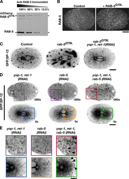
Activated RAB-5 potentiates the formation of mitotic ER clusters. (A) An anti–RAB-5 immunoblot of a serially diluted extract prepared from embryos stably expressing RAB-5Q78L fused to RFPmCherry. (B) Metaphase control (left) and RFP:RAB-5Q78L–expressing (right) embryos were fixed and stained with antibodies to RAB-5. Projections of deconvolved 3D datasets are shown. Bar, 10 μm. (C) Embryos expressing GFP:SP-12 alone (n = 18), GFP:SP-12 and RFP:RAB-5Q78L (n = 15), or GFP:SP-12 and RFP:RAB-5Q78L that were also depleted of YOP-1 and RET-1 by RNAi (n = 11) were imaged using spinning-disk confocal optics. Representative images of a central plane are shown. Bar, 10 μm. (D) Embryos expressing GFP:SP-12 that were depleted of YOP-1 and RET-1 (n = 13); RAB-5 (n = 21); or YOP-1, RET-1, and RAB-5 (n = 10) were imaged by spinning-disc confocal microscopy. Representative images of a single central section are shown. Bar, 10 μm. (E) Higher magnification (2×) views of a portion of the images in D. Arrowheads highlight aberrant ER loops present during interphase and mitosis in the triple-depleted embryos. Bar, 5 μm.


