| PMC full text: | Dev Dyn. Author manuscript; available in PMC 2008 Oct 5. Published in final edited form as: Dev Dyn. 2006 Mar; 235(3): 633–645. doi: 10.1002/dvdy.20672 |
Figure 6
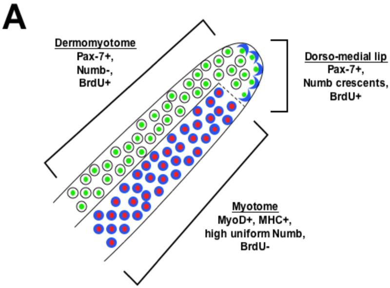
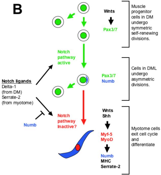
(A) Distribution of Numb protein reveals three somite compartments: Pax-7 expressing (green) dermomyotomal cells which lack Numb protein (blue), Pax-7 expressing cells of the dorso-medial lip of the dermomyotome in which Numb is localized asymmetrically in cortical crescents on the basal side of dividing cells, and MyoD/Myf-5 expressing (red) myotomal cells in which Numb protein accumulates uniformly. (B) A speculative model of some of the signals that modulate myogenesis in the somite. Wnt signals from the surface ectoderm lead to expression of Pax-3/7 in the dermomyotome (DM). Because cells in this compartment lack Numb protein, we speculate that they are sensitive to Notch signals and therefore undergo symmetric cell division. In the dorso-medial lip (DML) of the dermomyotome, localized Numb protein results in differential sensitivity to Notch ligands. We speculate that daughter cells that lack Numb remain in the dorso-medial lip of dermomyotome continue to proliferate, while those that contain Numb join the myotome and withdraw from the cell cycle. Muscle inducing cues from either within the somite (i.e., Wnt5a; (Linker et al., 2003)), the axial tissues (Wnts and Shh; (Munsterberg et al., 1995; Stern et al., 1995; Ikeya and Takada, 1998; Borycki et al., 1999)) or the surface ectoderm (Wnts; (Tajbakhsh et al., 1998)) induce expression of MyoD in Pax7-positive cells that contain a cortical crescent of Numb. The resultant differentiated myotomal cells contain uniformly high levels of Numb protein throughout their cytoplasm and secrete Serrate-2. We propose that Numb blocks the inhibitory effects of Notch signaling on muscle differentiation in the myotome, while Serrate-2 secreted by the myotome may promote the continued proliferation of dermomyotomal progenitors.
