| PMC full text: |
|
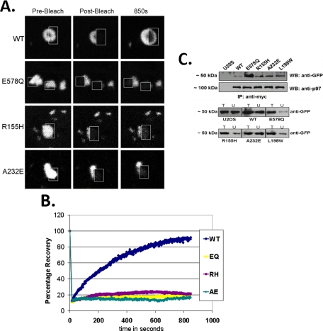
FIGURE 5.

IBMPFD mutants are “trapped” on aggregated polyglutamine. A, FRAP analysis of the molecular interaction between polyQ80-CFP and p97/VCP-DsReds (WT, E305Q/E578Q, R155H, or A232E) co-localizing inclusions. The relative fluorescence intensity was determined for each time point and is represented as the percentage of the prebleaching relative fluorescence intensity value. FRAP analysis was typically performed on >2 cells/field and >3 fields/experimental condition. Each experiment was repeated three times with different transfectants for each experiment. B, representative FRAP analysis images of co-localized p97/VCP-DsRed/polyQ80-CFP inclusions before bleaching, immediately after photobleaching and after recovery. Inclusions within cells were photobleached in a small region of interest (outlined box) and monitored for recovery of fluorescence into the region of interest. C, U20S control and U20S cells stably expressing tetracycline-inducible p97/VCP-WT-Myc, dominant negative p97/VCP-E578Q-Myc or IBMPFD mutant p97/VCP-R155H-Myc, -A232E-Myc, and -L198W-Myc were transfected with polyQ80-CFP. 36 h later lysates were collected, and p97/VCP was immunoprecipitated with an anti-Myc. The immunoprecipitate was subsequently immunoblotted for associated polyQ80-CFP with anti-GFP. Similar amounts of p97/VCP were immunoprecipitated. Total lysates (T) and unbound (U) fractions were separated by SDS-PAGE and immunoblotted with anti-GFP antibody. All of the blots are representative of three independent experiments.


