| PMC full text: | J Comp Neurol. Author manuscript; available in PMC 2010 Jan 15. Published in final edited form as: J Comp Neurol. 1995 May 1; 355(2): 296–315. doi: 10.1002/cne.903550208 |
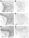
Fig. 5
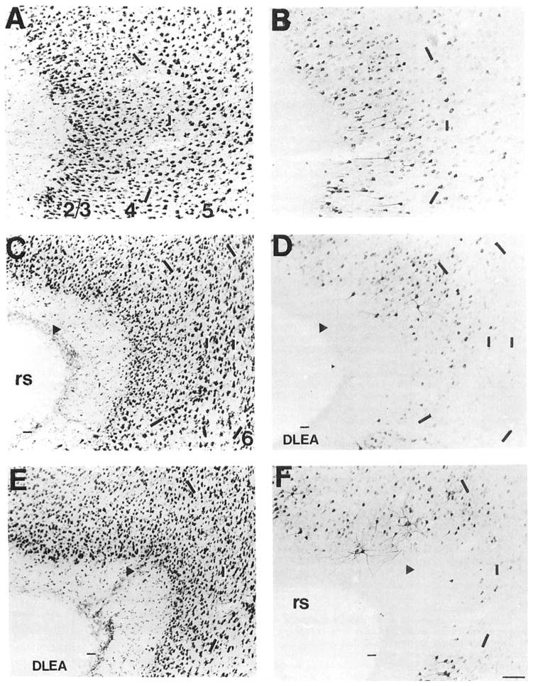
A series of brightfield photomicrographs demonstrate the laminar organization of COX 2-ir neurons in several levels of the perirhinal cortex (A,C,E). Photomicrographs are oriented in the normal transverse view. The arrowheads denote the deepest portion of the invagination of the rhinal sulcus, which we have taken as the division of the dorsal and ventral portions of this cortical field. Laminae are indicated in the adjacent Nissl-stained sections (B,D,F). A,B: Rostral perirhinal cortex, approximately at the level of Figure 19 of Zilles (1985). C,D: Intermediate perirhinal cortex, approximately at the level of Figure 23 of Zilles (1985). E,F: Caudal perirhinal cortex, approximately at the level of Figure 25 of Zilles (1985). Dashes in A, C, and E correspond to the lamina boundaries in B, D, and F. Scale bar = 100 μm.


