| PMC full text: | Published online 2011 Apr 14. doi: 10.1093/brain/awr063
|
Figure 8

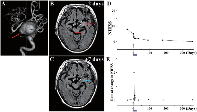
Case 8: (A) 3D-CT angiography showed the large aneurysm (red arrow) in the left internal carotid artery. (B) MRI 2 days before cell injection and (C) 7 days after cell injection. Red arrows in B indicate the infarcted lesions before cell injection, and blue arrow in C shows the reduced lesion volume and lower signal intensity after cell injection. (D) NIHSS scores for 1 year and (E) rate of change in NIHSS.


