| PMC full text: |
|
Figure 5
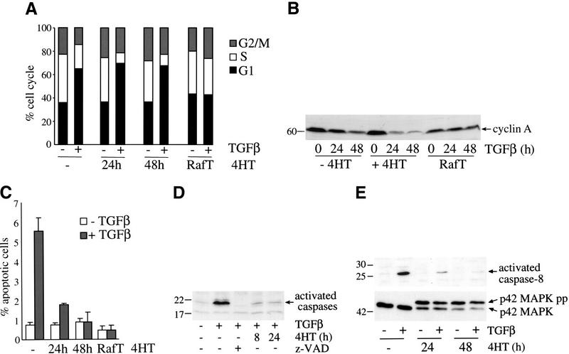
Short-term activation of Raf does not prevent TGFβ-induced cell cycle arrest but blocks TGFβ-induced apoptosis. (A) MDCK Raf–ER cells either untreated or treated with 100 nM 4HT for 24 or 48 h and MDCK Raf–ER cells transformed by long-term exposure to 4HT (RafT) were stimulated with TGFβ (7.5 ng/mL) for 24 h. The medium was changed to DMEM +2% FCS 24 h before TGFβ treatment. The cell cycle distribution was assayed by flow cytometry following propidium iodide staining. (B) MDCK Raf–ER cells either untreated or pretreated with 100 nM 4HT and MDCK Raf–ER cells transformed by long-term treatment with 4HT (RafT) were stimulated with 7.5 ng/mL TGFβ for the indicated times. Cells were cultured in DMEM +2% FCS for 24 h before TGFβ stimulation and assayed for cyclin A expression by Western blotting. (C) MDCK Raf–ER cells transformed by long-term exposure to 4HT (RafT) and MDCK Raf–ER cells either untreated or pretreated with 100 nM 4HT for the indicated periods of time were stimulated with 7.5 ng/mL TGFβ for 24 h. The percentage of apoptotic cells was determined by Hoechst staining. Data shown are the means ±
± standard deviations of three independent experiments performed in duplicate. (D) Effect of activated Raf on TGFβ-induced caspase activation. MDCK Raf–ER cells either untreated or pretreated with 100 nM 4HT for 8 h or 24 h were stimulated with 7.5 ng/mL TGFβ for 24 h. To inhibit caspase activation cells were pretreated with 100 μM z-VAD for 20 min. Cytosolic lysates were incubated with ZEK(bio)D-aomk peptide for 5 min at 37°C and assayed by Western blotting. (E) Activation of caspase-8 and p42MAPK was detected in MDCK Raf–ER cells either untreated or pretreated with 100 nM 4HT for 24 or 48 h. After stimulation with TGFβ (7.5 ng/mL) for 24 h, total cell lysates were analyzed by Western blotting.
