| PMC full text: |
|
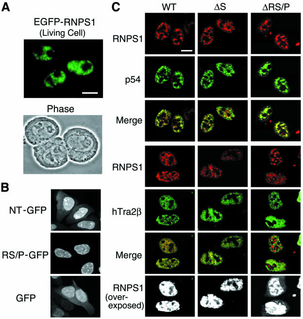
FIG. 3.

Subcellular localization of WT RNPS1 and deletion mutants, p54, and hTra2β. (A) EGFP-fused mouse RNPS1 was transiently transfected into living Ehrlich ascites tumor cells and analyzed by fluorescence microscopy (24 h after transfection). A phase contrast micrograph taken at the same time is shown below. (B) The GFP fusion constructs with either NT or RS/P domain (GFP alone as a control) were transiently transfected into HeLa cells. The cells were fixed and permeabilized at 12 h after transfection and analyzed by fluorescence microscopy. (C) The DsRed fusion proteins of human RNPS1 (WT) and two deletion mutants, RNPS1-ΔS and RNPS1-ΔRS/P, were transiently cotransfected into HeLa cells with FLAG-tagged p54 and FLAG-tagged hTra2β. The cells were fixed and permeabilized at 15 h after transfection, treated with an anti-FLAG antibody, and analyzed by confocal microscopy. Both images were also superimposed, and the yellow color indicates colocalization of the two proteins in nuclear speckles (Merge). Overexposed images (black and white) are also shown to highlight the cytoplasmic localization that is specifically observed in RNPS1-ΔRS/P protein. Bar, 10 μm.


