| PMC full text: | Cancer Cell. Author manuscript; available in PMC 2011 Dec 2. Published in final edited form as: Cancer Cell. 2011 Jan 18; 19(1): 17–30. doi: 10.1016/j.ccr.2010.12.014 |
Figure 2
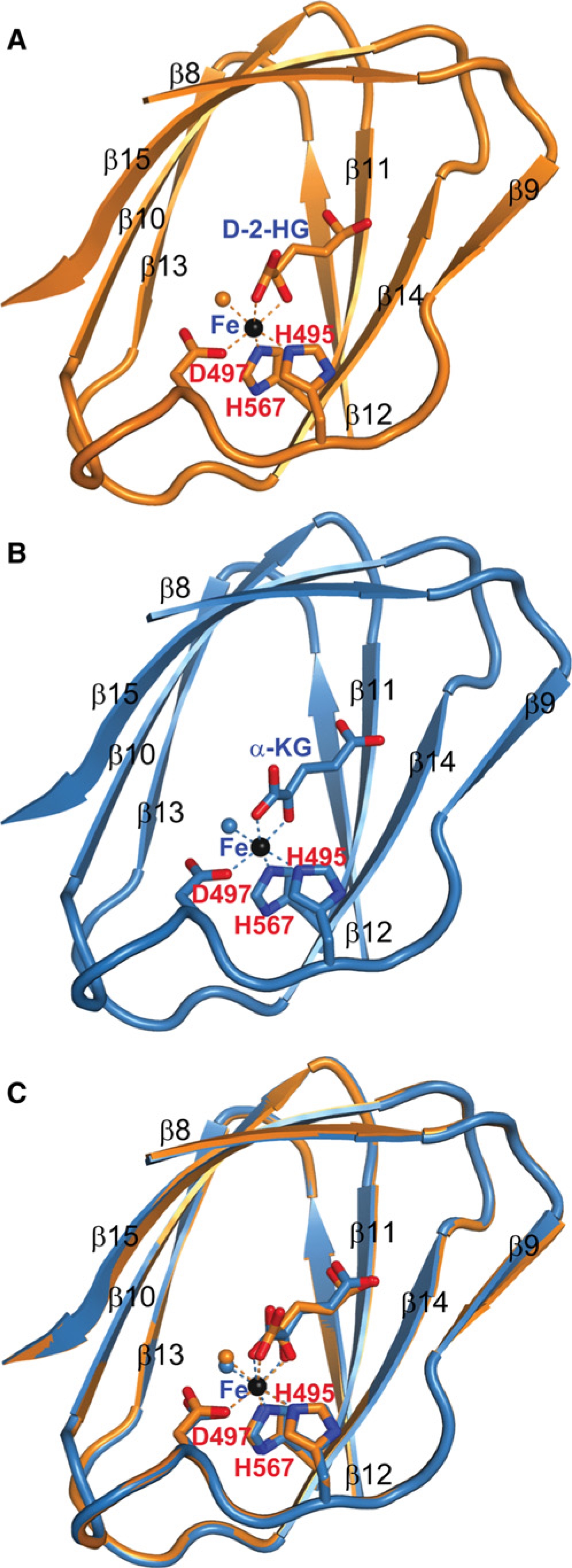
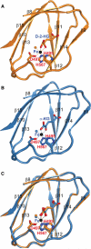
2-HG and α-KG Bind to the Same Site in Histone Demethylases
(A) The structure of D-2-HG bound to CeKDM7A JmjC domain. D-2-HG and CeKDM7A are shown in stick and cartoon representation, respectively. Secondary structural elements of CeKDM7A are indicated. Fe (II) is colored in black, Fe (II) coordination is represented by dotted lines and water molecule is shown as orange ball.
(B) The structure of α-KG bound to CeKDM7A JmjC domain, illustrated as in (A).
(C) Superimposition of structures shown in (A) and (B). See also Figure S2.
