| PMC full text: | Nat Immunol. Author manuscript; available in PMC 2012 Aug 1. Published in final edited form as:
|
Figure 1

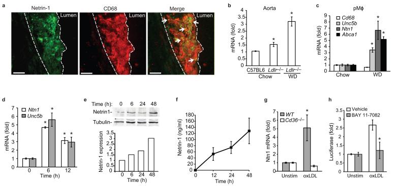
(a) Immunofluorescent staining of netrin-1 (green) and CD68 (red), and their colocalization (yellow, arrows), in aortic sinus atherosclerotic plaques of Ldlr−/− mice fed a WD. Dashed line indicates lesion border (scale bar= 50 μm). Staining is representative of plaques from 4 mice. (b) qPCR analysis of Ntn1 mRNA isolated from the aortic arch of C57BL/6 or Ldlr−/− mice fed a chow or WD. (c) qPCR analysis of Ntn1, Unc5b, Cd68 and Abca1 mRNA in pMø isolated from Ldlr−/− mice fed a chow or WD. (d) qPCR analysis of Ntn1 and Unc5b in pMø treated with 50 μg/ml oxLDL, and corresponding expression of netrin-1 protein measured in (e) cell lysates by immunoblot or (f) conditioned media by ELISA. (g) qPCR analysis of Ntn1 mRNA in wild-type (WT) or Cd36−/− pMø stimulated with 50 μg/ml oxLDL for 6 h. (h) Ntn1 promoter-luciferase reporter activity in HEK293 cells treated with oxLDL in the presence or absence of the NF-κB inhibitor BAY 11-7082 (20 μM). (b-h) Data are mean ± s.d. of triplicate samples in a single experiment and are representative of 3 independent experiments. *P<0.05.


