Abstract
Background
Accurate reduction of rotational displacement for transverse or comminute fracture of humeral shaft fracture is difficult during operation. The purpose of this study was to evaluate the reliability of the bicipital groove as a point of reference for the prediction of the rotational state of the humerus on two dimensional images of C-arm image intensifier during operation for humeral shaft fractures.Materials and methods
One hundred subjects, 62 male, 38 female, aged 22-53 years were recruited contralateral bicipital groove on the 45 degrees externally rotational standard anterior-posterior view recorded before surgery. Three observers, watched only contour of bicipital groove in monitor of C-arm image intensification with naked eye without looking at the subject and predicted rotational state of the humerus by comparing the contour of the opposite side of bicipital groove. The angle of discrepancy from real rotational position was then assessed.Results
The mean (SD), angular discrepancy between the neutral point and the predicted angle was 3.4°(±2.7°). A value within 5° was present in 72% of cases. All observations were within 15°. There was no interobserver variation (P = 0.47). The intraclass correlation coefficient (ICC) was 0.847.Conclusion
Contour of the bicipital groove on simple radiograph was a useful landmark. Comparing the contour of the bicipital groove in the 45 degrees externally rotational standard view bilaterally, was an effective method for reduction of rotational displacement of the humerus.Free full text

Prediction of the rotational state of the humerus by comparing the contour of the contralateral bicipital groove: Method for intraoperative evaluation
Abstract
Background:
Accurate reduction of rotational displacement for transverse or comminute fracture of humeral shaft fracture is difficult during operation. The purpose of this study was to evaluate the reliability of the bicipital groove as a point of reference for the prediction of the rotational state of the humerus on two dimensional images of C-arm image intensifier during operation for humeral shaft fractures.
Materials and Methods:
One hundred subjects, 62 male, 38 female, aged 22-53 years were recruited contralateral bicipital groove on the 45 degrees externally rotational standard anterior-posterior view recorded before surgery. Three observers, watched only contour of bicipital groove in monitor of C-arm image intensification with naked eye without looking at the subject and predicted rotational state of the humerus by comparing the contour of the opposite side of bicipital groove. The angle of discrepancy from real rotational position was then assessed.
Results:
The mean (SD), angular discrepancy between the neutral point and the predicted angle was 3.4°(±2.7°). A value within 5° was present in 72% of cases. All observations were within 15°. There was no interobserver variation (P = 0.47). The intraclass correlation coefficient (ICC) was 0.847.
Conclusion:
Contour of the bicipital groove on simple radiograph was a useful landmark. Comparing the contour of the bicipital groove in the 45 degrees externally rotational standard view bilaterally, was an effective method for reduction of rotational displacement of the humerus.
INTRODUCTION
Orthopaedic surgeons may find it difficult to get rotationally anatomical reduction of a shaft fracture of the humerus. Li et al.1 reported 27.2% of malrotation of 20° or more after intramedullary nailing. If rotational control is not achieved, nonunion is more likely to occur.2 In addition, malrotation of the humeral component of total elbow replacement influences laxity and causes maltracking.3 The rotational deformity of the distal humerus may limit the motion of the ipsilateral shoulder joint.4 Few technical tip or study have introduced to prevent malrotation of the humerus. The authors tried to find a simple landmark for prediction of rotational status during the operation. The anatomical structure of the bicipital groove varies among individuals. However, the position and shape are similar when the left and the right bicipital groove structures are compared in each individual.5–7 The objective of this study was to evaluate the reliability of the bicipital groove as a point of reference for the prediction of the rotational state of the humerus, by comparing the contralateral bicipital groove images on standard radiographs.
MATERIALS AND METHODS
One hundred volunteers were included in this study. The mean age of the 62 men was 34 ± 7.5 years (range, 24-53), and the mean age of the 38 women was 33 ± 6.8 years (range, 22-48). Subject who had a history of shoulder disease or fracture of the humerus were excluded. All volunteers gave informed consent about the radiation. The college and hospital institutional review board approved the protocols of this study.
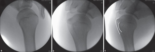
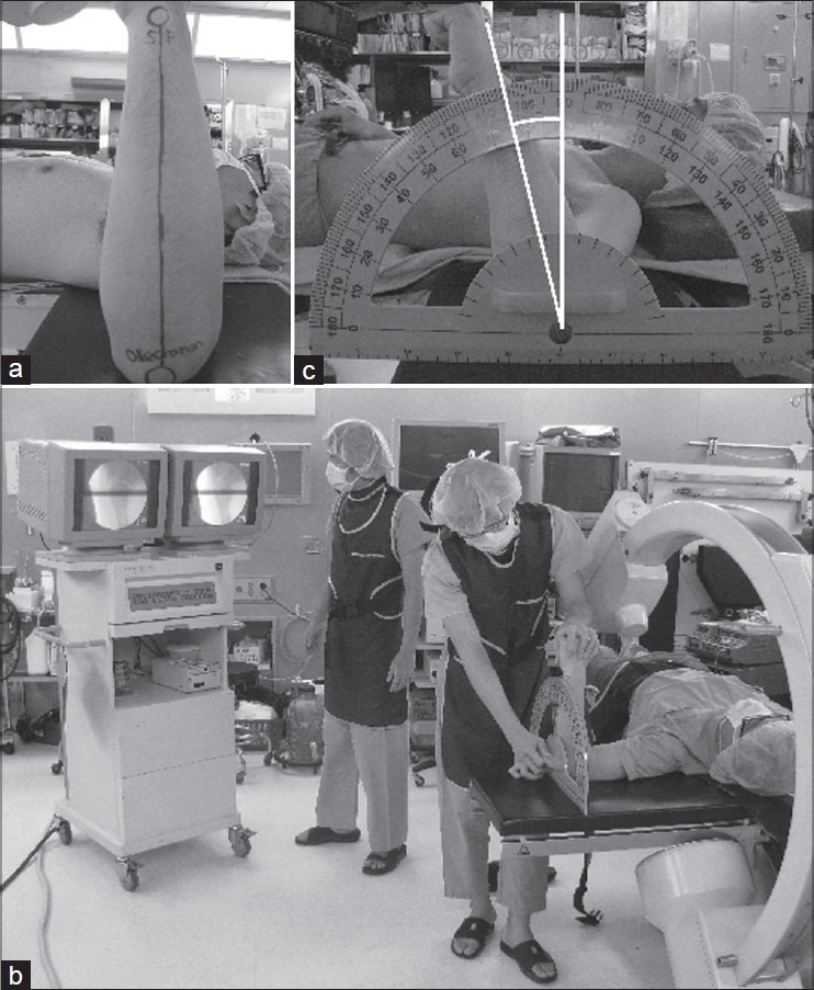
First, the subject was placed in the supine position with the shoulder abducted to 90°; the elbow flexion was to the same at 90° when the forearm was pronated fully. The images were obtained in the cephalic view at a 45° angle, a point with a clear outline of the lesser and greater tuberosity [Figure 1]. Images of the bicipital groove, at 45° of external rotation, were used in this study [Figure 2]. The angular orientation of the bicipital groove has been referenced to the transepicondylar axis at about 55° in prior studies.8,9 Then, standard line were drawn on the medial border of ulna from the tip of olecranon to the styloid process of the forearm [Figure 3a]. At the neutral rotation point, the shape of the proximal humerus was recorded and transferred to the right monitor of a C-arm image intensifier (OEC series 9800; OEC Medical Systems, Salt Lake City, Utah, USA) as a reference point for the rotational state. The contralateral (left) arm was taken position in the same posture. The subject arm was in a random position, rotationally, with the shoulder abduction at 90°. The observer group was composed of one expert surgeon (A), one orthopaedic resident (B), and one medical student (C). All three observers stood in front of the monitor, where they could not see the individual. They compared the images of the bilateral bicipital groove on the monitors with regard to the contour and proximal and distal width of the bicipital groove, distance of the interval from the lateral cortex of the proximal humerus to the lesser tuberosity, with the naked eyes [Figure 2c]. An assistant rotated the arm being examined inward or outward, and the point where the subjective image of the arm showed a similar and symmetrical shape to the image of the bicipital groove, previously recorded on the right side of the monitor [Figure 3b], was noted. The angular divergence was measured from the neutral point [Figure 3c]. The distribution and mean angular discrepancy were calculated using the values obtained from the three observers. The data were not normally distributed and therefore were logarithmically transformed. The Interobserver variation was evaluated statistically using the analysis of variance. The interobserver reliability was evaluated by calculating the Intraclass Correlation Coefficient (ICC) using PASW 17.0 statistical software (SPSS Inc, Chicago, IL, USA) for windows (Microsoft, Redmond, WA, USA).

Preparing the image of the contralateral shoulder. (The individual is in the supine position with the shoulder abducted at 90° (a), with a fully pronated forearm (b), and the image intensifier is at 45° in the cephalad view (c))

Fluoroscopic images of the proximal humerus. (Internal rotation at 45° (a), Neutral (b), External rotation at 45° which the line of the greater and lesser tuberosity make clear contour of the bicipital groove on radiograph in 45 degree rotated externally (c))

Comparing the subjective shoulder with the contralateral shoulder. (Lines are drawn on the medial border of the ulna from the tip of the olecranon to the styloid process on the subjective arm (a). An assistant rotate the subjective arm with random rotation being examined inward or outward until an observer select the similar image with the contralateral one (b) and measure the angular difference from the neutral point (c))
RESULTS
All observations which were expressed as an absolute value were placed within 15° of the neutral point. The discrepancy for the mean angular measurements were from -4° to +4° in 72% of the assessments for each observer and 99% of the assessments were within 10° [Table 1]. The total mean angular discrepancy was 3.4° (±2.7°). Those of each observer was 3.6° (±3.0°) in observer A, 3.2° (±2.3°) in observer B and 3.4° (±2.7°) in observer C. These differences were not statistically significant based on analysis of variance (ANOVA) (P = 0.495) [Table 2]. In addition, the intraclass correlation coefficient (ICC) was calculated to evaluate the interobserver reliability. The ICC value was calculated to be 0.847, which indicates a high interobserver reliability (>0.70). There was no interobserver variation (P = 0.47).
Table 1
Discrepancy in the angular distribution by the observers

Table 2
Measured angular differences

DISCUSSION
The bicipital groove of the proximal humerus has been extensively studied and its anatomy well documented.8,10,11 Some investigators have used the bicipital groove as a reference point with computed tomography.9,12 The relationship of the bicipital groove with humeral retroversion in cadavers was studied.13,14 Several investigators have suggested that the position of the bicipital groove relative to the humeral head is comparable.13–15 However, most investigations of the bicipital groove focused on retroversion of the humerus for the prevention of malrotation, which was suggested as a cause of hemiarthroplasty failure.15 The purpose was to evaluate the reliability of the bicipital groove as a point of reference for the prediction of the rotational state of the humerus on two-dimensional radiograph of C-arm image intensifier during operation especially for humeral shaft fractures.
This study assumed that the anatomical structures of the bicipital grooves were the same on both the right and left sides. Robertson et al.16 found that paired humeri have similar anthropometric features. DeLude et al.6 and Hernigou et al.7 reported no meaningful difference in comparisons of the left side and right side. Boileau et al.5 suggested that assessment of the rotational state of the humerus may be most accurately achieved based on the contralateral bicipital groove. Cassagnaud et al.,17 by contrast, reported a considerable difference between right and left side measurements.
At a rotation of 45° externally, the humeral head was rotated until the base of the head was perpendicular to the axis of the proximal humerus.15 In addition, the greater tuberosity was brought into relative clear prominence.18 The 45° externally rotated view showed relatively clear outlines of the contours of the bicipital groove that could be easily compared to the contralateral side with the naked eye. There is a relatively wide range of variation in the bicipital groove angle from 5 to 97, with a mean value of 55.5°.9 In cadavers, the measured angle of the bicipital groove was about 55.8°. Hempfing et al.15 suggested that the centre of the bicipital groove differs from the epicondylar axis by about -10°. This means that the epicondyles of the distal humerus are approximately 45° in relation to the internal rotation. Thus, this position could show a relatively neutral rotational state of the humerus, hypothetically. In this study, the C-arm image intensifier was at a 45° in the cephalad view, instead of rotating the arm. It was difficult for the assistant to rotate the shoulder of an individual more externally.
The purpose of this study was to determine an accurate and simple method that could be applied in the operating room setting. Similar to Evans and Wales19 evaluation of the rotational state, using the radial tubercle as a land mark or as Kim et al.20 used the lesser trochanter of the femur as a landmark for the correction of the rotational status during surgery for a fracture of the shaft of the femur. To the best of our knowledge, this is the first study to consider the bicipital groove as a useful landmark for estimating the rotational state of the humeral shaft fracture in the operating room setting. This methodology also could benefit for operating the corrective osteotomy and malunion of humeral shaft fractures. Li et al.1 found that the degree of malrotation correlated with a decreased range of motion in patents who underwent intramedullary nailing. Rotational deformities were took place according to patient's position during surgery.21
In the operating room, the procedures were performed by comparison of the injured side with the asymptomatic, contralateral side that was evaluated before surgery using the C-arm image intensifier. First, a 90° degree abduction and 45° external rotation antero posterior view of standard radiographs of the shoulder was performed. The shape of the bicipital groove, of the contralateral side, was then stored preoperatively in the C-arm image intensifier's second monitor. Before inserting the device, the proximal portion was rotated while the elbow was extended fully and the forearm pronated exactly until the shape of the greater tuberosity and interval of the bicipital groove was identical on both sides. This might be an effective method for successful surgical treatment of humeral shaft fractures with plate osteosynthesis or intramedullary nailing. This can be performed before the insertion of the locking screws, the surgeon controls the rotation of the proximal fragment.
There are limitations in our study. The two dimensional radiograph of C-arm image intensifier obtained was less precise than a three dimensional image and the comparison of the contour of the bilateral bicipital groove with the naked eyes of the observers was not objective. However, our methodology is cost effective, give less radiation exposure than the preoperative CT evaluation. This technique could use simply at the operating field. There was also no meaningful difference between the three observers. The study does not include clinical outcome. Further observation of clinical outcome and confirmation of these findings will be required in a larger study.
CONCLUSION
A simple radiological contour of the bicipital groove is a useful landmark. Comparing the shape of the bicipital groove in the 45 degrees externally rotated standard view bilaterally, was an effective method for estimating the rotational state of the humerus, intraoperatively.
ACKNOWLEDGMENT
The author Eugene Kim and Shinsuk Park were supported by the National Research Foundation grant funded by the Korea government (MEST) (2010-0027294). The authors thank to Jang Hwan Kim, M.D. and Miyeon Lee, Medical information library, Kangbuk Sung Hospital for their statistical assistance of the work.
Footnotes
Source of Support: The author Eugene Kim and Shinsuk Park were supported by the National Research Foundation grant funded by the Korea government (MEST) (2010-0027294)
Conflict of Interest: None.
REFERENCES
Articles from Indian Journal of Orthopaedics are provided here courtesy of Indian Orthopaedic Association
Full text links
Read article at publisher's site: https://doi.org/10.4103/0019-5413.104210
Read article for free, from open access legal sources, via Unpaywall:
https://www.ncbi.nlm.nih.gov/pmc/articles/PMC3543886
Citations & impact
Impact metrics
Alternative metrics

Discover the attention surrounding your research
https://www.altmetric.com/details/113485770
Article citations
The greater tuberosity version angle: a novel method of acquiring humeral alignment during intramedullary nailing.
Bone Jt Open, 5(10):929-936, 22 Oct 2024
Cited by: 0 articles | PMID: 39433305 | PMCID: PMC11493473
Geometrical analysis for assessing torsional alignment of humerus.
BMC Musculoskelet Disord, 21(1):92, 10 Feb 2020
Cited by: 4 articles | PMID: 32041587 | PMCID: PMC7011366
Similar Articles
To arrive at the top five similar articles we use a word-weighted algorithm to compare words from the Title and Abstract of each citation.
Analysis of the bicipital groove as a landmark for humeral head replacement.
J Shoulder Elbow Surg, 11(4):322-326, 01 Jul 2002
Cited by: 15 articles | PMID: 12195248
The bicipital groove as a landmark for humeral version reference during shoulder arthroplasty: a computed tomography study of normal humeral rotation.
J Shoulder Elbow Surg, 30(10):e613-e620, 03 Mar 2021
Cited by: 1 article | PMID: 33675970
A Novel Method for the Approximation of Humeral Head Retrotorsion Based on Three-Dimensional Registration of the Bicipital Groove.
J Bone Joint Surg Am, 100(15):e101, 01 Aug 2018
Cited by: 5 articles | PMID: 30063597
Lesser tuberosity is more reliable than bicipital groove when determining orientation of humeral head in primary shoulder arthroplasty.
Surg Radiol Anat, 32(1):31-37, 20 Aug 2009
Cited by: 4 articles | PMID: 19693428


