| PMC full text: | Immunity. Author manuscript; available in PMC 2014 Sep 19. Published in final edited form as: Immunity. 2013 Sep 19; 39(3): 10.1016/j.immuni.2013.08.007. Published online 2013 Sep 5. doi: 10.1016/j.immuni.2013.08.007 |
Figure 7

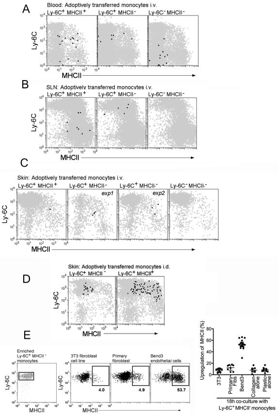
CD115+GFP+ blood monocyte subsets Ly-6C+MHCII+, Ly-6C+MHCII−, and Ly-6C−MHCII− were sorted from CD45.2 Cx3cr1gfp/gfp mice. Sorted monocytes 1×10⁶ Ly-6C+MHCII−, 3×10⁵ Ly-6C+MHCII+ or 1×10⁶ Ly-6C−MHCII− were injected i.v. into CD45.1+ WT mice, followed by i.d. injection of 2 μg LPS. At 18 h, (A) blood, (B) skin-draining LNs (SLN), and (C) skin were analyzed for monocytes migration and differentiation. Dot plot overlays display total CD11b+ cells (gray) and CD45.2 GFP+ adoptively transferred cells (black). C) Skin illustrates two separate experiments (exp 1, exp 2) adoptively transferring Ly-6C+MHCII− monocytes. D) Purified Ly-6C+MHCII− and Ly-6C+MHCII+ blood monocytes were injected intradermally into skin and recovered 18 h later to assess MHC II expression. E) Splenic Ly-6C+MHCII− monocytes, depleted completely of MHC II+ cells, were enriched by negative selection and cocultured with various cell types as shown. Ly-6C+ monocytes were gated before and after the cocultures and the induction of surface MHC II was assessed and plotted (on the right). Each data point represents one experiment.


