| PMC full text: | J Comp Neurol. Author manuscript; available in PMC 2013 Dec 9. Published in final edited form as: J Comp Neurol. 2012 May 1; 520(7): 10.1002/cne.22806. doi: 10.1002/cne.22806 |
Figure 3
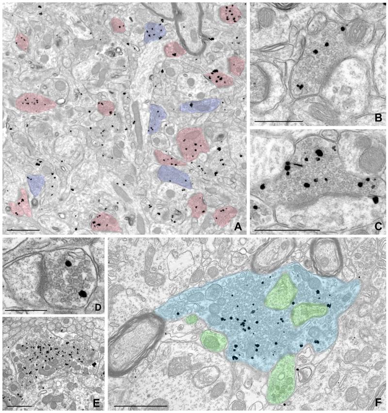
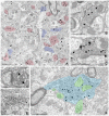
Pre-embedding immunogold labeling for NDCBE in forebrain. A: Low-magnification electron micrograph from cerebral cortex (layer II/III); labeling for NDCBE concentrates in presynaptic terminals. Terminals with prominent postsynaptic densities are colorized in pink; terminals lacking prominent PSDs and apposed to dendritic shafts are colorized in blue. B,C: Higher magnification views of neocortex, showing labeled presynaptic terminals that make axospinous synaptic contacts. D-F: NBCDE labeling in hippocampus. D: Small terminal in stratum radiatum of CA1. E,F: Large, probable mossy fiber terminals in stratum lucidum of CA3. Terminal in F is colorized blue, and postsynaptic elements are colorized green. Note that labeling is associated with the pool of synaptic vesicles, and is excluded from vesicle-poor zones of the terminal. Scale bar = 1 μm in A,E,F; 0.5 μm in B,C; 250 nm in D.


