| PMC full text: | J Immunol. Author manuscript; available in PMC 2015 Apr 1. Published in final edited form as: J Immunol. 2014 Apr 1; 192(7): 3029–3042. Published online 2014 Mar 3. doi: 10.4049/jimmunol.1302911 |
FIGURE 6

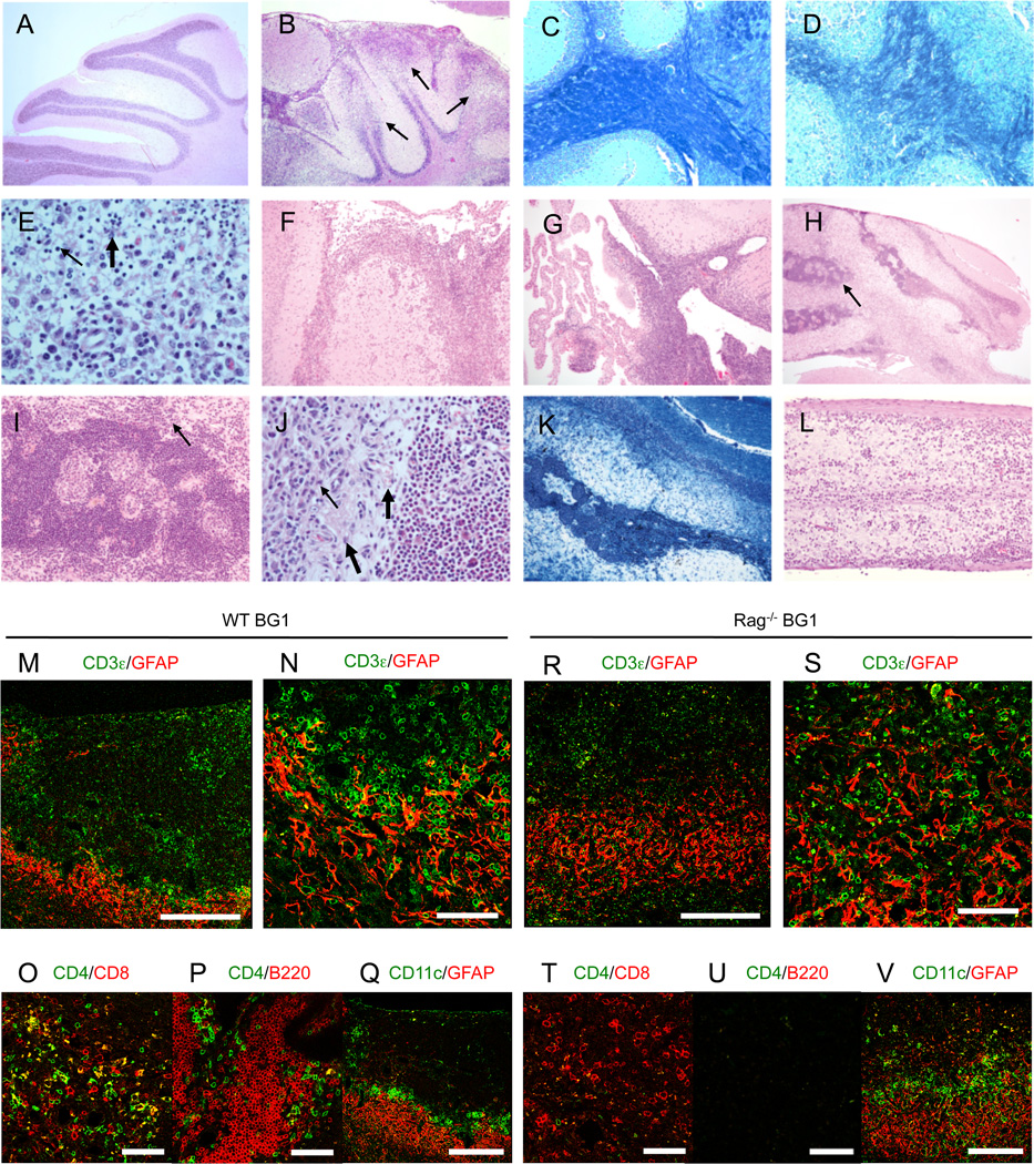
BG1 mice develop lesions within the gray and white matter of the brain and spinal cord. (A) H&E stain of cerebellum of a healthy C57BL/6 mouse. (B) Earlier phases of disease in BG1 mice are characterized by involvement of broad areas of cerebellar meninges and gray and white matter with necrosis and apoptosis accompanied by a dense glial and inflammatory response (thin arrows). (C) Luxol Fast Blue stain of healthy and (D) diseased area of the cerebellum of a BG1 mouse shows that the inflammatory response is intermixed with necrotic and apoptotic cells and results in loss of myelin (loss of dark blue stain) along with other elements of the neuropil. (E) CNS lesions show focal extensive regions of necrosis and apoptosis accompanied by mixed inflammatory cell populations. The inflammatory cellular component can vary within the site but is typically composed of mixtures of neutrophils (thick arrow), lymphocytes (thin arrow) and microglial elements. At this stage it is common to find extension of the inflammatory response from (F) colliculus meninges and (G) choroid plexus into adjacent gray matter nervous tissue. In late stages of disease, (H, I, J) the cerebellum and other affected sites become distinguished by the presence of dense accumulations of lymphocytes (H, thin arrows) and the replacement of nervous tissue elements by a vascular rich stroma (thick arrows) containing scattered inflammatory cells (thin arrows in I, J). (K) The extensive loss of myelin within the cerebellum is visualized by a lack of Luxol Fast Blue stain. (L) Lesions of the spinal cord are less dramatic but consistent with the pattern where meningeal and perivascular accumulations of predominately lymphocytes extend into the adjacent neuropil. Images are representative examples of six WT BG1 and Rag−/− BG1 mice with early onset or late spontaneous CNS disease. (M–Q) Diseased WT BG1 and (R–V) Rag−/− BG1 mice develop T cell accumulations surrounded and intermixed with GFAP+ astrocytes. Lymphocyte aggregates in WT BG1 Tg mice contain (M) CD8, CD4 T cells and (P) B cells, and are (Q) ringed with CD11c+ cells. The yellow stained cells in (O) are not T cells, as no CD4+ CD8+ double positive T cells are observed in BG1 mice, and likely represents either non-specific binding or staining of cellular debris (R–V) Lesions in diseased Rag−/− BG1 mice contain (R–U) CD8 T cells, without CD4 T cells or B cells. Lesions are ringed with (V) CD11c+ and GFAP+ astrocytes. Images are representative of three WT BG1 and four Rag−/− BG1 mice analyzed. Scale bar; 250µm on A, E, F and J, 60µm on the others.


