| PMC full text: | Published online 2015 Jun 8. doi: 10.2147/DDDT.S82724
|
Figure 7
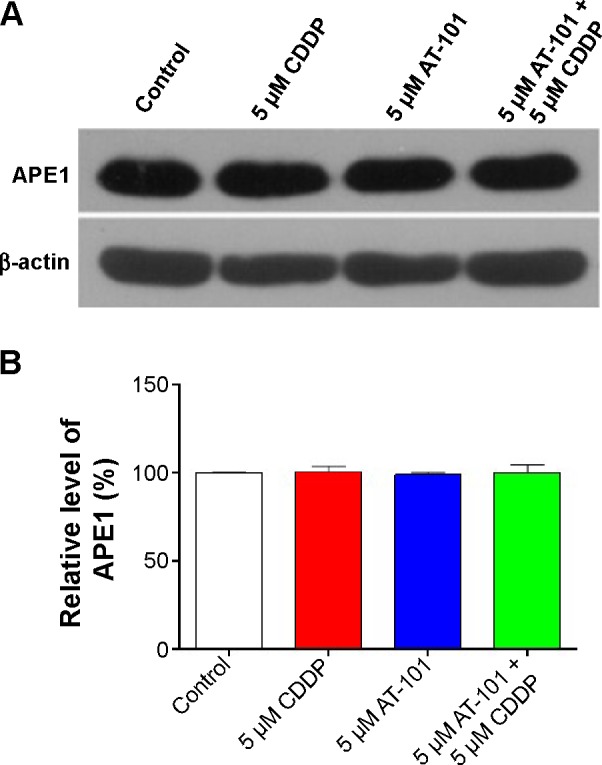
AT-101 plus CDDP does not affect expression levels of APE1/Ref-1 in A549 cells determined by Western blotting assay.
Notes: (A) Representative gel blots showing expression levels of APE1/Ref-1 in A549 cells treated with vehicle control, 5 μM CDDP, 5 μM AT-101, or 5 μM AT-101 plus 5 μM CDDP. (B) Bar graph showing the relative level of APE1/Ref-1 in A549 cells treated with vehicle control, 5 μM CDDP, 5 μM AT-101, or 5 μM AT-101 plus 5 μM CDDP. An equal amount of protein samples was separated by sodium dodecyl sulfate polyacrylamide gel electrophoresis and then transferred onto a polyvinylidene difluoride membrane. APE1/Ref-1 was probed using the primary antibody and visualized using the enhanced chemiluminescence. β-actin was used as the internal control for blot densitometric normalization. Data are the mean ± standard deviation of three independent experiments and analyzed by one-way analysis of variance.
Abbreviations: APE1, apurinic/apyrimidinic endonuclease 1; CDDP, cisplatin; Ref-1, redox effector factor 1.
