| PMC full text: | Published online 2017 Oct 30. doi: 10.1038/s41598-017-14696-z
|
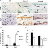
Figure 7

Assessment of leukocyte infiltration in the AVF outflow veins. (A) Staining for CD45 (leukocytes) from outflow vein of controls (C, first column) or with (S, second column) CorMatrix wrap. CD45-positive cells have cytoplasmic red staining. Images were captured at 40× magnification and the scale bar is 50 µm. (B) Quantitative analysis for CD45 staining in the outflow vein is demonstrated. There is a significant reduction in CD45 staining in Group S compared to Group C at day 7 (P
 <
< 0.05) and day 21 (P
 <
< 0.05). Each bar represents mean
 ±
± SEM (n
 =
= 6 per group). *
P
 <
< 0.05.
0.05.


