| PMC full text: | Published online 2017 Sep 8. doi: 10.1182/bloodadvances.2017007021
|
Figure 1.
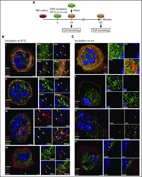
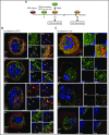
MKs endocytose OVA. As described in the timeline (A), MKs were pulsed with fluorescent OVA for 15 minutes at 37°C (B) or on ice (C) and the cells were either fixed immediately or incubated for an additional 60 minutes. After pulse, OVA was tested for colocalization with the endosomal marker early endosome antigen 1 (EEA1) (I), lysosomal marker lysosome-associated membrane glycoprotein 1 (LAMP1) (II), and multivesicular body marker CD63 (III), and, after 60 minutes, with α-granule marker CD62P (IV). The colocalization (Coloc.) channel was reconstructed in silico based on the colocalizing voxels (3-dimensional pixels) of OVA and each marker and summarizes the colocalization in each condition. Yellow arrows highlight some of the colocalizations in each panel. (Zeiss LSM 700 confocal microscope, oil-immersion objective 63×; whole cell scale bar = 10 µm, region of interest scale bar = 2 µm.)

