| PMC full text: |
|
Figure 1.
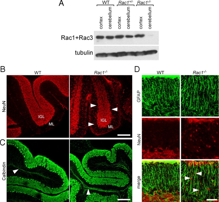
Loss of Rac1 from the nervous system results in defects in the migration of CGNs. A, Depletion of Rac1 from Rac1−/− cerebellum. P9 cerebellar and cortical protein extracts were prepared from wild-type, Rac1+/−, and Rac1−/− brains. Equal amounts of protein were subjected to Western blot analysis using an antibody recognizing Rac1 and cross-reacting with Rac3. The signal detected in cortical extracts of Rac1−/− brains represents Rac3, which is absent from cerebellar extracts. B, Ablation of Rac1 causes aberrant distribution of NeuN-positive neurons. Sagittal sections from P18 cerebella were immunostained with NeuN. Many CGNs stacked in the ML of the Rac1-null cerebellum can be observed (arrowheads). In contrast, in the wild type, the majority of CGNs have reached the IGL. Scale bar, 150 μm. C, Sagittal sections from P18 cerebella immunostained with calbindin (axonal tracts of Purkinje neurons, arrowheads). The Rac1-null cerebellum shows the typical monolayer of Purkinje neurons. Scale bar, 200 μm. D, Sagittal sections from P18 cerebella coimmunostained with NeuN and GFAP. The Rac1−/− cerebellum exhibits a normal organization of GFAP-positive Bergmann glia cells that are in proximity to NeuN-positive CGNs (arrowheads). Scale bar, 40 μm.

