| PMC full text: | Published online 2021 Feb 18. doi: 10.1002/advs.202004635
|
Figure 1
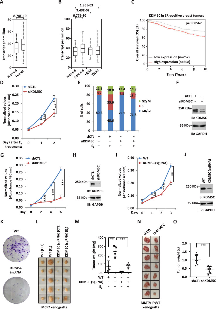
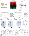
KDM5C is required for ERα‐positive breast cancer cell growth and tumorigenesis. A) The expression of KDM5C in a cohort of normal (n = 114) and clinical breast cancer (n = 1097) samples from TCGA (The Cancer Genome Atlas). B) The expression of KDM5C in different subtypes of clinical breast cancer samples from TCGA. (Normal, n = 114; Luminal, n = 566; HER2, n = 37; TNBC (triple‐negative breast cancer), n = 116). C) Kaplan–Meier survival analyses for OS (overall survival) of ER‐positive breast cancer patients using KDM5C as input (n = 560). D) MCF7 cells were transfected with control siRNA (siCTL) or siRNA specific against KDM5C (siKDM5C) in stripping medium for three days, and then treated with or without estrogen (E2, 10−7 m) for different duration as indicated followed by cell proliferation assay (± SD, ** p < 0.01). E) MCF7 cells were transfected with siCTL or siKDM5C in stripping medium for three days, and then treated with or without estrogen (E2, 10−7 m) for 24 h followed by FACS analysis. F) MCF7 cells transfected with siCTL or siKDM5C were subjected to immunoblotting (IB) using antibodies as indicated. G) MCF7 cells were infected with control shRNA (shCTL) or shRNA specific against KDM5C (shKDM5C) lenti‐virus for duration as indicated followed by cell proliferation assay (± SD, *** p < 0.001). H) MCF7 cells infected with shCTL or shKDM5C were subjected to immunoblotting (IB) using antibodies as indicated. I,K) Wild type (WT) and KDM5C knockdown (KDM5C (sgRNA)) MCF7 cells generated by CRISPR/Cas9 were subjected to cell proliferation assay (I) and colony formation assay (K) (± SD, ** p < 0.01, *** p < 0.001). J) WT and KDM5C (sgRNA) MCF7 cells were subjected to immunoblotting using antibodies as indicated. L) Xenograft experiments were performed by injecting WT and KDM5C (sgRNA) MCF7 cells subcutaneously into female BALB/C nude mice (5 mice per group) and then treated with or without estrogen (E2). Tumors were then excised, photographed, and weighted four weeks after subcutaneous injection. M) Weight of tumors as shown in (L) (± SD, ** p < 0.01, *** p < 0.001). N) Xenograft experiments were performed by injecting shRNA or shKDM5C lenti‐virus‐infected MMTV‐PyVT cells into female FVB mice (7 mice per group). Tumors were then excised, photographed, and weighted 23 days after subcutaneous injection. O) Weight of tumors as shown in (N) (± SD, *** p < 0.001).

