| PMC full text: | Published online 2021 Apr 12. doi: 10.1093/brain/awab153
|
Figure 2

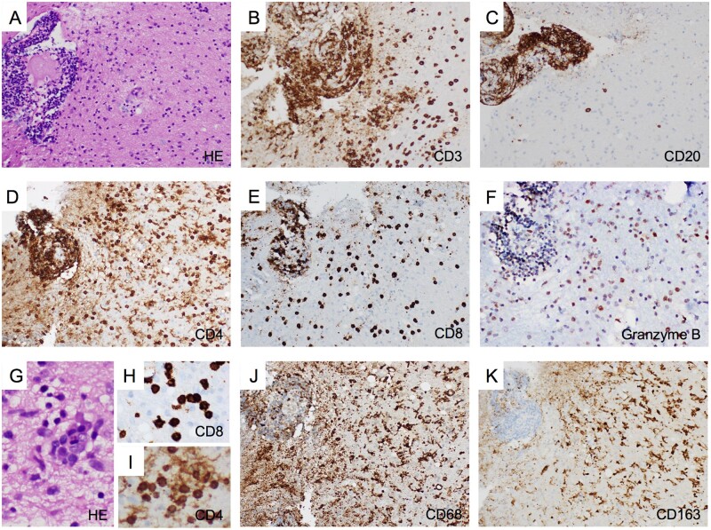
Histopathological characteristics of anti-AK5 limbic encephalitis. Right uncal biopsy from Patient 10 showing intense perivascular and parenchymal inflammatory infiltrates (A, haematoxylin and eosin staining, ×200), which was dominated by T cells that were abundant in both the perivascular space and the parenchyma (B, CD3 immunostaining, ×200). Conversely, B cells, though highly present in the perivascular space, were scattered in the parenchyma (C, CD20 immunostaining, ×200). The T-cell infiltrate included both CD4+ (D, ×200) and CD8+ T cells (E, ×200), which were especially abundant in the perivascular space but also present diffusely in the parenchyma. Cytotoxic CD8+ T cells expressing granzyme B (a protein stored in the granules of cytotoxic cells that induces apoptosis) were detected in perivascular and parenchymal infiltrates (F, ×200). Nodular clusters of inflammatory cells were detected in the parenchyma using haematoxylin and eosin staining (G, ×400) and CD8+ and CD4+ T-cell clusters were also observed (H and I, CD8 and CD4 immunostainings, respectively, ×400). Additionally, macrophages/microglia diffusely infiltrated the tissue, as shown by CD68 (J, ×100) and CD163 (K, ×100) immunostainings, the latter being associated with M2 differentiation related to an anti-inflammatory response including clearance of apoptotic bodies.


