| PMC full text: | Published online 2022 Apr 16. doi: 10.4103/joco.joco_93_21
|
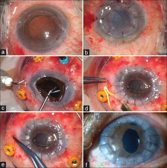
Figure 2

Intraoperative photographs. (a) Right eye. Transparent cornea trephination. (b) Opaque cornea transplantation. (c) Left eye. Angled sclerotomy 2 mm from the limbus with a 27-gauge needle, followed by open-sky intraocular lens haptic threading into the lumen of the needle. (d) After penetrating keratoplasty, cauterization of the externalized haptics to make flanges. (e) Flanges pushed back and fixed into the scleral tunnels. (f) Slit-lamp biomicroscopy. 7-day postoperative showing a clear graft in the left eye

