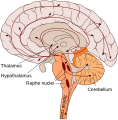
Category:SVG human brain (sagittal section)
Jump to navigation
Jump to search
Media in category "SVG human brain (sagittal section)"
The following 60 files are in this category, out of 60 total.
-
202102 Mid-sagittal plane of the brain.svg 512 × 512; 2.72 MB
-
Arbor Vitae in the Human Cerebellum.svg 512 × 399; 91 KB
-
Blank Diagram of the Human Cerebellum.svg 512 × 399; 92 KB
-
Brain anatomy.svg 512 × 494; 1.61 MB
-
Brain bulbar region Ar.svg 295 × 299; 217 KB
-
Brain bulbar region as.svg 295 × 299; 196 KB
-
Brain bulbar region IT.svg 295 × 299; 195 KB
-
Brain bulbar region ja.svg 295 × 299; 111 KB
-
Brain bulbar region ml.svg 295 × 299; 195 KB
-
Brain bulbar region ta.svg 295 × 299; 196 KB
-
Brain bulbar region-es.svg 295 × 299; 134 KB
-
Brain bulbar region-gu.svg 295 × 299; 195 KB
-
Brain bulbar region-mr.svg 295 × 299; 196 KB
-
Brain bulbar region-te.svg 295 × 299; 194 KB
-
Brain bulbar region-ur.svg 295 × 299; 194 KB
-
Brain bulbar region.PNG 295 × 299; 51 KB
-
Brain bulbar region.svg 295 × 299; 194 KB
-
Brain bulbar region.svg-kn.svg 295 × 299; 208 KB
-
Brain human sagittal section.svg 295 × 326; 145 KB
-
Brain logo.svg 612 × 792; 38 KB
-
Brain sagittal section stem highlighted.svg 295 × 326; 152 KB
-
Brain stem sagittal section.svg 566 × 639; 355 KB
-
Cerebral vascular territories midline.svg 512 × 481; 74 KB
-
CorpusCallosum.svg 1,025 × 598; 25 KB
-
Dopaminergic pathways.svg 446 × 307; 185 KB
-
Encephalon human sagittal section multilingual.svg 401 × 374; 158 KB
-
Fourth Ventricle in the Human Cerebellum.svg 512 × 399; 129 KB
-
Gehirn, medial - beschriftet lat-rus.svg 632 × 477; 499 KB
-
Gehirn, medial - beschriftet lat.svg 632 × 477; 307 KB
-
Gehirn, medial - Lobi ar.svg 724 × 482; 96 KB
-
Gehirn, medial - Lobi deu.svg 739 × 482; 121 KB
-
Gehirn, medial - Lobi en.svg 724 × 482; 120 KB
-
Gehirn, medial - Lobi es.svg 724 × 482; 55 KB
-
Gehirn, medial - Lobi ja.svg 724 × 482; 53 KB
-
Gray727 latin.svg 1,025 × 598; 23 KB
-
Gray727.svg 1,025 × 598; 18 KB
-
Head anatomy lateral view.svg 1,344 × 1,360; 57 KB
-
Human brain - Sagittal section.svg 380 × 400; 170 KB
-
Inferior colliculus of the human midbrain.svg 512 × 399; 92 KB
-
Medulla Oblongata and Cerebellum.svg 512 × 399; 91 KB
-
Mesocortical pathway.svg 446 × 307; 167 KB
-
Mesolimbic pathway.svg 446 × 307; 167 KB
-
Midbrain of the Human Brainstem.svg 512 × 399; 91 KB
-
Nigrostriatal pathway.svg 446 × 307; 167 KB
-
Pituitary gland representation cs.svg 261 × 236; 6 KB
-
Pituitary gland representation es.svg 261 × 236; 8 KB
-
Pituitary gland representation fr.svg 261 × 236; 5 KB
-
Pituitary gland representation.svg 261 × 236; 6 KB
-
PTSD brain.svg 530 × 342; 149 KB
-
Serotonergic neurons Ar.svg 295 × 299; 229 KB
-
Serotonergic neurons.svg 295 × 299; 205 KB
-
Skull and brain sagittal uk.svg 510 × 402; 470 KB
-
Skull and brain sagittal.svg 218 × 219; 240 KB
-
Skull and brainstem inner ear.svg 574 × 612; 702 KB
-
Skull and sagittal brain.svg 411 × 537; 157 KB
-
Superior colliculus of the human midbrain.svg 512 × 399; 92 KB
-
Transsphenoidaler Zugang.svg 617 × 524; 330 KB
-
Tuberoinfundibular pathway.svg 446 × 307; 169 KB
-
Ventral tegmental area.svg 446 × 307; 174 KB
-
ヒトの脳 模式図ii.svg 1,052 × 744; 41 KB


