Europe PMC

This website requires cookies, and the limited processing of your personal data in order to function. By using the site you are agreeing to this as outlined in our privacy notice and cookie policy.

Abstract 


Malaria remains a highly prevalent disease in more than 90 countries and accounts for at least 1 million deaths every year. Plasmodium falciparum infection is often associated with a procoagulant tonus characterized by thrombocytopenia and activation of the coagulation cascade and fibrinolytic system; however, bleeding and hemorrhage are uncommon events, suggesting that a compensated state of blood coagulation activation occurs in malaria. This article (i) reviews the literature related to blood coagulation and malaria in a historic perspective, (ii) describes basic mechanisms of coagulation, anticoagulation, and fibrinolysis, (iii) explains the laboratory changes in acute and compensated disseminated intravascular coagulation (DIC), (iv) discusses the implications of tissue factor (TF) expression in the endothelium of P. falciparum infected patients, and (v) emphasizes the procoagulant role of parasitized red blood cells (RBCs) and activated platelets in the pathogenesis of malaria. This article also presents the Tissue Factor Model (TFM) for malaria pathogenesis, which places TF as the interface between sequestration, endothelial cell (EC) activation, blood coagulation disorder, and inflammation often associated with the disease. The relevance of the coagulation-inflammation cycle for the multiorgan dysfunction and coma is discussed in the context of malaria pathogenesis.

Free full text 


Logo of nihpaLink to Publisher's site
Microcirculation. Author manuscript; available in PMC 2010 Jun 23.
Published in final edited form as:
PMCID: PMC2892216
NIHMSID: NIHMS207790
PMID: 18260002

Blood Coagulation, Inflammation and Malaria

I. ABSTRACT

Malaria remains a highly prevalent disease in more than 90 countries and accounts for at least 1 million deaths every year. Plasmodium falciparum infection is often associated with a procoagulant tonus characterized by thrombocytopenia and activation of the coagulation cascade and fibrinolytic system; however, bleeding and hemorrhage are uncommon events, suggesting that a compensated state of blood coagulation activation occurs in malaria. This article i) reviews the literature related to blood coagulation and malaria in a historic perspective, ii) describes basic mechanisms of coagulation, anticoagulation, and fibrinolysis, iii) explains the laboratory changes in acute and compensated disseminated intravascular coagulation (DIC), iv) discusses the implications of tissue factor (TF) expression in the endothelium of P. falciparum-infected patients, and v) emphasizes the pro-coagulant role of parasitized erythrocytes (pRBC) and activated platelets in the pathogenesis of malaria. This article also presents the ‘Tissue Factor Model’ (TFM) for malaria pathogenesis, which places TF as the interface between sequestration, endothelial cell activation, blood coagulation disorder and inflammation often associated with the disease. The relevance of the coagulation-inflammation cycle for the multiorgan dysfunction and coma is discussed in the context of malaria pathogenesis.

Keywords: endothelium, tissue factor, prothrombinase, apoptosis, sepsis

II. MALARIA: GENERAL ASPECTS

Malaria remains a major cause of morbidity and mortality in more than 90 countries and accounts for at least 1 million deaths every year and 450 million disease episodes annually [15]. Malaria is caused by Plasmodium sp. transmitted to the host by bite of the blood-feeding Anopheles sp. mosquitoes, which inject sporozoite-infected saliva [610]. There are four different types of Plasmodia (P. falciparum, P. vivax, P. ovale, P. malariae) that infect humans but P. falciparum causes almost all death [15]. Plasmodium parasite invades erythrocytes as part of its asexual life cycle within the human host [15, 8, 9]. As the P. falciparum parasites mature within the red blood cells (pRBC) to trophozoite and schizont stages [11, 12], they disappear from the peripheral circulation and localize specifically in the vascular beds of organs such as the brain, a process named sequestration [15]. Sequestration aids in parasite survival by avoiding clearance by the spleen and appears to be an important factor in malaria pathogenesis.

Clinically, malaria infection causes a range of symptoms from asymptomatic or mild flu-like illness (uncomplicated malaria)—particularly in immune individuals in endemic areas—to the uncommom complications of severe disease. This can present with a number of different syndromes, alone or in combination, including severe malarial anemia, respiratory complications and acidosis, renal failure, pulmonary edema, hepatic dysfunction, multiorgan failure, and cerebral malaria (CM) [15, 1324]. Actually, CM is the most-studied complication, and it is usually described as a syndrome consisting of decreased consciousness and unrousable coma not attributable to other causes in a patient with P. falciparum parasitemia [15, 1324]. When the patients survive, recovery is usually complete, but it can also leave some patients with long-term neurologic deficits [17]. The disease has a high mortality even when treated with appropriate antiparasite medication and intensive supportive care [24]. Because of the intensity of coma and the rapidity of its resolution, most authors agree that the underlying mechanisms are not compatible with primary vasocclusive pathology only, but resemble the picture of a toxic or metabolic encephalopathy [15].

While different theories have been proposed to explain the disease in humans [25, 26], including the sequestration and cytokine hypothesis, the mechanisms of malaria pathogenesis remain incompletely understood [15]. Nevertheless, a number of pathology studies in Southeast Asian adults and in African children have described an association between sequestration and coma in patients dying from malaria [2732]. More recently, disease severity in P. falciparum malaria patients was demonstrated to be directly associated with the biomass of pRBC as estimated by plasma levels of a parasite-specific histidine-rich protein (PfHRP2). PfHRP2 is a marker of total population of pRBC undergoing schizogony [33] and accordingly has been used as an indirect marker for sequestration [34, 35]. In agreement with a prominent role of cytoadherence to EC in disease pathogenesis is the fact that P. vivax malaria is not consistently accompanied by sequestration and/or severe disease [30, 36, 37]. Moreover, in some hemoglobinopathies (e.g. hemoglobinopathy C), the host polymorphism that affects P. falciparum erythrocyte membrane protein-1 (PfEMP-1) may protect against malaria by impairing the parasite’s ability to cytoadhere to microvessels [3840].

PfEMP-1 is the name for various antigenically diverse parasite-derived molecules located on the erythrocyte membrane mediating sequestration [4143]. PfEMP-1 interacts with several endothelial cell (EC)-surface receptors such as CD36 [4447], intercellular adhesion molecule 1 (ICAM-1) [48], thrombospondin [49], vascular cell adhesionmolecule 1 [50], E-selectin [50], and CD31 [51]. In addition, chondroitin sulfate A is bound by pRBC [52]and supports pRBC adhesion to both placental syncytiotrophoblast [53] and endothelium [54, 55]. Expression of some of these receptors appears tobe modulated, at least in part, by cytokines such as TNF-α, which is known to be increased in the plasma of malaria patients [56, 57]. Excess production of TNF-α is likely to be involved in the appearance of such symptoms as fever and headache associated with P. falciparum infection [58]. Actually, a systemic response characterized by increased levels of circulating cytokines such as IL-1β, IL-6, IL-8, IL-10, and HMGB-1 is observed in malaria in general and in cerebral malaria (CM) in particular [5965]. Furthermore, postmortem studies of human brain tissue in patients who died from CM display expression of TNF-α and IL-1β [66, 67]. Of note, some reports have linked cytokine levels to disease severity and complications [56, 57, 64, 6871]. It has been suggested that TNF-α and other cytokines up regulate adhesion molecules (e.g. ICAM-1) on vascular endothelium and thus modulate cytoadhrence [50, 72]. However, a relationship between in vitro cytoadhrence to CD36 and/or ICAM-1 and disease severity or cerebral symptoms has not been formally demonstrated [7375].

Malaria pathogenesis has been associated with both localized and widespread activation of EC in uncomplicated and fatal cases (e.g. cerebral malaria). Accordingly, molecules such as ICAM-1, VCAM-1, E-selectin, MIF, iNOS, uPAR and TF have been identified in the endothelium of P. falciparum-infected patients, according to immunohistochemistry (IHC) studies [30, 7682]. Increased plasma levels of soluble adhesion molecules sICAM-1, sVCAM-1, and sE-selectin [76, 83] and other markers of EC activation such as thrombomodulin [79, 84] and von Willebrand factor [85, 86] have also been reported in the same population. Further, both mild and severe cases of P. falciparum malaria have been found to be associated with thrombocytopenia [8691], hemostatic alterations [86106], and EC microparticle production [107], indicating a procoagulant state. It has thus been suggested that hemostatic alterations could be important in the disease progression and organ failure observed in malaria [92, 94, 98, 103, 104]. Of note, this concept has been applied in sepsis, a condition that shares with malaria a number of physiopathological mechanisms [108113]. The potential role of the coagulation cascade in Malaria pathogenesis is discussed below. A revision of the literature related to blood coagulation in malaria, in a historic perspective, is found in the Supplementary Material (Section A, “Malaria and Blood Coagulation: Historic Perspective”).

III-THE ROLE OF THE COAGULATION CASCADE IN P FALCIPARUM MALARIA PATHOGENESIS: STILL A CONTROVERSIAL ISSUE IN 2007

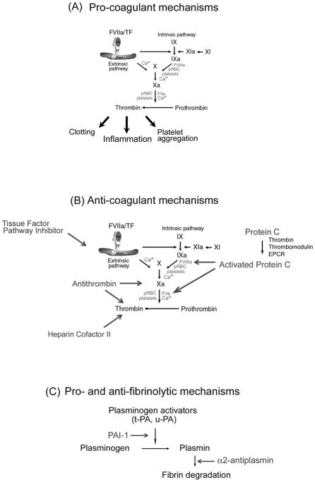
This section attempts to explain why the coagulation cascade could be regarded as a critical component in falciparum infection [15]. A description of basic mechanisms of coagulation cascade, anticoagulation and fibrinolysis is discussed in the Supplementary Material (Section B, “Tissue Factor and the Blood Coagulation Cascade”) and presented diagrammatically in Figure 1 (see Figure Legends). Likewise, the role of protease-activated receptors (PAR), and the participation of blood-borne TF and microparticles in inflammation is described as a supplement to this article, and has been comprehensively reviewed elsewhere [108113].

An external file that holds a picture, illustration, etc.
Object name is nihms207790f1.jpg

Coagulation cascade and its regulation. A) Pro-coagulant mechanisms. TF: a critical initiator of coagulation. Formation of a complex with Factor VIIa (FVIIa) leads to activation of FIX and FX. FXa in the presence of phosphatidyl serine and Ca2+ (prothrombinase complex) amplifies the coagulation cascade through conversion of prothrombin to thrombin, resulting in platelet aggregation, fibrin formation, and inflammation. Thrombin also activates FXI to XIa, which activates FIX to FIXa. FIXa in the presence of phosphatidyl serine and Ca2+ converts FX to FXa, consolidating the coagulation cascade. pRBC, parasitized red blood cells. B) Anticoagulant mechanism. TF pathway inhibitor (TFPI) binds to FXa and inhibits FVIIa/TF complex. Protein C is activated by thrombin (in the presence of thrombomodulin and EPCR), and APC inhibits the coagulation cascade through cleavage of cofactors FVa and FVIIIa. Antithrombin in the presence of heparin sulphate specifically interacts with and inhibits FXa and thrombin. Heparin cofactor II (in the presence of dermatan sulphate) inhibits thrombin. C). Pro- and antifibrinolytic mechanism. PAI-1, plasminogen activator inhibitor-1. The zymogen plasminogen is converted to the active serine protease, plasmin, through the action primarily of two-chain tissue plasminogen activator (tc-tPA) or two-chain urokinase (tc-uPA). Both tPA and uPA can be inhibited by plasminogen activator inhibitor-1 (PAI), while plasmin is inhibited by its major inhibitor, α2-antiplasmin, and to a lesser extent by α2-macroglobulin (not shown). For details, see Supplementary Material (Section B, “Tissue Factor and the Blood Coagulation Cascade”).

Controversy exists as to whether a coagulopathy is primarily or partially involved in the pathogenesis of malaria [94, 106]. In other words, a common question is whether coagulopathy has any “functional” importance in vivo in causing morbidity and/or fatal outcome. In actuality, controversy exists for a number of reasons: i) typical DIC (e.g., bleeding) is not present in most P. falciparum-infected patients [86, 92, 94, 9699, 102104] and occurs in 5% to 10% of severe malaria cases (1% of all cases) [15]; ii) thrombocytopenia in falciparum malaria [8890] was indicative of prognosis in one study [88] but also occurs in vivax malaria [96, 101, 104, 114116]; iii) fibrin and/or thrombus indicative of in vivo activation of the coagulation cascade have been clearly reported in some studies [99, 103, 117122] but infrequently found [29, 80] or undetectable in others [123, 124]; iv) experimental malaria treated with anticoagulants such as heparin does not change the morbidity associated with Plasmodium infection in mice or rhesus monkeys [125, 126], with one exception [127]. In addition, case reports have shown that use of anticoagulants such as heparin to treat severe malaria in humans has not changed outcome for most patients [128130]. Furthermore, a prospective and randomized study involving 97 humans infected with P. falciparum indicated that heparin did not affect the clinical course of the disease [131], and iv) laboratory changes reported in P. falciparum infection varies considerably from one study to another making it difficult for all authors to reach similar conclusions [86106].

Despite the above, a careful study of the literature devoted to malaria in particular, and vascular biology in general, indicate otherwise, i.e., activation of the coagulation cascade could be involved in malaria pathogenesis.

Coagulation activity in malaria

Numerous studies with human and animals indicate that there is undoubtedly increased coagulation activity in malaria. It occurs in uncomplicated (mild) cases [14, 15] and, although clinically not significant, can be confirmed as a compensated state according to in vivo coagulation tests [86, 87, 92101]. Most important, careful studies distinguishing mild, moderate, and severe P. falciparum infection as related to parasitemia have been reported in at least ten papers. These studies have demonstrated that the degree of coagulation derangement was often associated with disease severity [79, 86, 92, 94, 9698, 101104, 132]. Further, in some studies, it correlates with levels of parasitemia [86, 94, 97, 102, 104]. The fact that overt DIC (e.g., bleeding) is not present in severe malaria patients does not exclude an active role for the coagulation cascade in malaria pathogenesis. It is well known that severe organ dysfunction, including that seen in sepsis, is not necessarily accompanied by bleeding and/or hemorrhage [133136] but associated with a certain degree of activation of coagulation, fibrinolytic systems, and inflammation [108113].

Thrombocytopenia in malaria

The fact that platelet numbers are low in benign vivax malaria [96, 101, 104, 114, 115, 137], confirms rather than refutes, a role of the coagulation cascade in P. falciparum malaria pathogenesis because the mechanisms for thrombocytopenia in each infection differs. Thrombocytopenia in vivax malaria appears to be immune mediated [114] and occurs in the absence of blood coagulation activation [96, 101, 104]. In contrast, thrombocytopenia in falciparum malaria is most often accompanied by activation of the coagulation cascade [86, 87, 92101], among other mechanisms (e.g. immune-mediated lyses, peripheral destruction) [91, 106, 138141].

Fibrin and malaria

The fact that fibrin has been demonstrated in some cases [99, 103, 117122] but infrequently found [29, 80] or undetectable [123, 124] in others could be explained by the fact that coagulation activation is accompanied by compensatory fibrinolysis [142, 143]. Other mechanisms may also be involved such as high levels of neutrophil elastase (which degrade fibrin-stabilization Factor XIII) found in the plasma of malaria patients [97] and/or high levels of (pro-fibrinolytic) urokinase-type plasminogen activator (uPAR) expression detected in the endothelium of the same population [81]. In fact, several reports indicate that D-dimer levels are increased in the circulation of many cases of P. falciparum-infected patients, in complicated or mild cases [86, 92, 96, 100104, 144]. D-dimers are sensitive and specific in vivo markers of intravascular fibrin formation and fibrinolysis [142, 143]; this indicates that fibrin has been formed and digested intravascularly by plasmin in patients infected with P. falciparum. Of note, in 1972, O’Learly [93], studying DIC in monkeys, described: “Fibrin strands were sometimes seen in the engorged capillaries and veins, but organized thrombus were extremely rare. The most plausible explanation for this repeated observation is that the brisk fibrinolysis occurring secondary to DIC effectively digests the fibrin as it is elaborated and thus preventing the development of organized thrombus.” It is important to recognize that detection of fibrin in sepsis has also been reported in some studies [145147], although undetectable in others [148]. Nevertheless, most authors will agree that the coagulation cascade contributes to sepsis pathogenesis, as discussed in the Supplementary Material (Part C) and comprehensively reviewed elsewhere [108113].

Anticoagulants

The fact that anticoagulants such as heparin dos not affect the outcome of malaria is not an entirely unexpected finding based on the increasingly recognized cross-talk between blood coagulation and inflammation [108113]. In contrast to inhibition of thrombin by heparin [149, 150] or thrombin generation by active site-blocked Xa, which fails to reduce mortality [151], inhibitors of the TF initiation complex such as antibodies to TF [152], TFPI [153], or active site-blocked VIIa [154] reduce inflammatory responses in addition to their antithrombotic effects at least in some experimental models (e.g., sepsis). In other words, proximal inhibition of the coagulation cascade by TF blockade prevents coagulopathy, reduces inflammation, and attenuates lethality [152156]. It has been suggested that signaling by the ternary FVIIa/TF/FXa initiation complex provides a mechanism to activate protease-activated receptors (PAR; see below) without requiring massive activation of fluid-phase proteases that drive DIC. Accordingly, blockade of ternary initiation complex by molecules such as TF inhibitors appears to be effective in the treatment of experimental sepsis [152156].

Laboratory changes

The fact that laboratory changes are not the same in all studies dealing with the hemostatic disorder that follows P. falciparum infection [86104, 132] could be the result of the selection of patient group and/or severity of the infection or because these hemostatic elevations are inherently fluctuant. Sensitivity of specific coagulation assays employed by each lab may have also introduced additional variability into the analysis.

IV. ACUTE VERSUS “COMPENSATED” DISSEMINATED INTRAVASCULAR COAGULATION (DIC): AN IMPORTANT DISTINCTION

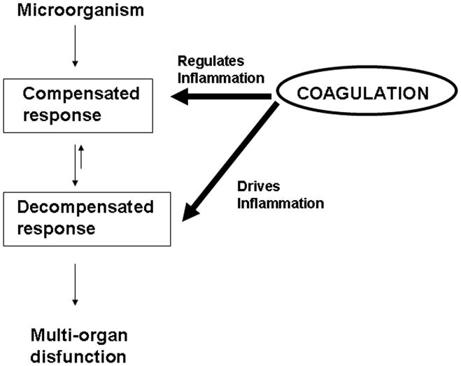
Activation of the coagulation cascade as a consequence of inflammation is well known event and can be viewed as an essential part of the host defense of the body, for example, to infectious agents. While activation of the coagulation cascade is a physiologic response triggered in an effort to contain the invading entity and to keep the consequent inflammatory response to a limited area, an exaggerated or uncontrolled response may lead to a situation in which coagulation and microthrombosis contribute to disease. This is illustrated by the occurrence of systemic coagulation activation in combination with microvascular failure, which results from the systemic inflammatory response to severe infection or sepsis and contributes to multiple organ dysfunction [108113]. Figure 2 summarizes this concept emphasizing the role of the coagulation cascade in regulating or driving inflammation [108]. It is remarkable that these two situations appear to occur in malaria.
An external file that holds a picture, illustration, etc.
Object name is nihms207790f2.jpg

Compensated and decompensated responses and modulation by the coagulation cascade. Activation of coagulation cascade as a consequence of inflammation is an essential part of the host defense of the body. This physiologic response is triggered in an effort to contain the invading entity and to keep the consequent inflammatory response to a limited area. An exaggerated or uncontrolled response, however, may lead to a situation in which coagulation and microthrombosis contribute to disease. This is illustrated by the occurrence of systemic coagulation activation in combination with microvascular failure, which results from the systemic inflammatory response to severe infection or sepsis, and that contributes to multiple organ dysfunction. Modified from Ruf, 2004 [108].

Thus, in uncomplicated malaria, a coagulation disorder is a common laboratory finding, but bleeding and hemorrhage are not observed [14, 15, 19, 21, 22]. Coagulation disorder is also commonly observed in severe malaria (~ 1% of all cases), but only in 5% to 10% of these cases it is associated with bleeding [15, 86, 94, 9698, 102, 104]. Therefore, an important distinction should be made between these two pathologic states, one where typical DIC is encountered (e.g., bleeding), and the other where a compensated state (e.g., laboratory changes only) is detectable.

DIC is a life-threatening acute, subacute, or chronic coagulation disorder occurring as a secondary complication in a variety of diseases. It is characterized by activation of the coagulation cascade, which leads to the formation of microthrombin through the microcirculation of the body. Sometimes the coagulopathy is localized to a specific organ, but it often presents an uneven distribution. Two major mechanism trigger DIC: release of TF into the circulation or endothelial injury [142, 143].

While acute DIC is the terminal phase of the coagulation disorder, it is often preceded by a period during which the coagulation cascade is already activated but the increased activation can be compensated for by the natural inhibitor systems, a state referred to as compensated DIC. In some cases, a continuous or intermittent slow rate of initiation of intravascular coagulation occurs and may or may never undergo decompensation to acute DIC. Under these conditions, the control mechanisms may effectively prevent severe clinical manifestations such as bleeding and hemorrhage by neutralizing active enzymes and/or by increasing the synthesis of the consumed hemostatic components. As the trigger for coagulation activation persists in DIC, inhibitors will be gradually exhausted, leading to more coagulation. In this process, many clotting factors—most notably, fibrinogen and platelets—are consumed, resulting eventually in complete impairment of the hemostasis system. This is why the term ‘consumptive coagulopathy’ is often used to denote this process. This results in bleeding tendency or decompensated DIC. In addition, activation of the coagulation cascade is accompanied by compensatory fibrinolysis where increase in plasminogen-dependent plasmin activity is detectable using markers such as D-dimers, as shown in Table 1. Table 1 also depicts the laboratory profile of acute and ‘compensated’ DIC, which has been comprehensively discussed in excellent reviews [142, 143].

TABLE 1

Profile of ‘compensated’ and acute DIC

DIC
CharacteristicAcuteCompensated
Prothombin timeakN or S
Partial thrombosplatin timebN or S
Platelet countc↓ or N
Fibrinogen leveld↓ or N or ↑
D-dimere↑ or N
Fibrin(ogen) split productsfN or ↑
Fibrin monomergN or ↑
F 1+2 fragmenth
Thrombin-antithrombin complexi
Plasmin-antiplasmin complexj
Bleeding and hemorrhage+
aEstimates the extrinsic and common pathway of the coagulation cascade.
bEstimates the intrinsic and common pathway of the coagulation cascade.
cBlood platelet count.
dPlasma fibrinogen concentration.
eD-dimers, crosslinked fibrin-derived products produced by plasmin, confirm that both the clotting cascade and the fibrinolytic system have been activated.
fFibrin(ogen) split product (FSP) orfibrin(ogen) degradation products (FDP) are produced when plasmin proteolytically degrades fibrinogen and/or fibrin. FDP and FSP indicate activation of the fibrinolytic system.
gFibrinogen molecules from which fibrinopeptides A and B have been cleaved by thrombin; elevated levels thus suggest the presence of thrombin or an activated clotting system.
hProthrombin fragment 1+2 (F 1+2) are generated when FXa proteolytically cleaves prothrombin to form thrombin.
iComplex formed when thrombin irreversibly binds AT. TAT and F 1+2 are markers of in vivo activation of the coagulation cascade.
jComplex formation occurs when plasmin binds to its natural inhibitor α2-antiplasmin. Plasma levels signify in vivo plasmin generation or an activated fibrinolytic system.
k↑, increased, prolonged, or elevated; N, normal; S, shortened; ↓, decreased; +, present/common; −, absent/uncommon. Modified from Mammen, 2000 [142].

Clinically, the symptoms of acute DIC differ among individuals but fever, dyspnea, cyanosis, and extreme respiratory difficulty may predominate due to pulmonary dysfunction (acute respiratory distress syndrome). Neurologic signs and symptoms including convulsion and coma represent another pattern. Renal changes such as oliguria and acute renal failure may dominate, while circulatory failure and shock appear suddenly or develop progressively. Many of these symptoms are present in severe malaria [15]. In contrast, compensated DIC [142, 143] is not accompanied by bleeding or hemorrhage as is the case in uncomplicated malaria [14, 22].

A final important aspect related to the blood coagulation cascade in the context of malaria pathogenesis is physiologic changes in the plasma level of coagulation factors and inhibitors that occur in children [157] and in pregnancy [158]. In these populations, which are at high risk for developing severe malaria [23, 159], there is decreased fibrinolytic activity resulting in a hypercoagulable state. While these are considered to be physiologic changes [157, 158], they may nonetheless predispose children and women to a procoagulant tonus that may contribute to severe disease [24, 159].

V. TISSUE FACTOR AND THE COAGULATION-INFLAMMATION CYCLE

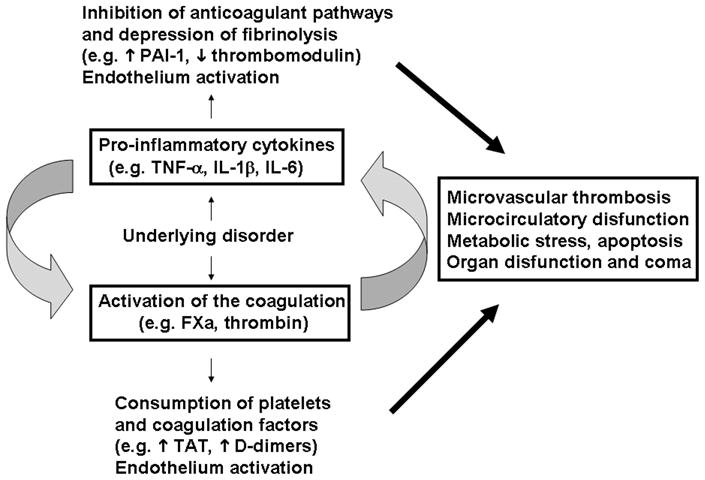
The TF pathway is the principal activator of clotting under physiologic conditions and in the presence of systemic inflammation and generalized infection [108113]. In fact, accumulating evidence indicates that TF drives the coagulation-inflammation circuit through generation of coagulation factor, which induces cytokine production and expression of adhesion molecules. Conversely, and equally true, cytokine-driven inflammation leads to activation of coagulation. As an example, the best known contribution of inflammation to coagulation is probably induction of TF by TNF-α on the cell surface of monocytes and EC [160, 161]. Accordingly, evidence indicates that TF expression drives the vicious coagulation-inflammation cycle. The role of this cycle in multiorgan dysfunction is depicted in Figure 3, and has been comprehensive reviewed elsewhere [108113].

An external file that holds a picture, illustration, etc.
Object name is nihms207790f3.jpg

The coagulation-inflammation cycle. Diagrammatic representation of activation of coagulation and inflammation in response to an underlying disorder (e.g., infection). Infection is potentially associated with induction of pro-inflammatory cytokines and TF expression. Exposure of TF-bearing inflammatory cells (e.g., EC, monocytes) to blood results in thrombin generation, platelet aggregation, and conversion of fibrinogen to fibrin. Thrombin and other activated coagulation factors activate protease-activated receptors on inflammatory cells, inducing release of proinflammatory cytokines, which will further modulate coagulation through induction of TF, on one hand, and prevention of fibrinolysis through decrease in PAI-1 and thrombomodulin function, on the other. The vicious cycle between coagulation-inflammation leads to microvascular thrombosis, EC activation, increased vascular permeability, ROS generation, metabolic stress, apoptosis and ultimately to organ dysfunction. For details, see text and Supplementary Material (Section C, “Coagulation-inflammation cycle and the Relevance for Multiorgan Dysfunction and Coma in Malaria and Sepsis”).

Inflammatory cytokines induce Tissue Factor expression

Cytokines such as TNF-α, IL-1β, IL-2, IL-6, IFN-γ, VEGF, MCP-1, or PDGF are capable of inducing TF expression in vitro. P-selectin, E-selectin, and CD40 are other examples of positive modulators of TF synthesis in EC [108113, 162, 163]. In vivo occurrence of coagulation/cytokine-dependent inflammation has also been demonstrated. Intramuscular injection of IL-6 results in thrombin generation in baboons, while infusion of TNF-α in humans produces a procoagulant state accompanied by inhibition of fibrinolysis [164, 165].

Blood coagulation factors induce cytokine expression

In vitro, FXa induces IL-6, IL-8, and MCP-1 production from EC and monocytes and promotes expression of E-selectin, ICAM-1, and VCAM-1, whereas thrombin induces IL-8 and IL-6 in mononuclear cells [108113, 162, 163]. In vivo, it is well known that intravascular FXa and thrombin generation induces inflammation within the microcirculation via interaction with specific receptors on platelets, EC, and white blood cells. Accordingly, administration of FVIIa enhances IL-6 and IL-8 production in health volunteers [166]. In addition, thrombin, FXa, FVIIa/TF or peptides that activate PARs induce leukocyte rolling, adhesion, and extravasation in vitro or in vivo [166176]. These effects are mediated by PAR, which induce gene expression, production of cytokines, and expression of adhesion molecules [167].

These adhesion molecules promote the initial rolling and tethering interactions between circulating granulocytes, monocytes, and lymphocytes to EC at sites of tissue injury. Interaction of monocytes with perturbed EC and the synergistic action [163, 177180] of pro-inflammatory molecules potentially lead to exacerbated TF expression and further EC activation, which sustains the coagulation-inflammation cycle. Therefore, interplay between cytokines and coagulation factors promotes the “vicious” cycle of coagulation-inflammation of sepsis which appears to be critical in malaria pathogenesis, as well. Actually, severe malaria and sepsis share physiopathological mechanism and both cases are associated, to a certain degree, with i) uncontrolled infection, ii) systemic inflammation syndrome and cytokinemia, iii) increased capillary permeability, iv) hypoxia, v) acidosis, vi) EC activation, vii) microvascular coagulopathy, and viii) multiorgan dysfunction [15, 108113]. The relevance of the coagulation-inflammation cycle for multiorgan dysfunction and coma in malaria, and sepsis is discussed in the Supplementary Material (Section C).

VI. TISSUE FACTOR: THE INTERFACE BETWEEN SEQUESTRATION, ENDOTHELIUM ACTIVATION, BLOOD COAGULATION DISORDER, AND INFLAMMATION IN MALARIA

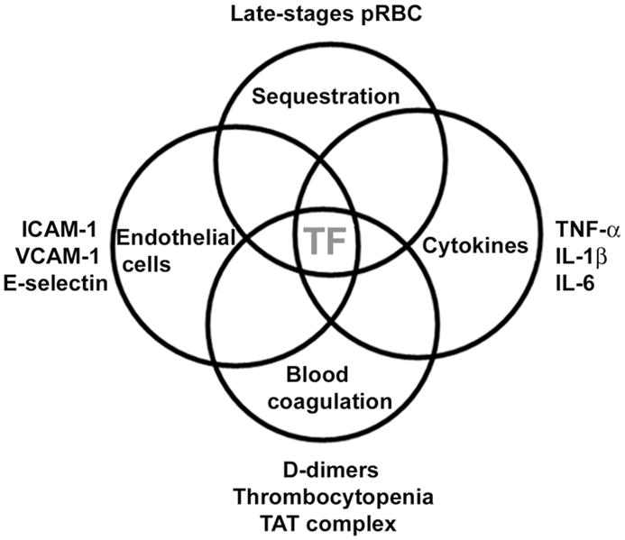
Taking into consideration the concept that blood coagulation is initiated by TF expression [181184], it was presumed that in vivo expression of TF was required to explain the coagulation disorder observed in malaria. TF expression by EC also appears to be important in the disease pathogenesis, because it is the only molecule that can be placed in the interface of all four pathologic features reportedly found in malaria, as shown in Figure 4.
An external file that holds a picture, illustration, etc.
Object name is nihms207790f4.jpg

TF and the interface of pathologic features observed in malaria. Sequestration is associated with endothelial cell activation (estimated by an increase in ICAM-1, VCAM-1, E-selectin, and TF expression). TF expression is needed for activation of the coagulation cascade, which promotes thrombocytopenia, increase in TAT levels, and D-dimers. Coagulation factors also induce secretion of inflammatory cytokines (TNF-α, IL-1β, and IL-6), which in turn induce TF expression in different cell types promoting and sustaining a coagulation-inflammation cycle (for details, see text). TF, Tissue Factor.

The potential role of pRBC (sequestration) in TF expression by EC

We have recently demonstrated that expression of TF occurs in EC exposed to pRBC in vitro [82]. Expression of TF was blocked by TF inhibitor [185, 186] and was dependent on physical contact between pRBC and EC, as is apoptosis [187] and EC activation [188]. How pRBC induce TF expression remains to be understood at the molecular level, although it may involve receptor-mediated signaling [189], reactive oxygen species (ROS) [190], and NF-κB, which is nuclear translocated in brain EC incubated in vitro with pRBC [191]. Consistent with in vitro findings described above, ultrastructural studies using electron microscopy have shown that direct contact between pRBC and EC occurs in vivo by strictures called knobs [192]; presumably, this interaction is associated with EC activation in vivo [193, 194]. Moreover, sequestration of pRBC in the brain appears to be associated with coma according to studies on Southeast Asian adults and African children with fatal CM [2731]. These studies imply that pRBC cytoadherence to cerebral EC may be an important and indispensable factor in P. falciparum malaria pathogenesis.

Strong support for the central role of parasite sequestration in disease severity has been recently published in a study involving 337 P. falciparum malaria patients. It was demonstrated that disease severity was associated with the number of sequestered pRBC as indirectly estimated by plasma levels of a parasite-specific, histidine-rich protein (PfHRP2) [30, 34, 35]. Further, a large trial on severe malaria showed that artesunate reduces mortality by 34% compared with quinine [35]. Both artesunate and quinine are active against sequestered parasites, preventing their development to schizonts, but artesunate is also active against the younger forms of the parasite, preventing their maturation and sequestration in the microcirculation of vital organs. Of note, the greatest mortality benefit in this trial compared with quinine was in patients with high parasitemias, indicating that prevention of sequestration, rather than prevention of schizont rupture, decreased mortality [35].

Also, host polymorphism that affects PfEMP-1 display (e.g. hemoglobinopathy C) may protect against malaria by impairing the parasite’s ability to adhere to microvessels and induce inflammation [3840]. Likewise, CM is not observed in malaria caused by P. vivax, a condition where sequestration is not consistently observed [30, 36, 37]. Collectively, these in vitro and in vivo data support the view that sequestration is necessary for disease pathogenesis. It is clear, however, that cytoadherence per se cannot explain the coagulation disorder and neurologic syndrome observed during infection. Therefore, elements other than sequestration itself might account for malaria pathogenesis.

The role of pRBC in the amplification step of the coagulation cascade

We have recently demonstrated that late-stage pRBC inherently support the productive assembly of both intrinsic Xnase and prothrombinase complexes in vitro [82], amplifying by thousands of times the generation of FXa and thrombin, respectively [195, 196]. These findings are consistent with the fact that pRBC present changes in phospholipid composition as it matures and display PS, a negatively charged phospholipid that supports assembly of coagulation complexes [197]. Assemblies were attained at a remarkably low parasite concentration in vitro (0.025% hematocrit; 5% parasitemia); indicating that minute amounts of pRBC may successfully propagate the blood coagulation cascade in vivo when initiated by TF expression [82]. It should be emphasized, however, that platelets become pro-coagulant only when primed by low amounts of thrombin [195]. In contrast, pRBC are already ‘primed’ and as a consequence can immediately amplify blood coagulation independently of previous thrombin production and by a mechanism that is insensitive to platelet aggregation inhibitors such as aspirin. Of interest, only mature forms of pRBC are pro-hemostatic. These experimental data are important, as late- but not early-stage pRBC are found sequestered in the brain of most postmortem studies described so far [2731].

It is noteworthy that while our coagulation studies used a laboratory-adapted parasite strain (3D7) [82], pRBC obtained directly from parasitized individuals had shortened recalcification time [198] and clotting time [199]. Both coagulation tests are sensitive to surface-mediated amplification of the coagulation cascade, indicating that pRBC are indeed procoagulant in vivo. Therefore, it is plausible to suggest that a procoagulant and proinflammatory microenvironment is encountered at sequestration sites and that this response may be overamplified by pRBC and activated platelets, leading to explosive thrombin generation if uncontrolled by coagulation inhibitors such TFPI [200], antithrombin [201], and the APC/thrombomodulin pathway [202]. It is likely that this reaction is particularly amplified in the vessels of the brain, a tissue where EC trombomodulin is not found [203, 204]. Thrombomodulin is a critical anticoagulant. Through interaction with thrombin, it changes the specificity of the enzyme that activates PC. APC has pleiotropic effects, among them inactivation of FVa and FVIIIa, and a direct antiinflammatory effect on EC [205]. Thus, lack of thrombomodulin in the brain could contribute to the enhanced blood coagulation and platelet aggregation observed in CM cases. More recenlty, it has been suggested that tissues which display low levels of thrombomodulin (e.g. brain and placenta) are prone to sequestration, as opposed to sites where higher levels of thrombomodulin/APC are found such as the heart and skeletal muscle [206].

The role of platelets in malaria pathogenesis

Platelets are considered crucial to malaria pathogenesis [15] and to other syndromes [207]. We suggest that amplification of the coagulation cascade through formation of coagulation complexes [195, 196] is the primary mechanism by which it contributes to disease pathogenesis. Amplification results in additional thrombin formation, which promotes recruitment of activated platelets to the site of injury [195, 196]. Activated platelets secrete a number of proinflammatory molecules through the so-called ‘release reaction’ and may contribute to local inflammatory processes by a number of mechanisms [208, 209]. Among these, platelets secrete such chemokines as TGF-β, which displays pro-apoptotic properties, and IL-1β, which activates white blood cells, induces TF expression, and promotes neutrophil and monocyte adherence [208, 209]. Platelets are a source of PAI-1, an important inhibitor of fibrinolysis [210]. Platelets also express P-selectin and promote neutrophil platelet EC interactions. Moreover, platelets are a major source of soluble CD40 ligand [211]. CD40 ligand belongs to the TNF superfamily of molecules and has multiple actions that may be of significance in microcirculation [211]. These actions include upregulation of cytokine expression on vascular smooth muscle cells and EC, increased expression of surface adhesion molecules on EC, and upregulation of TF synthesis on macrophages [108113]. Finally, it has been recognized that thrombocytopenia in malaria is present in uncomplicated, mild, and severe cases [8691] and is predictive of fatal outcome in one study [88].

TF Expression in vivo in Cerebral Malaria Cases

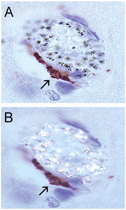
We have recently demonstrated that sequestration is associated with TF expression by brain EC of P. falciparum-infected children [82]. A typical staining for TF in EC from a pediatric CM case is shown in Figure 5. This finding strongly suggests that activation of the coagulation cascade took place in vivo, a contention corroborated by histopathologic findings of platelet and monocyte accumulation and fibrin deposition in the brain of some CM patients [29, 212, 213]. Notably, in our series of IHC studies TF expression in EC was associated with sequestration of mature forms in some cases, while in others, the presence of hemozoin (malaria pigments) indicative of schizont rupture (previous sequestration) was observed [82]. In still others, no pRBC or hemozoin were found. The events associated with malaria in general and CM in particular thus appear to be a highly dynamic process where sequestration occurs and is followed by schizont rupture and then clearance. Although the pathologic studies [2732, 7678] are only a snapshot of this dynamic and complex process, they clearly indicate that TF was present in EC at the time of death of all CM patients [82]. Finally, while it is not known whether TF-bearing microparticles contribute to TF staining in EC [82], it is known that uncomplicated cases of malaria—in contrast to CM cases—do not display circulating EC microparticles [107]. Because 4 of 13 P. falciparum patients we studied died from other causes (nonmalarial coma) but were positive for TF staining [82], it is concluded that, at least in these cases, TF was indeed expressed by EC.
An external file that holds a picture, illustration, etc.
Object name is nihms207790f5.jpg

TF staining of EC associated with sequestration in a pediatric CM case. IHC was performed with anti-TF monoclonal antibodies. A) Regular light showing positive staining in the EC. B) Polarized light that detects hemozoin, indicative of sequestration. Staining was negative in the absence of primary antibody. For details, see text and [82].

Our IHC studies show that some vessels contain sequestration and no TF staining [82], suggesting that the tightly regulated mechanism of TF expression [182], among other mechanisms [214], may play a role in preventing uncontrolled EC activation with widespread TF expression. Of note, in vivo TF staining in human EC has been reported in only a few studies [203, 215219], and it is remarkable that our studies consistently show positive staining and even, in some cases, at sites of sequestration [82]. Thus, a picture is emerging where relevant processes described in malaria presumably trigger TF expression in EC (e.g., sequestration and/or sequestration-associated events) while others may converge to express TF (e.g., cytokines) and vice-versa.

TF Expression in vivo in Parasitemic Patients Who Died from Other Causes

Our IHC studies also show variable degrees of TF staining in the five of six parasitemic children who died from nonmalarial causes [82]. This suggests that uncomplicated cases display TF expression in EC but formal evidence is still needed using appropriate samples. The one asymptomatic ‘parasitemic control’ [220] with no evidence of TF expression died from direct head trauma [82]. Therefore, TF expression in EC appears to be a general feature of malaria, and it presumably occurs in uncomplicated cases and it is not specific for CM. Consistent with this notion, widespread EC activation has been reported in uncomplicated malaria [76, 78], a condition where sequestration [76, 78], thrombocytopenia [8691], and activation of blood coagulation are generally observed [86, 94, 9698, 104]. These noncomatose patients, however, present no or few clinical manifestations compatible with a coagulation disorder (e.g., bleeding and hemorrhage) and their laboratory profiles [142, 143] resembles compensated DIC (Table I). Accordingly, most of these patients display normal or close to normal PT and PTT, while PC and PC-1 levels are low and D-dimer and TAT levels high in the absence of bleeding [86, 94, 9698, 104].

Compensated versus Non-compensated States

It remains to be revealed why some patients develop CM while others present ‘uncomplicated’ malaria, a condition where sequestration and activation of blood coagulation also occurs. It may be that activation of blood coagulation, while normally homeostatic, may contribute to disease when uncontrolled or undergoing decompensation [108, 112]. Consistent with this view, platelet accumulation in the vessels of the brain [212], apoptosis [221], and formation of EC microparticles [107] have been specifically reported in CM (or complicated malaria) but not in uncomplicated cases. The finding that microparticles known to express TF [222] and PS and to participate in inflammation [223, 224]—are present in severe cases suggests that it may contribute to the inflammation-coagulation cycle at late stages of the disease. Microparticle formation could be also regarded as detectable markers of decompensation in P. falciparum-infected patients. Other reports have demonstrated that high levels of soluble uPAR [225], thrombomodulin [79], HMGB1 [226] and overall activation of the coagulation cascade [98] occurs in severe (sometimes lethal) malaria as opposed to healthy or less severe cases.

Considering that compensated states occur in malaria, specific risk factors for decompensation could be identified, in theory, in parasitemic patients. Numerous possibilities can be listed including patient immune status, polymorphism of pro- and/or anticoagulant inflammatory molecules [227, 228], PfEMP-1 and/or other parasite molecules [39, 229]. Heterogeneity of platelets [230], EC [231], and parasite population [232], associated diseases and/or a combination of factors may play a role in decompensation. Consistent with the notion that compensated states do occur in malaria is the fact that it shares physiopathologic mechanisms with sepsis [5], a condition in which compensatory mechanisms of the coagulation cascade have been studied in in vivo escalation models after E. coli challenge in baboons [112, 233]. These models have allowed investigators to conclude that the TF pathway of coagulation drives inflammation and that blockade of TF reduces lethality [152156]. It is thus possible to envision TF inhibitors as prototypes to prevent development of CM or perhaps as drugs to be used in treatment of established CM.

Sequestration in Other Vascular Beds

Sequestration in malaria occurs (although not equally distributed) in sites other than the brain [2732] including the eye, heart, liver, kidney, intestine and lung [27, 32], placenta [159], adipose tissue [27], dermis [76], and skeletal muscle [78] with widespread EC activation [76, 77]. Of note, pathology studies have demonstrated signs of inflammation in some of these tissues. For example, inflammatory infiltrates and fibrin have been detected in the lung of malaria patients who develop acute respiratory distress syndrome [15, 234, 235], a pathology where the role for TF has been described [236]. Also, “placental malaria”, a disease often associated with poor birth outcomes [237], is accompanied by TF expression by macrophages [238] with fibrin deposition and monocyte accumulation in the villae [239]. Placenta is a tissue particularly rich in TF [240] and presents a hemostatic system that appears to play a critical role in maternal-fetal homeostasis [241, 242]. At last, it remains to be investigated whether other cerebral and non-cerebral tissues display TF expression in the endothelium, as reported for the frontal cortex of patients who died from malaria [82].

VII. THE “TISSUE FACTOR MODEL” OF HUMAN MALARIA PATHOGENESIS

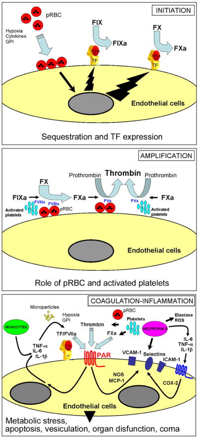
Previous models, namely the ‘sequestration’ and ‘cytokine’ hypotheses, have tried to explain malaria pathogenesis [25, 26]; immunopathologic mechanisms have also been described [243]. The ‘sequestration’ hypothesis suggests that “the presence of infected erythrocytes is an essential event” [26]. The ‘cytokine’ hypothesis proposes that, “while significant cerebral sequestration is often present and sometimes block vessels (with predictable sequelae), it is not essential for the onset of the cerebral manifestation of malaria” [25]. We propose an alternative model, here named the “Tissue Factor Model” (TFM) for human malaria pathogenesis. This model, summarized in Figure 6 (see Figure Legends), attempts to explain the disease by combining concepts evolved from both ‘sequestration’ and ‘cytokine’ hypotheses. In contrast to previous models, however, it classifies TF expression in the endothelium, and the amplification of the coagulation cascade by pRBC and platelets, as critical steps in disease pathogenesis. The TFM is based on experimental and clinical findings and on concepts from vascular biology.
An external file that holds a picture, illustration, etc.
Object name is nihms207790f6.jpg

The TF model of human CM: Initiation: Normal, quiescent endothelium does not express TF in the absence of biologic stimulation. According to this model, sequestration and/or sequestration-associated events (e.g. cytokines, fibrin, hypoxia, apoptosis, proinflammatory molecules released by pRBC which activate TLRs such as GPI and plasmodium DNA-containing hemozoin) primarily induces EC activation in the microvessels of the brain and in other vascular beds. This contributes to TF expression, at sequestration sites, and/or possibly paracrinally. Monocytes may also be a source of TF in malaria. Mechanistically, TF initiates the coagulation cascade through binding to coagulation FVIIa and the substrate FIX and FX (extrinsic Xase). In this ternary initiation complex, FIXa and FXa are generated. Amplification: FXa, FVa, and prothrombin assemble in the pRBC surface and/or activated platelets, with formation of the prothrombinase complex leading to explosive thrombin formation and amplification of the coagulation cascade. Thrombin thus formed promotes fibrin deposition and induces platelet aggregation. Thrombin also activates FXI to FXIa (not shown), which activates FIX to FIXa. FIXa, FVIIIa, and FX assemble in the membrane of activated platelets or pRBC with formation of the intrinsic Xnase complex required for production of additional FXa owing to feedback inhibition of the FVIIa/TF complex by TFPI(not shown). Therefore, the TF model of human CM proposes that initiation of blood coagulation by TF expression and the amplification phase supported by pRBC (and/or activated platelets) - particularly at sequestration sites where the concentration of pRBC-derived phosphatydilserine is presumably very high - are critical for disease pathogenesis. Coagulation-inflammation: TF/VIIa, TF/FVIIa/FXa, and thrombin activate protease-activated receptors (PARs) in different cell types including mononuclear cells and EC at sequestration sites and/or paracrinally. PAR activation in EC is accompanied by upregulation of molecules (e.g., ICAM-1, VCAM-1, E-selectin, COX-2, NO synthase) and production of proinflammatory cytokines (e.g., TNF-α, IL-1β, IL-6) reportedly found in CM. Because cytokines act synergistically with coagulation factors and induce upregulation of TF and adhesion molecules, they both are critical to perpetuate the inflammatory response that promotes increased interaction of monocytes, platelets, and/or pRBC with activated EC. Hypoxia, GPI, DNA-containing hemozoin and other events may also contribute to the coagulation-inflammation cycle. The result is a convergence of signals leading to exacerbated TF expression that sustains the coagulation-inflammatory cycle. This cycle may leads to microvascular thrombosis, additional EC activation, increased vascular permeability, ROS generation, metabolic stress, apoptosis and ultimately to organ dysfunction (e.g. SARS) that is often associated with poor prognosis in malaria. In extreme cases, death occurs. For details, see text.

Specifically, the TFM takes into account the following assumptions: i) sequestration is associated with disease pathogenesis [31, 32, 34, 35]; ii) P. falciparum infection is accompanied by EC activation [30, 7682] and correlates with disease severity according to some studies [76, 78]; iii) malaria patients develop a coagulation disorder [86104, 132]; iv) the degree of hemostatic alterations is often associated with the severity of malaria [79, 86, 92, 94, 9698, 101104, 132] and correlate with parasitemia according to some studies [86, 94, 97, 102, 104]; v) antiparasitic treatment halts the coagulation disorder and improve clinical outcome [79, 92, 94, 9698, 132, 244]; vi) P. falciparum infection is accompanied by increased plasma concentration of various cytokines [5, 5964]; vii) TF is expressed in the endothelium of parasitemic patients [82], viii) pRBC, in addition to activated platelets, support the assembly of prothrombinase and intrinsic Xnase complexes in vitro [82] and are procoagulant in vivo [198, 199]; ix) TF is reportedly in the interface of blood coagulation and inflammation [108113]; x) similarities exist between malaria and other clinical conditions (e.g., sepsis) with abnormal expression of TF expression and sustained coagulation-inflammation cycle [111]; and finally, xi) TF can be placed in the interface of all four pathologic features reported in malaria (this article). These findings indicate that TF expression by endothelium is not an epiphenomenon but a major and differential component of the disease.

Favoring a prominent role for sequestration-associated TF expression by the endothelium in falciparum malaria pathogenesis is the fact that high levels of circulating cytokines (e.g. TNF-α) [245] and thrombocytopenia, apparently immune-mediated [114], are present in vivax malaria [101, 102, 115, 116]. However, vivax malaria is a benign condition where sequestration of pRBC is not a consistent finding and coagulation cascade activation or DIC does not take place [30, 36, 37, 96, 101]. Therefore, TNF-α and other cytokines do not account for TF expression in the endothelium of falciparum malaria patients, despite its pro-coagulant effect in vitro [160, 161]. In other words, it is not only the absence of consistent sequestration but the lack of blood coagulation activation as well two important factors that distinguishe vivax from falciparum malaria, pathobiologically. Finally, procoagulant activity of monocytes occurs in both P. falciparum and P. vivax malaria, indicating that events other than or besides monocyte procoagulant activity are critical for the genesis of severe malaria [96].

The TFM also differs from the previous hypothesis in five major respects: i) sequestration and/or sequestration-associated events is (are) the critical and perhaps the most important steps leading to disease pathogenesis primarily because it is accompanied by EC activation, TF expression, and initiation of the coagulation cascade [82]; ii) sequestered pRBC and activated platelets play an active role in amplifying, propagating, and consolidating the coagulation cascade initiated by TF expression, particularly at sequestration sites [82, 198, 199] where the kinetics of the coagulation reactions occur favorably because the concentration of phosphatydilserine is presumably very high; iii) the two previous steps are critical in mounting and sustaining a coagulation-inflammatory cycle that presumably leads to organ dysfunction by a combination of various molecular mechanisms [108113, 246]; iv) there are compensated and decompensated states in malaria. This supposition is based on numerous studies comparing the clinical profile and activation status of the coagulation cascade in uncomplicated, moderate, and severe cases of malaria [86, 94, 9698, 104] and experimental models of decompensation in sepsis [112, 233]; v) the TFM also introduces orderly steps to didactically explain the disease, namely initiation, amplification, and coagulation-inflammation steps. Figure 6 depicts these events diagrammatically.

The TFM model thus highlights the fundamental importance of sequestration-associated activation of EC to trigger the blood coagulation cascade, on one hand, and the role of pRBC and activated platelets to inherently amplify this response through formation of multimolecular blood coagulation complexes, on the other [82]. In other words, TF is needed but is not sufficient alone (as is the case for sequestration) to produce disease without an amplification step. It should be emphasized that the revised theory of the blood coagulation proposes that TF is critical but not enough to sustain normal hemostasis [182]. Under normal conditions, the level of FXa initially produced by the extrinsic pathway (FVIIa/TF) is feedback inhibited by TFPI [182]. Thus, the intrinsic Xnase complex (FXIa and FVIIIa, Ca2+ and PS) is required for production of additional FXa that amplifies and consolidate the coagulation cascade. In this context, lack of amplification by the intrinsic Xnase explains why hemophiliacs bleed despite normal levels of TF [247]. Likewise, TF expression by the endothelium and the amplification of the coagulation cascade by pRBC and/or activated platelets (particularly at sequestration sites) appears to be critical events in mounting and sustaining a coagulation-inflammation cycle which contributes to organ dysfunction and coma, in P. falciparum malaria.

The TFM also considers that events potentially associated with sequestration such as hypoxia [15], fibrin formation [99, 103, 117122], and apoptosis [221, 248250] may contribute to a procoagulant/-inflammatory cycle in malaria. Further, cytokine production [5] may play a critical role in the disease pathogenesis. TNF-α levels, for example, are increased in P. falciparum-infected patient plasma [56, 57], which has been reported to induce TF expression and NF-κB activation in EC in vitro [251]. Moreover, TNF-α upregulates expression of adhesion molecules on EC and suppresses thrombomodulin expression [252]. Thus, it favors adhesion of leukocytes which in turn damage EC by releasing radical oxygen species (ROS) and preformed proteases [111, 253]. It is also remarkable that TNF-α [254], and IL-1β [255] induce increased vascular permeability of EC through a FVIIa- and TF-dependent manner. It may be that the coagulation-inflammation cycle [108113, 256] contributes to the brain-barrier dysfunction observed in malaria [257]. One may also speculate that this cycle creates an appropriate environment that favors sequestration in vivo.

Because sequestration is followed by schizogony, it is plausible that molecules released by or leaked from pRBC [258] positively modulate inflammation in vivo. For example, the malaria toxin - glycosylphosphatidylinositol anchor (GPI) - is a potent pro-inflammatory stimulus known to induce adhesion molecule expression in ECs, and to activate macrophages through TLR-2/-4 [259262]. Of note, injection of GPI mimics several aspects of the pathology observed after infection of mice with P. berghei ANKA [263]. At last, plasmodium DNA-containing hemozoin activates TLR-9 and promotes IL-12p40 production by dendritic cells in vitro [264] and display pro-inflammatory effects [260, 265, 266]. The role of TLR, monocytes and the immune system in inflammation/coagulation is concisely discussed in the Supplementary Material (Section D, “Monocytes, Toll-like Receptors, Tissue Factor, and the Immune System”) and reviewed elsewhere [111].

It is important to recognize that host response such as activation of the “contact” pathway [95] and complement system reported in malaria patients [267, 268] and its crosstalk with the coagulation cascade [111, 269272] may contribute to the inflammatory tonus observed in the disease. Further, neutrophils should be regarded as effectors in malaria pathogenesis [273]. Neutrophils interact with coagulation proteins and plasma neutrophil elastase is increased in patients with severe falciparum infection [95, 97, 132] and correlates inversely with platelet counts, antithrombin [97] and FXIII levels [132]. Of note, elastase appears to account, at least in part, for the pivotal role of neutrophils in sepsis [109, 274, 275]. Elastase proteolytically inhibits thrombomodulin function and may therefore interfere with activation of PC [276]. Additionally, elastase damages EC by promoting EC detachment, and degrades fibrinolytic enzymes or anticoagulants such as TFPI [273, 274, 277], thus favoring a procoagulant tonus. Finally, ROS are involved in oxidative stress and apoptosis in malaria [278, 279] and in other conditions where the coagulation-inflammation cycle contributes to disease pathogenesis (e.g. SARS)[15, 111, 274, 280]. The molecular mechanisms by which activation of the coagulation cascade (and coagulation-inflammation cycle) is associated with metabolic stress and organ dysfunction is highly complex and discussed in the Supplementary Material (Part C, “Coagulation-inflammation Cycle and the Relevance for Multiorgan Dysfunction and Coma in Malaria and Sepsis”) [108113].

While the TFM for malaria pathogenesis should be regarded as an oversimplification of the pathologic processes involved in malaria, it should also be regarded as a flexible platform for future studies, and not a static model. Hopefully it will be modified, revised, and challenged as our understanding of malaria pathogenesis progresses. This model, however, identifies malaria as a disease with a strong vascular component where EC are the primary substrate of pathology. This is consistent with the fact that the endothelial surface area is vast and estimated to comprise 1012 EC in the adult human (there are ~1010 blood monocytes) [281]. It seems likely, therefore, that EC expression plays a prominent role in malaria and that one can consider malaria as a disease of the EC and/or microcirculation. Of interest, sickle-cell anemia, which shares many pathologic aspects with malaria such as activation of the endothelium (and coagulation cascade), is also considered an EC disease [282]. Whether the TF-bearing EC are apoptotic and vice versa is not yet known, but it is certainly an important issue that deserves further investigation [283]. Apoptosis occurs in experimental or falciparum malaria in vitro and in vivo [187, 221, 248250, 278]. It also occurs in sepsis and has been implicated as a critical mechanism associated with organ dysfunction [246]. Of note, apoptotic cells become pro-coagulany and display PS, which assembles the coagulation complexes [284287]. In other words, apoptotic cells potentially support the coagulation cascade and appear to participate in inflammation in vivo [284286]. Therefore, one may speculate that in malaria there is an apoptotic-coagulation-inflammation imbalance. Finally, monocytes may play an important role in the disease pathogenesis and/or contribute to the procoagulant/inflammatory tonus observed in falciparum malaria [96], as described for sepsis [5]. Not surprisingly, malaria has been regarded as an inflammatory sepsis-like syndrome [5], a condition where cytokines, TF expression by EC and/or monocytes reportedly plays a pivotal role [108113, 216].

VIII. CONCLUSIONS

The concept that TF is a potentially critical mediator of CM suggests that therapeutics targeting TF, and/or the endothelial cells may be successful in the treatment of malaria [154, 288, 289]. While anticoagulation in general might attenuate the local inflammatory escalation by reducing thrombin levels the antiinflammatory benefits of TF inhibition in particular are unique, as it blocks a proximal step of the coagulation cascade while levels of TNF-α—an important pathway for fighting infection—remains intact [154, 288]. Finally, identification of affordable therapeutics targeting EC and hemostatic components are particularly important vis-à-vis the increasingly reported resistance of Plasmodium sp. to antimalarial drugs [290].

Supplementary Material

supplemental data

Acknowledgments

This work was supported by the Division of Intramural Research, National Institute of Allergy and Infectious Diseases (NIAID), National Institutes of Health, USA. Because IMBF, is government employees and this is a government work, the work is in the public domain in the United States. Notwithstanding any other agreements, the NIH reserves the right to provide the work to PubMedCentral for display and use by the public, and PubMedCentral may tag or modify the work consistent with its customary practices. You can establish rights outside of the U.S. subject to a government use license.

We are grateful to Drs. Jose Marcos C. Ribeiro, Thomas E. Wellems, Robert W. Gwadz, and Kathryn C. Zoon (NIAID) for continuous encouragement and support. We express our thanks to Drs Richard O. Whitten, Jerrold M. Ward and Terrie E. Taylor for immunohistochemistry studies. We thank the Reviewers for their time, comments and suggestions. We acknowledge NIAID intramural editor Brenda Rae Marshall for assistance.

References

1. Miller LH, Baruch DI, Marsh K, Doumbo OK. The pathogenic basis of malaria. Nature. 2002;415:673–679. [Abstract] [Google Scholar]
2. Newton CR, Taylor TE, Whitten RO. Pathophysiology of fatal falciparum malaria in African children. Am J Trop Med Hyg. 1998;58:673–683. [Abstract] [Google Scholar]
3. Weatherall DJ, Miller LH, Baruch DI, Marsh K, Doumbo OK, Casals-Pascual C, Roberts DJ. Malaria and the red cell. Hematology Am Soc Hematol Educ Program. 2002:35–57. [Abstract] [Google Scholar]
4. White NJ, Breman JG. Malaria and babesiosis: diseases caused by red blood cell parasites. In: Kasper DL, Brawnwald E, Fauci AS, Hauser SL, Longo DL, Jameson DL, editors. Harrison`s Principles of Internal Medicine. Vol. 1. New York, NY: McGraw Hill; 2005. pp. 1218–1233. [Google Scholar]
5. Clark IA, Cowden WB. The pathophysiology of falciparum malaria. Pharmacol Ther. 2003;99:221–260. [Abstract] [Google Scholar]
6. Ribeiro JM, Francischetti IM. Role of arthropod saliva in blood feeding: sialome and post-sialome perspectives. Annu Rev Entomol. 2003;48:73–88. [Abstract] [Google Scholar]
7. Francischetti IM, Valenzuela JG, Pham VM, Garfield MK, Ribeiro JM. Toward a catalog for the transcripts and proteins (sialome) from the salivary gland of the malaria vector Anopheles gambiae. J Exp Biol. 2002;205:2429–2451. [Abstract] [Google Scholar]
8. Krettli AU, Miller LH. Malaria: a sporozoite runs through it. Curr Biol. 2001;11:R409–412. [Abstract] [Google Scholar]
9. Baldacci P, Menard R. The elusive malaria sporozoite in the mammalian host. Mol Microbiol. 2004;54:298–306. [Abstract] [Google Scholar]
10. Garcia JE, Puentes A, Patarroyo ME. Developmental biology of sporozoite-host interactions in Plasmodium falciparum malaria: implications for vaccine design. Clin Microbiol Rev. 2006;19:686–707. [Europe PMC free article] [Abstract] [Google Scholar]
11. Bozdech Z, Llinas M, Pulliam BL, Wong ED, Zhu J, DeRisi JL. The transcriptome of the intraerythrocytic developmental cycle of Plasmodium falciparum. PLoS Biol. 2003;1:E5. [Europe PMC free article] [Abstract] [Google Scholar]
12. Paul RE, Diallo M, Brey PT. Mosquitoes and transmission of malaria parasites -not just vectors. Malar J. 2004;3:39. [Europe PMC free article] [Abstract] [Google Scholar]
13. Maitland K. Severe malaria: lessons learned from the management of critical illness in children. Trends Parasitol. 2006;22:457–462. [Abstract] [Google Scholar]
14. Grobusch MP, Kremsner PG. Uncomplicated malaria. Curr Top Microbiol Immunol. 2005;295:83–104. [Abstract] [Google Scholar]
15. Anstey NM, Jacups SP, Cain T, Pearson T, Ziesing PJ, Fisher DA, Currie BJ, Marks PJ, Maguire GP. Pulmonary manifestations of uncomplicated falciparum and vivax malaria: cough, small airways obstruction, impaired gas transfer, and increased pulmonary phagocytic activity. J Infect Dis. 2002;185:1326–1334. [Abstract] [Google Scholar]
16. Turner G. Cerebral malaria. Brain Pathol. 1997;7:569–582. [Abstract] [Google Scholar]
17. Idro R, Jenkins NE, Newton CR. Pathogenesis, clinical features, and neurological outcome of cerebral malaria. Lancet Neurol. 2005;4:827–840. [Abstract] [Google Scholar]
18. Mackintosh CL, Beeson JG, Marsh K. Clinical features and pathogenesis of severe malaria. Trends Parasitol. 2004;20:597–603. [Abstract] [Google Scholar]
19. Phillips RE, Looareesuwan S, Warrell DA, Lee SH, Karbwang J, Warrell MJ, White NJ, Swasdichai C, Weatherall DJ. The importance of anaemia in cerebral and uncomplicated falciparum malaria: role of complications, dyserythropoiesis and iron sequestration. Q J Med. 1986;58:305–323. [Abstract] [Google Scholar]
20. Warrell DA. Cerebral malaria: clinical features, pathophysiology and treatment. Ann Trop Med Parasitol. 1997;91:875–884. [Abstract] [Google Scholar]
21. Molyneux ME, Taylor TE, Wirima JJ, Borgstein A. Clinical features and prognostic indicators in paediatric cerebral malaria: a study of 131 comatose Malawian children. Q J Med. 1989;71:441–459. [Abstract] [Google Scholar]
22. Klein Klouwenberg PM, Oyakhirome S, Schwarz NG, Glaser B, Issifou S, Kiessling G, Klopfer A, Kremsner PG, Langin M, Lassmann B, Necek M, Potschke M, Ritz A, Grobusch MP. Malaria and asymptomatic parasitaemia in Gabonese infants under the age of 3 months. Acta Trop. 2005;95:81–85. [Abstract] [Google Scholar]
23. Greenwood BM, Bojang K, Whitty CJ, Targett GA. Malaria. Lancet. 2005;365:1487–1498. [Abstract] [Google Scholar]
24. Marsh K, Forster D, Waruiru C, Mwangi I, Winstanley M, Marsh V, Newton C, Winstanley P, Warn P, Peshu N, et al. Indicators of life-threatening malaria in African children. N Engl J Med. 1995;332:1399–1404. [Abstract] [Google Scholar]
25. Clark IA, Rockett KA. The cytokine theory of human cerebral malaria. Parasitol Today. 1994;10:410–412. [Abstract] [Google Scholar]
26. Berendt AR, Tumer GD, Newbold CI. Cerebral malaria: the sequestration hypothesis. Parasitol Today. 1994;10:412–414. [Abstract] [Google Scholar]
27. MacPherson GG, Warrell MJ, White NJ, Looareesuwan S, Warrell DA. Human cerebral malaria. A quantitative ultrastructural analysis of parasitized erythrocyte sequestration. Am J Pathol. 1985;119:385–401. [Europe PMC free article] [Abstract] [Google Scholar]
28. Pongponratn E, Riganti M, Punpoowong B, Aikawa M. Microvascular sequestration of parasitized erythrocytes in human falciparum malaria: a pathological study. Am J Trop Med Hyg. 1991;44:168–175. [Abstract] [Google Scholar]
29. Pongponratn E, Turner GD, Day NP, Phu NH, Simpson JA, Stepniewska K, Mai NT, Viriyavejakul P, Looareesuwan S, Hien TT, Ferguson DJ, White NJ. An ultrastructural study of the brain in fatal Plasmodium falciparum malaria. Am J Trop Med Hyg. 2003;69:345–359. [Abstract] [Google Scholar]
30. Silamut K, Phu NH, Whitty C, Turner GD, Louwrier K, Mai NT, Simpson JA, Hien TT, White NJ. A quantitative analysis of the microvascular sequestration of malaria parasites in the human brain. Am J Pathol. 1999;155:395–410. [Europe PMC free article] [Abstract] [Google Scholar]
31. Taylor TE, Fu WJ, Carr RA, Whitten RO, Mueller JS, Fosiko NG, Lewallen S, Liomba NG, Molyneux ME. Differentiating the pathologies of cerebral malaria by postmortem parasite counts. Nat Med. 2004;10:143–145. [Abstract] [Google Scholar]
32. Seydel KB, Milner DA, Jr, Kamiza SB, Molyneux ME, Taylor TE. The distribution and intensity of parasite sequestration in comatose Malawian children. J Infect Dis. 2006;194:208–205. [Europe PMC free article] [Abstract] [Google Scholar]
33. Desakorn V, Dondorp AM, Silamut K, Pongtavornpinyo W, Sahassananda D, Chotivanich K, Pitisuttithum P, Smithyman AM, Day NP, White NJ. Stage-dependent production and release of histidine-rich protein 2 by Plasmodium falciparum. Trans R Soc Trop Med Hyg. 2005;99:517–524. [Abstract] [Google Scholar]
34. Dondorp AM, Desakorn V, Pongtavornpinyo W, Sahassananda D, Silamut K, Chotivanich K, Newton PN, Pitisuttithum P, Smithyman AM, White NJ, Day NP. Estimation of the total parasite biomass in acute falciparum malaria from plasma PfHRP2. PLoS Med. 2005;2:e204. [Europe PMC free article] [Abstract] [Google Scholar]
35. Dondorp A, Nosten F, Stepniewska K, Day N, White N. Artesunate versus quinine for treatment of severe falciparum malaria: a randomised trial. Lancet. 2005;366:717–725. [Abstract] [Google Scholar]
36. Sina B. Focus on Plasmodium vivax. Trends Parasitol. 2002;18:287–289. [Abstract] [Google Scholar]
37. Luxemburger C, Ricci F, Nosten F, Raimond D, Bathet S, White NJ. The epidemiology of severe malaria in an area of low transmission in Thailand. Trans R Soc Trop Med Hyg. 1997;91:256–262. [Abstract] [Google Scholar]
38. Agarwal A, Guindo A, Cissoko Y, Taylor JG, Coulibaly D, Kone A, Kayentao K, Djimde A, Plowe CV, Doumbo O, Wellems TE, Diallo D. Hemoglobin C associated with protection from severe malaria in the Dogon of Mali, a West African population with a low prevalence of hemoglobin S. Blood. 2000;96:2358–2363. [Abstract] [Google Scholar]
39. Fairhurst RM, Wellems TE. Modulation of malaria virulence by determinants of Plasmodium falciparum erythrocyte membrane protein-1 display. Curr Opin Hematol. 2006;13:124–130. [Abstract] [Google Scholar]
40. Williams TN. Human red blood cell polymorphisms and malaria. Curr Opin Microbiol. 2006;9:388–394. [Abstract] [Google Scholar]
41. Su XZ, Heatwole VM, Wertheimer SP, Guinet F, Herrfeldt JA, Peterson DS, Ravetch JA, Wellems TE. The large diverse gene family var encodes proteins involved in cytoadherence and antigenic variation of Plasmodium falciparum-infected erythrocytes. Cell. 1995;82:89–100. [Abstract] [Google Scholar]
42. Baruch DI, Pasloske BL, Singh HB, Bi X, Ma XC, Feldman M, Taraschi TF, Howard RJ. Cloning the P. falciparum gene encoding PfEMP1, a malarial variant antigen and adherence receptor on the surface of parasitized human erythrocytes. Cell. 1995;82:77–87. [Abstract] [Google Scholar]
43. Smith JD, Chitnis CE, Craig AG, Roberts DJ, Hudson-Taylor DE, Peterson DS, Pinches R, Newbold CI, Miller LH. Switches in expression of Plasmodium falciparum var genes correlate with changes in antigenic and cytoadherent phenotypes of infected erythrocytes. Cell. 1995;82:101–110. [Europe PMC free article] [Abstract] [Google Scholar]
44. Barnwell JW, Asch AS, Nachman RL, Yamaya M, Aikawa M, Ingravallo P. A human 88-kD membrane glycoprotein (CD36) functions in vitro as a receptor for a cytoadherence ligand on Plasmodium falciparum-infected erythrocytes. J Clin Invest. 1989;84:765–772. [Europe PMC free article] [Abstract] [Google Scholar]
45. Oquendo P, Hundt E, Lawler J, Seed B. CD36 directly mediates cytoadherence of Plasmodium falciparum parasitized erythrocytes. Cell. 1989;58:95–101. [Abstract] [Google Scholar]
46. Ockenhouse CF, Tandon NN, Magowan C, Jamieson GA, Chulay JD. Identification of a platelet membrane glycoprotein as a falciparum malaria sequestration receptor. Science. 1989;243:1469–1471. [Abstract] [Google Scholar]
47. Baruch DI, Gormely JA, Ma C, Howard RJ, Pasloske BL. Plasmodium falciparum erythrocyte membrane protein 1 is a parasitized erythrocyte receptor for adherence to CD36, thrombospondin, and intercellular adhesion molecule 1. Proc Natl Acad Sci U S A. 1996;93:3497–3502. [Europe PMC free article] [Abstract] [Google Scholar]
48. Berendt AR, Simmons DL, Tansey J, Newbold CI, Marsh K. Intercellular adhesion molecule-1 is an endothelial cell adhesion receptor for Plasmodium falciparum. Nature. 1989;341:57–59. [Abstract] [Google Scholar]
49. Roberts DD, Sherwood JA, Spitalnik SL, Panton LJ, Howard RJ, Dixit VM, Frazier WA, Miller LH, Ginsburg V. Thrombospondin binds falciparum malaria parasitized erythrocytes and may mediate cytoadherence. Nature. 1985;318:64–66. [Abstract] [Google Scholar]
50. Ockenhouse CF, Tegoshi T, Maeno Y, Benjamin C, Ho M, Kan KE, Thway Y, Win K, Aikawa M, Lobb RR. Human vascular endothelial cell adhesion receptors for Plasmodium falciparum-infected erythrocytes: roles for endothelial leukocyte adhesion molecule 1 and vascular cell adhesion molecule 1. J Exp Med. 1992;176:1183–1189. [Europe PMC free article] [Abstract] [Google Scholar]
51. Treutiger CJ, Heddini A, Fernandez V, Muller WA, Wahlgren M. PECAM-1/CD31, an endothelial receptor for binding Plasmodium falciparum-infected erythrocytes. Nat Med. 1997;3:1405–1408. [Abstract] [Google Scholar]
52. Rogerson SJ, Chaiyaroj SC, Ng K, Reeder JC, Brown GV. Chondroitin sulfate A is a cell surface receptor for Plasmodium falciparum-infected erythrocytes. J Exp Med. 1995;182:15–20. [Europe PMC free article] [Abstract] [Google Scholar]
53. Fried M, Duffy PE. Adherence of Plasmodium falciparum to chondroitin sulfate A in the human placenta. Science. 1996;272:1502–1504. [Abstract] [Google Scholar]
54. Robert C, Pouvelle B, Meyer P, Muanza K, Fujioka H, Aikawa M, Scherf A, Gysin J. Chondroitin-4-sulphate (proteoglycan), a receptor for Plasmodium falciparum-infected erythrocyte adherence on brain microvascular endothelial cells. Res Immunol. 1995;146:383–393. [Abstract] [Google Scholar]
55. Rogerson SJ, Novakovic S, Cooke BM, Brown GV. Plasmodium falciparum-infected erythrocytes adhere to the proteoglycan thrombomodulin in static and flow-based systems. Exp Parasitol. 1997;86:8–18. [Abstract] [Google Scholar]
56. Kwiatkowski D, Hill AV, Sambou I, Twumasi P, Castracane J, Manogue KR, Cerami A, Brewster DR, Greenwood BM. TNF concentration in fatal cerebral, non-fatal cerebral, and uncomplicated Plasmodium falciparum malaria. Lancet. 1990;336:1201–1204. [Abstract] [Google Scholar]
57. Grau GE, Taylor TE, Molyneux ME, Wirima JJ, Vassalli P, Hommel M, Lambert PH. Tumor necrosis factor and disease severity in children with falciparum malaria. N Engl J Med. 1989;320:1586–1591. [Abstract] [Google Scholar]
58. Kwiatkowski D, Molyneux ME, Stephens S, Curtis N, Klein N, Pointaire P, Smit M, Allan R, Brewster DR, Grau GE, et al. Anti-TNF therapy inhibits fever in cerebral malaria. Q J Med. 1993;86:91–98. [Abstract] [Google Scholar]
59. Friedland JS, Ho M, Remick DG, Bunnag D, White NJ, Griffin GE. Interleukin-8 and Plasmodium falciparum malaria in Thailand. Trans R Soc Trop Med Hyg. 1993;87:54–55. [Abstract] [Google Scholar]
60. Mshana RN, Boulandi J, Mshana NM, Mayombo J, Mendome G. Cytokines in the pathogenesis of malaria: levels of IL-I beta, IL-4, IL-6, TNF-alpha and IFN-gamma in plasma of healthy individuals and malaria patients in a holoendemic area. J Clin Lab Immunol. 1991;34:131–139. [Abstract] [Google Scholar]
61. Lyke KE, Burges R, Cissoko Y, Sangare L, Dao M, Diarra I, Kone A, Harley R, Plowe CV, Doumbo OK, Sztein MB. Serum levels of the proinflammatory cytokines interleukin-1 beta (IL-1beta), IL-6, IL-8, IL-10, tumor necrosis factor alpha, and IL-12(p70) in Malian children with severe Plasmodium falciparum malaria and matched uncomplicated malaria or healthy controls. Infect Immun. 2004;72:5630–5637. [Europe PMC free article] [Abstract] [Google Scholar]
62. Harpaz R, Edelman R, Wasserman SS, Levine MM, Davis JR, Sztein MB. Serum cytokine profiles in experimental human malaria. Relationship to protection and disease course after challenge. J Clin Invest. 1992;90:515–523. [Europe PMC free article] [Abstract] [Google Scholar]
63. Brown H, Turner G, Rogerson S, Tembo M, Mwenechanya J, Molyneux M, Taylor T. Cytokine expression in the brain in human cerebral malaria. J Infect Dis. 1999;180:1742–1746. [Abstract] [Google Scholar]
64. Day NP, Hien TT, Schollaardt T, Loc PP, Chuong LV, Chau TT, Mai NT, Phu NH, Sinh DX, White NJ, Ho M. The prognostic and pathophysiologic role of pro- and antiinflammatory cytokines in severe malaria. J Infect Dis. 1999;180:1288–1297. [Abstract] [Google Scholar]
65. Ho M, Schollaardt T, Snape S, Looareesuwan S, Suntharasamai P, White NJ. Endogenous interleukin-10 modulates proinflammatory response in Plasmodium falciparum malaria. J Infect Dis. 1998;178:520–525. [Abstract] [Google Scholar]
66. Porta J, Carota A, Pizzolato GP, Wildi E, Widmer MC, Margairaz C, Grau GE. Immunopathological changes in human cerebral malaria. Clin Neuropathol. 1993;12:142–146. [Abstract] [Google Scholar]
67. Udomsangpetch R, Chivapat S, Viriyavejakul P, Riganti M, Wilairatana P, Pongponratin E, Looareesuwan S. Involvement of cytokines in the histopathology of cerebral malaria. Am J Trop Med Hyg. 1997;57:501–506. [Abstract] [Google Scholar]
68. Kern P, Hemmer CJ, Van Damme J, Gruss HJ, Dietrich M. Elevated tumor necrosis factor alpha and interleukin-6 serum levels as markers for complicated Plasmodium falciparum malaria. Am J Med. 1989;87:139–143. [Abstract] [Google Scholar]
69. Urquhart AD. Putative pathophysiological interactions of cytokines and phagocytic cells in severe human falciparum malaria. Clin Infect Dis. 1994;19:117–131. [Abstract] [Google Scholar]
70. Wenisch C, Linnau KF, Looaresuwan S, Rumpold H. Plasma levels of the interleukin-6 cytokine family in persons with severe Plasmodium falciparum malaria. J Infect Dis. 1999;179:747–750. [Abstract] [Google Scholar]
71. Shaffer N, Grau GE, Hedberg K, Davachi F, Lyamba B, Hightower AW, Breman JG, Phuc ND. Tumor necrosis factor and severe malaria. J Infect Dis. 1991;163:96–101. [Abstract] [Google Scholar]
72. Ho M, White NJ. Molecular mechanisms of cytoadherence in malaria. Am J Physiol. 1999;276:C1231–1242. [Abstract] [Google Scholar]
73. Ringwald P, Peyron F, Lepers JP, Rabarison P, Rakotomalala C, Razanamparany M, Rabodonirina M, Roux J, Le Bras J. Parasite virulence factors during falciparum malaria: rosetting, cytoadherence, and modulation of cytoadherence by cytokines. Infect Immun. 1993;61:5198–5204. [Europe PMC free article] [Abstract] [Google Scholar]
74. Ho M, Singh B, Looareesuwan S, Davis TM, Bunnag D, White NJ. Clinical correlates of in vitro Plasmodium falciparum cytoadherence. Infect Immun. 1991;59:873–878. [Europe PMC free article] [Abstract] [Google Scholar]
75. Ockenhouse CF, Ho M, Tandon NN, Van Seventer GA, Shaw S, White NJ, Jamieson GA, Chulay JD, Webster HK. Molecular basis of sequestration in severe and uncomplicated Plasmodium falciparum malaria: differential adhesion of infected erythrocytes to CD36 and ICAM-1. J Infect Dis. 1991;164:163–169. [Abstract] [Google Scholar]
76. Turner GD, Ly VC, Nguyen TH, Tran TH, Nguyen HP, Bethell D, Wyllie S, Louwrier K, Fox SB, Gatter KC, Day NP, Tran TH, White NJ, Berendt AR. Systemic endothelial activation occurs in both mild and severe malaria. Correlating dermal microvascular endothelial cell phenotype and soluble cell adhesion molecules with disease severity. Am J Pathol. 1998;152:1477–1487. [Europe PMC free article] [Abstract] [Google Scholar]
77. Turner GD, Morrison H, Jones M, Davis TM, Looareesuwan S, Buley ID, Gatter KC, Newbold CI, Pukritayakamee S, Nagachinta B, et al. An immunohistochemical study of the pathology of fatal malaria. Evidence for widespread endothelial activation and a potential role for intercellular adhesion molecule-1 in cerebral sequestration. Am J Pathol. 1994;145:1057–1069. [Europe PMC free article] [Abstract] [Google Scholar]
78. Garcia F, Cebrian M, Dgedge M, Casademont J, Bedini JL, Neves O, Filella X, Cinta Cid M, Corachan M, Grau JM. Endothelial cell activation in muscle biopsy samples is related to clinical severity in human cerebral malaria. J Infect Dis. 1999;179:475–483. [Abstract] [Google Scholar]
79. Hemmer CJ, Bierhaus A, von Riedesel J, Gabat S, Liliensiek B, Pitronik P, Lin J, Grauer A, Amiral J, Ziegler R, et al. Elevated thrombomodulin plasma levels as a result of endothelial involvement in plasmodium falciparum malaria. Thromb Haemost. 1994;72:457–464. [Abstract] [Google Scholar]
80. Clark IA, Awburn MM, Whitten RO, Harper CG, Liomba NG, Molyneux ME, Taylor TE. Tissue distribution of migration inhibitory factor and inducible nitric oxide synthase in falciparum malaria and sepsis in African children. Malar J. 2003;2:6. [Europe PMC free article] [Abstract] [Google Scholar]
81. Fauser S, Deininger MH, Kremsner PG, Magdolen V, Luther T, Meyermann R, Schluesener HJ. Lesion associated expression of urokinase-type plasminogen activator receptor (uPAR, CD87) in human cerebral malaria. J Neuroimmunol. 2000;111:234–240. [Abstract] [Google Scholar]
82. Francischetti IM, Seydel KB, Monteiro RQ, Whitten RO, Erexson CR, Noronha AL, Ostera GR, Kamiza SB, Molyneux ME, Ward JM, Taylor TE. Plasmodium falciparum-infected erythrocytes induce tissue factor expression in endothelial cells and support the assembly of multimolecular coagulation complexes. J Thromb Haemost. 2007;5:155–165. [Europe PMC free article] [Abstract] [Google Scholar]
83. Hviid L, Theander TG, Elhassan IM, Jensen JB. Increased plasma levels of soluble ICAM-1 and ELAM-1 (E-selectin) during acute Plasmodium falciparum malaria. Immunol Lett. 1993;36:51–58. [Abstract] [Google Scholar]
84. Boehme MW, Werle E, Kommerell B, Raeth U. Serum levels of adhesion molecules and thrombomodulin as indicators of vascular injury in severe Plasmodium falciparum malaria. Clin Investig. 1994;72:598–603. [Abstract] [Google Scholar]
85. Hollestelle MJ, Donkor C, Mantey EA, Chakravorty SJ, Craig A, Akoto AO, O’Donnell J, van Mourik JA, Bunn J. von Willebrand factor propeptide in malaria: evidence of acute endothelial cell activation. Br J Haematol. 2006;133:562–569. [Abstract] [Google Scholar]
86. Horstmann RD, Dietrich M. Haemostatic alterations in malaria correlate to parasitaemia. Blut. 1985;51:329–335. [Abstract] [Google Scholar]
87. Horstmann RD, Dietrich M, Bienzle U, Rasche H. Malaria-induced thrombocytopenia. Blut. 1981;42:157–164. [Abstract] [Google Scholar]
88. Gerardin P, Rogier C, Ka AS, Jouvencel P, Brousse V, Imbert P. Prognostic value of thrombocytopenia in African children with falciparum malaria. Am J Trop Med Hyg. 2002;66:686–691. [Abstract] [Google Scholar]
89. Ladhani S, Lowe B, Cole AO, Kowuondo K, Newton CR. Changes in white blood cells and platelets in children with falciparum malaria: relationship to disease outcome. Br J Haematol. 2002;119:839–847. [Abstract] [Google Scholar]
90. Jadhav UM, Patkar VS, Kadam NN. Thrombocytopenia in malaria--correlation with type and severity of malaria. J Assoc Physicians India. 2004;52:615–618. [Abstract] [Google Scholar]
91. Scott CS, Van Zyl D, Ho E, Ruivo L, Mendelow B, Coetzer TL. Thrombocytopenia in patients with malaria: automated analysis of optical platelet counts and platelet clumps with the Cell Dyn CD4000 analyser. Clin Lab Haematol. 2002;24:295–302. [Abstract] [Google Scholar]
92. Dennis LH, Eichelberger JW, Inman MM, Conrad ME. Depletion of coagulation factors in drug-resistant Plasmodium falciparum malaria. Blood. 1967;29:713–721. [Abstract] [Google Scholar]
93. O’Leary DS, Barr CF, Wellde BT, Conrad ME. Experimental infection with Plasmodium falciparum in Aotus monkeys. 3. The development of disseminated intravascular coagulation. Am J Trop Med Hyg. 1972;21:282–287. [Abstract] [Google Scholar]
94. Pukrittayakamee S, White NJ, Clemens R, Chittamas S, Karges HE, Desakorn V, Looareesuwan S, Bunnag D. Activation of the coagulation cascade in falciparum malaria. Trans R Soc Trop Med Hyg. 1989;83:762–766. [Abstract] [Google Scholar]
95. Clemens R, Pramoolsinsap C, Lorenz R, Pukrittayakamee S, Bock HL, White NJ. Activation of the coagulation cascade in severe falciparum malaria through the intrinsic pathway. Br J Haematol. 1994;87:100–105. [Abstract] [Google Scholar]
96. Mohanty D, Ghosh K, Nandwani SK, Shetty S, Phillips C, Rizvi S, Parmar BD. Fibrinolysis, inhibitors of blood coagulation, and monocyte derived coagulant activity in acute malaria. Am J Hematol. 1997;54:23–29. [Abstract] [Google Scholar]
97. Holst FG, Hemmer CJ, Foth C, Seitz R, Egbring R, Dietrich M. Low levels of fibrin-stabilizing factor (factor XIII) in human Plasmodium falciparum malaria: correlation with clinical severity. Am J Trop Med Hyg. 1999;60:99–104. [Abstract] [Google Scholar]
98. Hemmer CJ, Kern P, Holst FG, Radtke KP, Egbring R, Bierhaus A, Nawroth PP, Dietrich M. Activation of the host response in human Plasmodium falciparum malaria: relation of parasitemia to tumor necrosis factor/cachectin, thrombin-antithrombin III, and protein C levels. Am J Med. 1991;91:37–44. [Abstract] [Google Scholar]
99. Jaroonvesama N. Intravascular coagulation in falciparum malaria. Lancet. 1972;1:221–223. [Abstract] [Google Scholar]
100. Sucharit P, Chongsuphajaisiddhi T, Harinasuta T, Tongprasroeth N, Kasemsuth R. Studies on coagulation and fibrinolysis in cases of Falciparum malaria. Southeast Asian J Trop Med Public Health. 1975;6:33–39. [Abstract] [Google Scholar]
101. Jaroonvesama N, Harinasuta T, Muangmanee L, Asawapokee N. Coagulation studies in falciparum and vivax malaria. Southeast Asian J Trop Med Public Health. 1975;6:419–424. [Abstract] [Google Scholar]
102. Rojanasthien S, Surakamollert V, Isarangkura P, Boonpucknavig S. A new method for factor VII deficient substrate preparation and coagulation studies in malaria. Southeast Asian J Trop Med Public Health. 1993;24(Suppl 1):225–228. [Abstract] [Google Scholar]
103. Punyagupta S, Srichaikul T, Nitiyanant P, Petchclai B. Acute pulmonary insufficiency in falciparum malaria: summary of 12 cases with evidence of disseminated intravascular coagulation. Am J Trop Med Hyg. 1974;23:551–559. [Abstract] [Google Scholar]
104. Rojanasthien S, Surakamolleart V, Boonpucknavig S, Isarangkura P. Hematological and coagulation studies in malaria. J Med Assoc Thai. 1992;75(Suppl 1):190–194. [Abstract] [Google Scholar]
105. Butler T, Tong MJ, Fletcher JR, Dostalek RJ, Robbins TO. Blood coagulation studies in Plasmodium falciparum malaria. Am J Med Sci. 1973;265:63–67. [Abstract] [Google Scholar]
106. Kelton JG, Keystone J, Moore J, Denomme G, Tozman E, Glynn M, Neame PB, Gauldie J, Jensen J. Immune-mediated thrombocytopenia of malaria. J Clin Invest. 1983;71:832–836. [Europe PMC free article] [Abstract] [Google Scholar]
107. Combes V, Taylor TE, Juhan-Vague I, Mege JL, Mwenechanya J, Tembo M, Grau GE, Molyneux ME. Circulating endothelial microparticles in malawian children with severe falciparum malaria complicated with coma. Jama. 2004;291:2542–2544. [Abstract] [Google Scholar]
108. Ruf W. Protease-activated receptor signaling in the regulation of inflammation. Crit Care Med. 2004;32:S287–292. [Abstract] [Google Scholar]
109. Slofstra SH, Spek CA, ten Cate H. Disseminated intravascular coagulation. Hematol J. 2003;4:295–302. [Abstract] [Google Scholar]
110. Marshall JC. Inflammation, coagulopathy, and the pathogenesis of multiple organ dysfunction syndrome. Crit Care Med. 2001;29:S99–106. [Abstract] [Google Scholar]
111. Opal SM, Esmon CT. Bench-to-bedside review: functional relationships between coagulation and the innate immune response and their respective roles in the pathogenesis of sepsis. Crit Care. 2003;7:23–38. [Europe PMC free article] [Abstract] [Google Scholar]
112. Taylor FB, Jr, Wada H, Kinasewitz G. Description of compensated and uncompensated disseminated intravascular coagulation (DIC) responses (non-overt and overt DIC) in baboon models of intravenous and intraperitoneal Escherichia coli sepsis and in the human model of endotoxemia: toward a better definition of DIC. Crit Care Med. 2000;28:S12–19. [Abstract] [Google Scholar]
113. Levi M, van der Poll T, Buller HR. Bidirectional relation between inflammation and coagulation. Circulation. 2004;109:2698–2704. [Abstract] [Google Scholar]
114. Yamaguchi S, Kubota T, Yamagishi T, Okamoto K, Izumi T, Takada M, Kanou S, Suzuki M, Tsuchiya J, Naruse T. Severe thrombocytopenia suggesting immunological mechanisms in two cases of vivax malaria. Am J Hematol. 1997;56:183–186. [Abstract] [Google Scholar]
115. Park JW, Park SH, Yeom JS, Huh AJ, Cho YK, Ahn JY, Min GS, Song GY, Kim YA, Ahn SY, Woo SY, Lee BE, Ha EH, Han HS, Yoo K, Seoh JY. Serum cytokine profiles in patients with Plasmodium vivax malaria: a comparison between those who presented with and without thrombocytopenia. Ann Trop Med Parasitol. 2003;97:339–344. [Abstract] [Google Scholar]
116. Lee SH, Looareesuwan S, Chan J, Wilairatana P, Vanijanonta S, Chong SM, Chong BH. Plasma macrophage colony-stimulating factor and P-selectin levels in malaria-associated thrombocytopenia. Thromb Haemost. 1997;77:289–293. [Abstract] [Google Scholar]
117. Rigdon H. The pathological lesions in the brain in malaria. Southern Medicine Journal. 1944;37:687–694. [Google Scholar]
118. Thomas JD. Clinical and histopathological correlation of cerebral malaria. Trop Geogr Med. 1971;23:232–238. [Abstract] [Google Scholar]
119. Chandrak P, Carr RA, Seed PT, Lucas SB, Liomba NG, Whitten RO, Grau GE, Mackenzie CD, Molyneux ME, Taylor TE. Fibrin thrombi in the brain in fatal pediatric malaria correlate with malarial pigment globules (abstract#297). 48th Meeting of the Am. Soc Trop. Med. Hyg; 1999. p. 61. [Google Scholar]
120. Boonpucknavig V, Boonpucknavig S, Udomsangpetch R, Nitiyanant P. An immunofluorescence study of cerebral malaria. A correlation with histopathology. Arch Pathol Lab Med. 1990;114:1028–1034. [Abstract] [Google Scholar]
121. Oo MM, Aikawa M, Than T, Aye TM, Myint PT, Igarashi I, Schoene WC. Human cerebral malaria: a pathological study. J Neuropathol Exp Neurol. 1987;46:223–231. [Abstract] [Google Scholar]
122. Dudgeon LS, Clarke C. A contribution to the microscopical histology of malaria, as occurring inte Salonica forces in 1916, and a comparison of these findings with certain clinical phenomena. The Lancet. 1917;190:153–156. [Google Scholar]
123. Edington GM. Pathology of malaria in West Africa. Br Med J. 1967;1:715–718. [Europe PMC free article] [Abstract] [Google Scholar]
124. Janota I, Doshi B. Cerebral malaria in the United Kingdom. J Clin Pathol. 1979;32:769–772. [Europe PMC free article] [Abstract] [Google Scholar]
125. Reid HA, Sucharit P. Ancrod, heparin, and -aminocaproic acid in simian Knowlesi malaria. Lancet. 1972;2:1110–1112. [Abstract] [Google Scholar]
126. Howard WA, Collins WE. Heparin therapy in simian Plasmodium knowlesi malaria. Lancet. 1972;2:738–739. [Abstract] [Google Scholar]
127. Dennis LH, Conrad ME. Anticoagulant and antimalarial action of heparin in simian malaria. Lancet. 1968;1:769–771. [Abstract] [Google Scholar]
128. Bergin JJ. Malaria and the lung. Mil Med. 1967;132:522–526. [Abstract] [Google Scholar]
129. Punyagupta S, Srichaikul T, Akarawong K. The use of heparin in fatal pulmonary edema due to acute falciparum malaria. J Med Assoc Thai. 1972;55:121–131. [Abstract] [Google Scholar]
130. von Sonnenburg F, Loscher T, Nothdurft HD, Prufer L. Complicated malaria tropica: specific and supportive therapy in the imported diseases. Dtsch Med Wochenschr. 1986;111:934–938. [Abstract] [Google Scholar]
131. Hemmer CJ, Kern P, Holst FG, Nawroth PP, Dietrich M. Neither heparin nor acetylsalicylic acid influence the clinical course in human Plasmodium falciparum malaria: a prospective randomized study. Am J Trop Med Hyg. 1991;45:608–612. [Abstract] [Google Scholar]
132. Pukrittayakamee S, Clemens R, Pramoolsinsap C, Karges HE, Vanijanonta S, Bunnag D, White NJ. Polymorphonuclear leucocyte elastase in Plasmodium falciparum malaria. Trans R Soc Trop Med Hyg. 1992;86:598–601. [Abstract] [Google Scholar]
133. Mavrommatis AC, Theodoridis T, Economou M, Kotanidou A, El Ali M, Christopoulou-Kokkinou V, Zakynthinos SG. Activation of the fibrinolytic system and utilization of the coagulation inhibitors in sepsis: comparison with severe sepsis and septic shock. Intensive Care Med. 2001;27:1853–1859. [Abstract] [Google Scholar]
134. Mavrommatis AC, Theodoridis T, Orfanidou A, Roussos C, Christopoulou-Kokkinou V, Zakynthinos S. Coagulation system and platelets are fully activated in uncomplicated sepsis. Crit Care Med. 2000;28:451–457. [Abstract] [Google Scholar]
135. Oren H, Cingoz I, Duman M, Yilmaz S, Irken G. Disseminated intravascular coagulation in pediatric patients: clinical and laboratory features and prognostic factors influencing the survival. Pediatr Hematol Oncol. 2005;22:679–688. [Abstract] [Google Scholar]
136. Bone RC, Balk RA, Cerra FB, Dellinger RP, Fein AM, Knaus WA, Schein RM, Sibbald WJ. Definitions for sepsis and organ failure and guidelines for the use of innovative therapies in sepsis. The ACCP/SCCM Consensus Conference Committee. American College of Chest Physicians/Society of Critical Care Medicine. Chest. 1992;101:1644–1655. [Abstract] [Google Scholar]
137. Makkar RP, Mukhopadhyay S, Monga A, Monga A, Gupta AK. Plasmodium vivax malaria presenting with severe thrombocytopenia. Braz J Infect Dis. 2002;6:263–265. [Abstract] [Google Scholar]
138. Karanikas G, Zedwitz-Liebenstein K, Eidherr H, Schuetz M, Sauerman R, Dudczak R, Winkler S, Pabinger I, Kletter K. Platelet kinetics and scintigraphic imaging in thrombocytopenic malaria patients. Thromb Haemost. 2004;91:553–557. [Abstract] [Google Scholar]
139. Sorensen PG, Mickley H, Schmidt KG. Malaria-induced immune thrombocytopenia. Vox Sang. 1984;47:68–72. [Abstract] [Google Scholar]
140. Skudowitz RB, Katz J, Lurie A, Levin J, Metz J. Mechanisms of thrombocytopenia in malignant tertian malaria. Br Med J. 1973;2:515–518. [Europe PMC free article] [Abstract] [Google Scholar]
141. Mohanty D, Marwaha N, Ghosh K, Sharma S, Garewal G, Shah S, Devi S, Das KC. Functional and ultrastructural changes of platelets in malarial infection. Trans R Soc Trop Med Hyg. 1988;82:369–375. [Abstract] [Google Scholar]
142. Mammen EF. Disseminated intravascular coagulation (DIC) Clin Lab Sci. 2000;13:239–245. [Abstract] [Google Scholar]
143. Dempfle CE. Coagulopathy of sepsis. Thromb Haemost. 2004;91:213–224. [Abstract] [Google Scholar]
144. Reid HA, Nkrumah FK. Fibrin-degradation products in cerebral malaria. Lancet. 1972;1:218–221. [Abstract] [Google Scholar]
145. Skjorten F. Hyaline microthrombi in an autopsy material. A quantitative study with discussion of the relationship to small vessel thrombosis. Acta Pathol Microbiol Scand. 1969;76:361–375. [Abstract] [Google Scholar]
146. Tanaka K, Imamura T. Incidence and clinicopathological significance of DIC in autopsy cases. Bibl Haematol. 1983:79–93. [Abstract] [Google Scholar]
147. Watanabe T, Imamura T, Nakagaki K, Tanaka K. Disseminated intravascular coagulation in autopsy cases. Its incidence and clinicopathologic significance. Pathol Res Pract. 1979;165:311–322. [Abstract] [Google Scholar]
148. Hersch M, Gnidec AA, Bersten AD, Troster M, Rutledge FS, Sibbald WJ. Histologic and ultrastructural changes in nonpulmonary organs during early hyperdynamic sepsis. Surgery. 1990;107:397–410. [Abstract] [Google Scholar]
149. Lasch HG, Heene DH. Heparin therapy of diffuse intravascular coagulation (DIC) Thromb Diath Haemorrh. 1975;33:105–106. [Abstract] [Google Scholar]
150. Corrigan JJ, Jr, Jordan CM. Heparin therapy in septicemia with disseminated intravascular coagulation. N Engl J Med. 1970;283:778–782. [Abstract] [Google Scholar]
151. Taylor FB, Jr, Chang AC, Peer GT, Mather T, Blick K, Catlett R, Lockhart MS, Esmon CT. DEGR-factor Xa blocks disseminated intravascular coagulation initiated by Escherichia coli without preventing shock or organ damage. Blood. 1991;78:364–368. [Abstract] [Google Scholar]
152. Taylor FB, Jr, Chang A, Ruf W, Morrissey JH, Hinshaw L, Catlett R, Blick K, Edgington TS. Lethal E. coli septic shock is prevented by blocking tissue factor with monoclonal antibody. Circ Shock. 1991;33:127–134. [Abstract] [Google Scholar]
153. Creasey AA, Chang AC, Feigen L, Wun TC, Taylor FB, Jr, Hinshaw LB. Tissue factor pathway inhibitor reduces mortality from Escherichia coli septic shock. J Clin Invest. 1993;91:2850–2860. [Europe PMC free article] [Abstract] [Google Scholar]
154. Taylor FB, Chang AC, Peer G, Li A, Ezban M, Hedner U. Active site inhibited factor VIIa (DEGR VIIa) attenuates the coagulant and interleukin-6 and -8, but not tumor necrosis factor, responses of the baboon to LD100 Escherichia coli. Blood. 1998;91:1609–1615. [Abstract] [Google Scholar]
155. Welty-Wolf KE, Carraway MS, Ortel TL, Ghio AJ, Idell S, Egan J, Zhu X, Jiao JA, Wong HC, Piantadosi CA. Blockade of tissue factor-factor X binding attenuates sepsis-induced respiratory and renal failure. Am J Physiol Lung Cell Mol Physiol. 2006;290:L21–31. [Abstract] [Google Scholar]
156. Geisbert TW, Hensley LE, Jahrling PB, Larsen T, Geisbert JB, Paragas J, Young HA, Fredeking TM, Rote WE, Vlasuk GP. Treatment of Ebola virus infection with a recombinant inhibitor of factor VIIa/tissue factor: a study in rhesus monkeys. Lancet. 2003;362:1953–1958. [Abstract] [Google Scholar]
157. Parmar N, Albisetti M, Berry LR, Chan AK. The fibrinolytic system in newborns and children. Clin Lab. 2006;52:115–124. [Abstract] [Google Scholar]
158. Franchini M. Haemostasis and pregnancy. Thromb Haemost. 2006;95:401–413. [Abstract] [Google Scholar]
159. Rogerson SJ, Grau GE, Hunt NH. The microcirculation in severe malaria. Microcirculation. 2004;11:559–576. [Abstract] [Google Scholar]
160. Bierhaus A, Zhang Y, Deng Y, Mackman N, Quehenberger P, Haase M, Luther T, Muller M, Bohrer H, Greten J, et al. Mechanism of the tumor necrosis factor alpha-mediated induction of endothelial tissue factor. J Biol Chem. 1995;270:26419–26432. [Abstract] [Google Scholar]
161. Parry GC, Mackman N. Transcriptional regulation of tissue factor expression in human endothelial cells. Arterioscler Thromb Vasc Biol. 1995;15:612–621. [Abstract] [Google Scholar]
162. Johnson K, Aarden L, Choi Y, De Groot E, Creasey A. The proinflammatory cytokine response to coagulation and endotoxin in whole blood. Blood. 1996;87:5051–5060. [Abstract] [Google Scholar]
163. Charo IF, Taubman MB. Chemokines in the pathogenesis of vascular disease. Circ Res. 2004;95:858–866. [Abstract] [Google Scholar]
164. van der Poll T, Buller HR, ten Cate H, Wortel CH, Bauer KA, van Deventer SJ, Hack CE, Sauerwein HP, Rosenberg RD, ten Cate JW. Activation of coagulation after administration of tumor necrosis factor to normal subjects. N Engl J Med. 1990;322:1622–1627. [Abstract] [Google Scholar]
165. van der Poll T, Levi M, Buller HR, van Deventer SJ, de Boer JP, Hack CE, ten Cate JW. Fibrinolytic response to tumor necrosis factor in healthy subjects. J Exp Med. 1991;174:729–732. [Europe PMC free article] [Abstract] [Google Scholar]
166. de Jonge E, Friederich PW, Vlasuk GP, Rote WE, Vroom MB, Levi M, van der Poll T. Activation of coagulation by administration of recombinant factor VIIa elicits interleukin 6 (IL-6) and IL-8 release in healthy human subjects. Clin Diagn Lab Immunol. 2003;10:495–497. [Europe PMC free article] [Abstract] [Google Scholar]
167. Coughlin SR. Protease-activated receptors in hemostasis, thrombosis and vascular biology. J Thromb Haemost. 2005;3:1800–1814. [Abstract] [Google Scholar]
168. Steinhoff M, Buddenkotte J, Shpacovitch V, Rattenholl A, Moormann C, Vergnolle N, Luger TA, Hollenberg MD. Proteinase-activated receptors: transducers of proteinase-mediated signaling in inflammation and immune response. Endocr Rev. 2005;26:1–43. [Abstract] [Google Scholar]
169. Riewald M, Kravchenko VV, Petrovan RJ, O’Brien PJ, Brass LF, Ulevitch RJ, Ruf W. Gene induction by coagulation factor Xa is mediated by activation of protease-activated receptor 1. Blood. 2001;97:3109–3116. [Abstract] [Google Scholar]
170. Camerer E, Huang W, Coughlin SR. Tissue factor- and factor X-dependent activation of protease-activated receptor 2 by factor VIIa. Proc Natl Acad Sci U S A. 2000;97:5255–5260. [Europe PMC free article] [Abstract] [Google Scholar]
171. Vergnolle N. Proteinase-activated receptor-2-activating peptides induce leukocyte rolling, adhesion, and extravasation in vivo. J Immunol. 1999;163:5064–5069. [Abstract] [Google Scholar]
172. Cirino G, Cicala C, Bucci M, Sorrentino L, Ambrosini G, DeDominicis G, Altieri DC. Factor Xa as an interface between coagulation and inflammation. Molecular mimicry of factor Xa association with effector cell protease receptor-1 induces acute inflammation in vivo. J Clin Invest. 1997;99:2446–2451. [Europe PMC free article] [Abstract] [Google Scholar]
173. Busch G, Seitz I, Steppich B, Hess S, Eckl R, Schomig A, Ott I. Coagulation factor Xa stimulates interleukin-8 release in endothelial cells and mononuclear leukocytes: implications in acute myocardial infarction. Arterioscler Thromb Vasc Biol. 2005;25:461–466. [Abstract] [Google Scholar]
174. Senden NH, Jeunhomme TM, Heemskerk JW, Wagenvoord R, van’t Veer C, Hemker HC, Buurman WA. Factor Xa induces cytokine production and expression of adhesion molecules by human umbilical vein endothelial cells. J Immunol. 1998;161:4318–4324. [Abstract] [Google Scholar]
175. Johnson K, Choi Y, DeGroot E, Samuels I, Creasey A, Aarden L. Potential mechanisms for a proinflammatory vascular cytokine response to coagulation activation. J Immunol. 1998;160:5130–5135. [Abstract] [Google Scholar]
176. Chu AJ. Tissue factor upregulation drives a thrombosis-inflammation circuit in relation to cardiovascular complications. Cell Biochem Funct. 2006;24:173–192. [Abstract] [Google Scholar]
177. Liu Y, Pelekanakis K, Woolkalis MJ. Thrombin and tumor necrosis factor alpha synergistically stimulate tissue factor expression in human endothelial cells: regulation through c-Fos and c-Jun. J Biol Chem. 2004;279:36142–36147. [Abstract] [Google Scholar]
178. Hezi-Yamit A, Wong PW, Bien-Ly N, Komuves LG, Prasad KS, Phillips DR, Sinha U. Synergistic induction of tissue factor by coagulation factor Xa and TNF: evidence for involvement of negative regulatory signaling cascades. Proc Natl Acad Sci U S A. 2005;102:12077–12082. [Europe PMC free article] [Abstract] [Google Scholar]
179. Lorant DE, Patel KD, McIntyre TM, McEver RP, Prescott SM, Zimmerman GA. Coexpression of GMP-140 and PAF by endothelium stimulated by histamine or thrombin: a juxtacrine system for adhesion and activation of neutrophils. J Cell Biol. 1991;115:223–234. [Europe PMC free article] [Abstract] [Google Scholar]
180. Shimizu T, Nishihira J, Watanabe H, Abe R, Honda A, Ishibashi T, Shimizu H. Macrophage migration inhibitory factor is induced by thrombin and factor Xa in endothelial cells. J Biol Chem. 2004;279:13729–13737. [Abstract] [Google Scholar]
181. Osterud B, Rapaport SI. Activation of factor IX by the reaction product of tissue factor and factor VII: additional pathway for initiating blood coagulation. Proc Natl Acad Sci U S A. 1977;74:5260–5264. [Europe PMC free article] [Abstract] [Google Scholar]
182. Broze GJ., Jr Tissue factor pathway inhibitor and the revised theory of coagulation. Annu Rev Med. 1995;46:103–112. [Abstract] [Google Scholar]
183. ten Cate H, Bauer KA, Levi M, Edgington TS, Sublett RD, Barzegar S, Kass BL, Rosenberg RD. The activation of factor X and prothrombin by recombinant factor VIIa in vivo is mediated by tissue factor. J Clin Invest. 1993;92:1207–1212. [Europe PMC free article] [Abstract] [Google Scholar]
184. Bauer KA, Kass BL, ten Cate H, Hawiger JJ, Rosenberg RD. Factor IX is activated in vivo by the tissue factor mechanism. Blood. 1990;76:731–736. [Abstract] [Google Scholar]
185. Francischetti IM, Valenzuela JG, Andersen JF, Mather TN, Ribeiro JM. Ixolaris, a novel recombinant tissue factor pathway inhibitor (TFPI) from the salivary gland of the tick, Ixodes scapularis: identification of factor X and factor Xa as scaffolds for the inhibition of factor VIIa/tissue factor complex. Blood. 2002;99:3602–3612. [Abstract] [Google Scholar]
186. Monteiro RQ, Rezaie AR, Ribeiro JM, Francischetti IM. Ixolaris: a factor Xa heparin-binding exosite inhibitor. Biochem J. 2005;387:871–877. [Europe PMC free article] [Abstract] [Google Scholar]
187. Pino P, Vouldoukis I, Kolb JP, Mahmoudi N, Desportes-Livage I, Bricaire F, Danis M, Dugas B, Mazier D. Plasmodium falciparum--infected erythrocyte adhesion induces caspase activation and apoptosis in human endothelial cells. J Infect Dis. 2003;187:1283–1290. [Abstract] [Google Scholar]
188. Viebig NK, Wulbrand U, Forster R, Andrews KT, Lanzer M, Knolle PA. Direct activation of human endothelial cells by Plasmodium falciparum-infected erythrocytes. Infect Immun. 2005;73:3271–3277. [Europe PMC free article] [Abstract] [Google Scholar]
189. Yipp BG, Robbins SM, Resek ME, Baruch DI, Looareesuwan S, Ho M. Src-family kinase signaling modulates the adhesion of Plasmodium falciparum on human microvascular endothelium under flow. Blood. 2003;101:2850–2857. [Abstract] [Google Scholar]
190. Taoufiq Z, Pino P, Dugas N, Conti M, Tefit M, Mazier D, Vouldoukis I. Transient supplementation of superoxide dismutase protects endothelial cells against Plasmodium falciparum-induced oxidative stress. Mol Biochem Parasitol. 2006;150:166–173. [Abstract] [Google Scholar]
191. Tripathi AK, Sullivan DJ, Stins MF. Plasmodium falciparum-infected erythrocytes increase intercellular adhesion molecule 1 expression on brain endothelium through NF-kappaB. Infect Immun. 2006;74:3262–3270. [Europe PMC free article] [Abstract] [Google Scholar]
192. Pologe LG, Ravetch JV. A chromosomal rearrangement in a P. falciparum histidine-rich protein gene is associated with the knobless phenotype. Nature. 1986;322:474–477. [Abstract] [Google Scholar]
193. Crabb BS, Cooke BM, Reeder JC, Waller RF, Caruana SR, Davern KM, Wickham ME, Brown GV, Coppel RL, Cowman AF. Targeted gene disruption shows that knobs enable malaria-infected red cells to cytoadhere under physiological shear stress. Cell. 1997;89:287–296. [Abstract] [Google Scholar]
194. Raventos-Suarez C, Kaul DK, Macaluso F, Nagel RL. Membrane knobs are required for the microcirculatory obstruction induced by Plasmodium falciparum-infected erythrocytes. Proc Natl Acad Sci U S A. 1985;82:3829–3833. [Europe PMC free article] [Abstract] [Google Scholar]
195. Monroe DM, Hoffman M, Roberts HR. Platelets and thrombin generation. Arterioscler Thromb Vasc Biol. 2002;22:1381–1389. [Abstract] [Google Scholar]
196. Mann KG, Butenas S, Brummel K. The dynamics of thrombin formation. Arterioscler Thromb Vasc Biol. 2003;23:17–25. [Abstract] [Google Scholar]
197. Eda S, Sherman IW. Cytoadherence of malaria-infected red blood cells involves exposure of phosphatidylserine. Cell Physiol Biochem. 2002;12:373–384. [Abstract] [Google Scholar]
198. Mohanty D, Marwaha N, Ghosh K, Chauhan AP, Shah S, Sharma S, Das KC. Vascular occlusion and disseminated intravascular coagulation in falciparum malaria. Br Med J (Clin Res Ed) 1985;290:115–116. [Europe PMC free article] [Abstract] [Google Scholar]
199. Udeinya IJ, Miller LH. Plasmodium falciparum: effect of infected erythrocytes on clotting time of plasma. Am J Trop Med Hyg. 1987;37:246–249. [Abstract] [Google Scholar]
200. Broze GJ, Jr, Warren LA, Novotny WF, Higuchi DA, Girard JJ, Miletich JP. The lipoprotein-associated coagulation inhibitor that inhibits the factor VII-tissue factor complex also inhibits factor Xa: insight into its possible mechanism of action. Blood. 1988;71:335–343. [Abstract] [Google Scholar]
201. Rezaie AR. Insight into the molecular basis of coagulation proteinase specificity by mutagenesis of the serpin antithrombin. Biochemistry. 2002;41:12179–12185. [Abstract] [Google Scholar]
202. Esmon CT. The protein C pathway. Chest. 2003;124:26S–32S. [Abstract] [Google Scholar]
203. Drake TA, Cheng J, Chang A, Taylor FB., Jr Expression of tissue factor, thrombomodulin, and E-selectin in baboons with lethal Escherichia coli sepsis. Am J Pathol. 1993;142:1458–1470. [Europe PMC free article] [Abstract] [Google Scholar]
204. Ishii H, Salem HH, Bell CE, Laposata EA, Majerus PW. Thrombomodulin, an endothelial anticoagulant protein, is absent from the human brain. Blood. 1986;67:362–365. [Abstract] [Google Scholar]
205. Joyce DE, Gelbert L, Ciaccia A, DeHoff B, Grinnell BW. Gene expression profile of antithrombotic protein c defines new mechanisms modulating inflammation and apoptosis. J Biol Chem. 2001;276:11199–11203. [Abstract] [Google Scholar]
206. Clark IA, Budd AC, Alleva LM, Cowden WB. Human malarial disease: a consequence of inflammatory cytokine release. Malar J. 2006;5:85. [Europe PMC free article] [Abstract] [Google Scholar]
207. Nguyen TC, Carcillo JA. Bench-to-bedside review: Thrombocytopenia-associated multiple organ failure - a newly appreciated syndrome in the critically ill. Crit Care. 2006;10:235. [Europe PMC free article] [Abstract] [Google Scholar]
208. Gawaz M, Langer H, May AE. Platelets in inflammation and atherogenesis. J Clin Invest. 2005;115:3378–3384. [Europe PMC free article] [Abstract] [Google Scholar]
209. Weyrich AS, Zimmerman GA. Platelets: signaling cells in the immune continuum. Trends Immunol. 2004;25:489–495. [Abstract] [Google Scholar]
210. Dellas C, Loskutoff DJ. Historical analysis of PAI-1 from its discovery to its potential role in cell motility and disease. Thromb Haemost. 2005;93:631–640. [Abstract] [Google Scholar]
211. Henn V, Slupsky JR, Grafe M, Anagnostopoulos I, Forster R, Muller-Berghaus G, Kroczek RA. CD40 ligand on activated platelets triggers an inflammatory reaction of endothelial cells. Nature. 1998;391:591–594. [Abstract] [Google Scholar]
212. Grau GE, Mackenzie CD, Carr RA, Redard M, Pizzolato G, Allasia C, Cataldo C, Taylor TE, Molyneux ME. Platelet accumulation in brain microvessels in fatal pediatric cerebral malaria. J Infect Dis. 2003;187:461–466. [Abstract] [Google Scholar]
213. Patnaik JK, Das BS, Mishra SK, Mohanty S, Satpathy SK, Mohanty D. Vascular clogging, mononuclear cell margination, and enhanced vascular permeability in the pathogenesis of human cerebral malaria. Am J Trop Med Hyg. 1994;51:642–647. [Abstract] [Google Scholar]
214. Ghigo D, Todde R, Ginsburg H, Costamagna C, Gautret P, Bussolino F, Ulliers D, Giribaldi G, Deharo E, Gabrielli G, Pescarmona G, Bosia A. Erythrocyte stages of Plasmodium falciparum exhibit a high nitric oxide synthase (NOS) activity and release an NOS-inducing soluble factor. J Exp Med. 1995;182:677–688. [Europe PMC free article] [Abstract] [Google Scholar]
215. Contrino J, Hair G, Kreutzer DL, Rickles FR. In situ detection of tissue factor in vascular endothelial cells: correlation with the malignant phenotype of human breast disease. Nat Med. 1996;2:209–215. [Abstract] [Google Scholar]
216. Lupu C, Westmuckett AD, Peer G, Ivanciu L, Zhu H, Taylor FB, Jr, Lupu F. Tissue factor-dependent coagulation is preferentially up-regulated within arterial branching areas in a baboon model of Escherichia coli sepsis. Am J Pathol. 2005;167:1161–1172. [Abstract] [Google Scholar]
217. Hebbel RP, Osarogiagbon R, Kaul D. The endothelial biology of sickle cell disease: inflammation and a chronic vasculopathy. Microcirculation. 2004;11:129–151. [Abstract] [Google Scholar]
218. Solovey A, Gui L, Key NS, Hebbel RP. Tissue factor expression by endothelial cells in sickle cell anemia. J Clin Invest. 1998;101:1899–1904. [Europe PMC free article] [Abstract] [Google Scholar]
219. Faulk WP, Labarrere CA, Carson SD. Tissue factor: identification and characterization of cell types in human placentae. Blood. 1990;76:86–96. [Abstract] [Google Scholar]
220. Vinetz JM, Gilman RH. Asymptomatic Plasmodium parasitemia and the ecology of malaria transmission. Am J Trop Med Hyg. 2002;66:639–640. [Abstract] [Google Scholar]
221. Hemmer CJ, Lehr HA, Westphal K, Unverricht M, Kratzius M, Reisinger EC. Plasmodium falciparum Malaria: reduction of endothelial cell apoptosis in vitro. Infect Immun. 2005;73:1764–1770. [Europe PMC free article] [Abstract] [Google Scholar]
222. Jin M, Drwal G, Bourgeois T, Saltz J, Wu HM. Distinct proteome features of plasma microparticles. Proteomics. 2005;5:1940–1952. [Abstract] [Google Scholar]
223. Furie B, Furie BC. Role of platelet P-selectin and microparticle PSGL-1 in thrombus formation. Trends Mol Med. 2004;10:171–178. [Abstract] [Google Scholar]
224. Giesen PL, Rauch U, Bohrmann B, Kling D, Roque M, Fallon JT, Badimon JJ, Himber J, Riederer MA, Nemerson Y. Blood-borne tissue factor: another view of thrombosis. Proc Natl Acad Sci U S A. 1999;96:2311–2315. [Europe PMC free article] [Abstract] [Google Scholar]
225. Ostrowski SR, Ullum H, Goka BQ, Hoyer-Hansen G, Obeng-Adjei G, Pedersen BK, Akanmori BD, Kurtzhals JA. Plasma concentrations of soluble urokinase-type plasminogen activator receptor are increased in patients with malaria and are associated with a poor clinical or a fatal outcome. J Infect Dis. 2005;191:1331–1341. [Abstract] [Google Scholar]
226. Alleva LM, Yang H, Tracey KJ, Clark IA. High mobility group box 1 (HMGB1) protein: possible amplification signal in the pathogenesis of falciparum malaria. Trans R Soc Trop Med Hyg. 2005;99:171–174. [Abstract] [Google Scholar]
227. Endler G, Mannhalter C. Polymorphisms in coagulation factor genes and their impact on arterial and venous thrombosis. Clin Chim Acta. 2003;330:31–55. [Abstract] [Google Scholar]
228. Mockenhaupt FP, Cramer JP, Hamann L, Stegemann MS, Eckert J, Oh NR, Otchwemah RN, Dietz E, Ehrhardt S, Schroder NW, Bienzle U, Schumann RR. Toll-like receptor (TLR) polymorphisms in African children: Common TLR-4 variants predispose to severe malaria. Proc Natl Acad Sci U S A. 2006;103:177–182. [Europe PMC free article] [Abstract] [Google Scholar]
229. Mayer DC, Mu JB, Feng X, Su XZ, Miller LH. Polymorphism in a Plasmodium falciparum erythrocyte-binding ligand changes its receptor specificity. J Exp Med. 2002;196:1523–1528. [Europe PMC free article] [Abstract] [Google Scholar]
230. Kempton CL, Hoffman M, Roberts HR, Monroe DM. Platelet heterogeneity: variation in coagulation complexes on platelet subpopulations. Arterioscler Thromb Vasc Biol. 2005;25:861–866. [Abstract] [Google Scholar]
231. Cines DB, Pollak ES, Buck CA, Loscalzo J, Zimmerman GA, McEver RP, Pober JS, Wick TM, Konkle BA, Schwartz BS, Barnathan ES, McCrae KR, Hug BA, Schmidt AM, Stern DM. Endothelial cells in physiology and in the pathophysiology of vascular disorders. Blood. 1998;91:3527–3561. [Abstract] [Google Scholar]
232. Montgomery J, Milner DA, Jr, Tse MT, Njobvu A, Kayira K, Dzamalala CP, Taylor TE, Rogerson SJ, Craig AG, Molyneux ME. Genetic analysis of circulating and sequestered populations of Plasmodium falciparum in fatal pediatric malaria. J Infect Dis. 2006;194:115–122. [Abstract] [Google Scholar]
233. Taylor FB., Jr Staging of the pathophysiologic responses of the primate microvasculature to Escherichia coli and endotoxin: examination of the elements of the compensated response and their links to the corresponding uncompensated lethal variants. Crit Care Med. 2001;29:S78–89. [Abstract] [Google Scholar]
234. Gachot B, Wolff M, Nissack G, Veber B, Vachon F. Acute lung injury complicating imported Plasmodium falciparum malaria. Chest. 1995;108:746–749. [Abstract] [Google Scholar]
235. Maguire GP, Handojo T, Pain MC, Kenangalem E, Price RN, Tjitra E, Anstey NM. Lung injury in uncomplicated and severe falciparum malaria: a longitudinal study in papua, Indonesia. J Infect Dis. 2005;192:1966–1974. [Europe PMC free article] [Abstract] [Google Scholar]
236. Ruf W, Riewald M. Tissue factor-dependent coagulation protease signaling in acute lung injury. Crit Care Med. 2003;31:S231–237. [Abstract] [Google Scholar]
237. Rogerson SJ, Pollina E, Getachew A, Tadesse E, Lema VM, Molyneux ME. Placental monocyte infiltrates in response to Plasmodium falciparum malaria infection and their association with adverse pregnancy outcomes. Am J Trop Med Hyg. 2003;68:115–119. [Abstract] [Google Scholar]
238. Imamura T, Sugiyama T, Cuevas LE, Makunde R, Nakamura S. Expression of tissue factor, the clotting initiator, on macrophages in Plasmodium falciparum-infected placentas. J Infect Dis. 2002;186:436–440. [Abstract] [Google Scholar]
239. Abrams ET, Brown H, Chensue SW, Turner GD, Tadesse E, Lema VM, Molyneux ME, Rochford R, Meshnick SR, Rogerson SJ. Host response to malaria during pregnancy: placental monocyte recruitment is associated with elevated beta chemokine expression. J Immunol. 2003;170:2759–2764. [Abstract] [Google Scholar]
240. Osterud B, Bjorklid E. Sources of tissue factor. Semin Thromb Hemost. 2006;32:11–23. [Abstract] [Google Scholar]
241. Isermann B, Sood R, Pawlinski R, Zogg M, Kalloway S, Degen JL, Mackman N, Weiler H. The thrombomodulin-protein C system is essential for the maintenance of pregnancy. Nat Med. 2003;9:331–337. [Abstract] [Google Scholar]
242. Lanir N, Aharon A, Brenner B. Procoagulant and anticoagulant mechanisms in human placenta. Semin Thromb Hemost. 2003;29:175–184. [Abstract] [Google Scholar]
243. Hunt NH, Golenser J, Chan-Ling T, Parekh S, Rae C, Potter S, Medana IM, Miu J, Ball HJ. Immunopathogenesis of cerebral malaria. Int J Parasitol. 2006;36:569–582. [Abstract] [Google Scholar]
244. Areekul S, Devakul K, Vivatanasesth P, Kanakakorn K, Kasemsuth R. Studies on fibrinolytic activity in patients with Plasmodium falcipalum malaria. Southeast Asian J Trop Med Public Health. 1972;3:198–204. [Abstract] [Google Scholar]
245. Karunaweera ND, Grau GE, Gamage P, Carter R, Mendis KN. Dynamics of fever and serum levels of tumor necrosis factor are closely associated during clinical paroxysms in Plasmodium vivax malaria. Proc Natl Acad Sci U S A. 1992;89:3200–3203. [Europe PMC free article] [Abstract] [Google Scholar]
246. Hotchkiss RS, Nicholson DW. Apoptosis and caspases regulate death and inflammation in sepsis. Nat Rev Immunol. 2006;6:813–822. [Abstract] [Google Scholar]
247. Broze GJ., Jr Why do hemophiliacs bleed? Hosp Pract (Off Ed) 1992;27:71–74. 79–82, 85–76. [Abstract] [Google Scholar]
248. Toure-Balde A, Sarthou JL, Aribot G, Michel P, Trape JF, Rogier C, Roussilhon C. Plasmodium falciparum induces apoptosis in human mononuclear cells. Infect Immun. 1996;64:744–750. [Europe PMC free article] [Abstract] [Google Scholar]
249. Kern P, Dietrich M, Hemmer C, Wellinghausen N. Increased levels of soluble Fas ligand in serum in Plasmodium falciparum malaria. Infect Immun. 2000;68:3061–3063. [Europe PMC free article] [Abstract] [Google Scholar]
250. Matsumoto J, Kawai S, Terao K, Kirinoki M, Yasutomi Y, Aikawa M, Matsuda H. Malaria infection induces rapid elevation of the soluble Fas ligand level in serum and subsequent T lymphocytopenia: possible factors responsible for the differences in susceptibility of two species of Macaca monkeys to Plasmodium coatneyi infection. Infect Immun. 2000;68:1183–1188. [Europe PMC free article] [Abstract] [Google Scholar]
251. Bierhaus A, Hemmer CJ, Mackman N, Kutob R, Ziegler R, Dietrich M, Nawroth PP. Antiparasitic treatment of patients with P. falciparum malaria reduces the ability of patient serum to induce tissue factor by decreasing NF-kappa B activation. Thromb Haemost. 1995;73:39–48. [Abstract] [Google Scholar]
252. Conway EM, Rosenberg RD. Tumor necrosis factor suppresses transcription of the thrombomodulin gene in endothelial cells. Mol Cell Biol. 1988;8:5588–5592. [Europe PMC free article] [Abstract] [Google Scholar]
253. Nawroth PP, Stern DM. Modulation of endothelial cell hemostatic properties by tumor necrosis factor. J Exp Med. 1986;163:740–745. [Europe PMC free article] [Abstract] [Google Scholar]
254. Friedl J, Puhlmann M, Bartlett DL, Libutti SK, Turner EN, Gnant MF, Alexander HR. Induction of permeability across endothelial cell monolayers by tumor necrosis factor (TNF) occurs via a tissue factor-dependent mechanism: relationship between the procoagulant and permeability effects of TNF. Blood. 2002;100:1334–1339. [Abstract] [Google Scholar]
255. Puhlmann M, Weinreich DM, Farma JM, Carroll NM, Turner EM, Alexander HR., Jr Interleukin-1beta induced vascular permeability is dependent on induction of endothelial tissue factor (TF) activity. J Transl Med. 2005;3:37. [Europe PMC free article] [Abstract] [Google Scholar]
256. Gingrich MB, Traynelis SF. Serine proteases and brain damage - is there a link? Trends Neurosci. 2000;23:399–407. [Abstract] [Google Scholar]
257. Medana IM, Turner GD. Human cerebral malaria and the blood-brain barrier. Int J Parasitol. 2006;36:555–568. [Abstract] [Google Scholar]
258. Huber SM, Duranton C, Lang F. Patch-clamp analysis of the “new permeability pathways” in malaria-infected erythrocytes. Int Rev Cytol. 2005;246:59–134. [Abstract] [Google Scholar]
259. Schofield L, Novakovic S, Gerold P, Schwarz RT, McConville MJ, Tachado SD. Glycosylphosphatidylinositol toxin of Plasmodium up-regulates intercellular adhesion molecule-1, vascular cell adhesion molecule-1, and E-selectin expression in vascular endothelial cells and increases leukocyte and parasite cytoadherence via tyrosine kinase-dependent signal transduction. J Immunol. 1996;156:1886–1896. [Abstract] [Google Scholar]
260. Gazzinelli RT, Denkers EY. Protozoan encounters with Toll-like receptor signalling pathways: implications for host parasitism. Nat Rev Immunol. 2006;6:895–906. [Abstract] [Google Scholar]
261. Krishnegowda G, Hajjar AM, Zhu J, Douglass EJ, Uematsu S, Akira S, Woods AS, Gowda DC. Induction of proinflammatory responses in macrophages by the glycosylphosphatidylinositols of Plasmodium falciparum: cell signaling receptors, glycosylphosphatidylinositol (GPI) structural requirement, and regulation of GPI activity. J Biol Chem. 2005;280:8606–8616. [Abstract] [Google Scholar]
262. Debierre-Grockiego F, Schofield L, Azzouz N, Schmidt J, Santos de Macedo C, Ferguson MA, Schwarz RT. Fatty acids from Plasmodium falciparum down-regulate the toxic activity of malaria glycosylphosphatidylinositols. Infect Immun. 2006;74:5487–5496. [Europe PMC free article] [Abstract] [Google Scholar]
263. Schofield L, Hewitt MC, Evans K, Siomos MA, Seeberger PH. Synthetic GPI as a candidate anti-toxic vaccine in a model of malaria. Nature. 2002;418:785–789. [Abstract] [Google Scholar]
264. Parroche P, Lauw FN, Goutagny N, Latz E, Monks BG, Visintin A, Halmen KA, Lamphier M, Olivier M, Bartholomeu DC, Gazzinelli RT, Golenbock DT. Malaria hemozoin is immunologically inert but radically enhances innate responses by presenting malaria DNA to Toll-like receptor 9. Proc Natl Acad Sci U S A. 2007;104:1919–1924. [Europe PMC free article] [Abstract] [Google Scholar]
265. Coban C, Ishii KJ, Kawai T, Hemmi H, Sato S, Uematsu S, Yamamoto M, Takeuchi O, Itagaki S, Kumar N, Horii T, Akira S. Toll-like receptor 9 mediates innate immune activation by the malaria pigment hemozoin. J Exp Med. 2005;201:19–25. [Europe PMC free article] [Abstract] [Google Scholar]
266. Jaramillo M, Plante I, Ouellet N, Vandal K, Tessier PA, Olivier M. Hemozoin-inducible proinflammatory events in vivo: potential role in malaria infection. J Immunol. 2004;172:3101–3110. [Abstract] [Google Scholar]
267. Wenisch C, Spitzauer S, Florris-Linau K, Rumpold H, Vannaphan S, Parschalk B, Graninger W, Looareesuwan S. Complement activation in severe Plasmodium falciparum malaria. Clin Immunol Immunopathol. 1997;85:166–171. [Abstract] [Google Scholar]
268. Perrin LH, Mackey LJ, Miescher PA. The hematology of malaria in man. Semin Hematol. 1982;19:70–82. [Abstract] [Google Scholar]
269. Saadi S, Holzknecht RA, Patte CP, Stern DM, Platt JL. Complement-mediated regulation of tissue factor activity in endothelium. J Exp Med. 1995;182:1807–1814. [Europe PMC free article] [Abstract] [Google Scholar]
270. Jansen PM, Pixley RA, Brouwer M, de Jong IW, Chang AC, Hack CE, Taylor FB, Jr, Colman RW. Inhibition of factor XII in septic baboons attenuates the activation of complement and fibrinolytic systems and reduces the release of interleukin-6 and neutrophil elastase. Blood. 1996;87:2337–2344. [Abstract] [Google Scholar]
271. Colman RW, Schmaier AH. Contact system: a vascular biology modulator with anticoagulant, profibrinolytic, antiadhesive, and proinflammatory attributes. Blood. 1997;90:3819–3843. [Abstract] [Google Scholar]
272. Schmaier AH. The kallikrein-kinin and the renin-angiotensin systems have a multilayered interaction. Am J Physiol Regul Integr Comp Physiol. 2003;285:R1–13. [Abstract] [Google Scholar]
273. Gillis S, Furie BC, Furie B. Interactions of neutrophils and coagulation proteins. Semin Hematol. 1997;34:336–342. [Abstract] [Google Scholar]
274. Marshall JC. Neutrophils in the pathogenesis of sepsis. Crit Care Med. 2005;33:S502–505. [Abstract] [Google Scholar]
275. Takahasi H, Urano T, Nagai N, Takada Y, Takada A. Neutrophil elastase may play a key role in developing symptomatic disseminated intravascular coagulation and multiple organ failure in patients with head injury. J Trauma. 2000;49:86–91. [Abstract] [Google Scholar]
276. Takano S, Kimura S, Ohdama S, Aoki N. Plasma thrombomodulin in health and diseases. Blood. 1990;76:2024–2029. [Abstract] [Google Scholar]
277. Higuchi DA, Wun TC, Likert KM, Broze GJ., Jr The effect of leukocyte elastase on tissue factor pathway inhibitor. Blood. 1992;79:1712–1719. [Abstract] [Google Scholar]
278. Guha M, Kumar S, Choubey V, Maity P, Bandyopadhyay U. Apoptosis in liver during malaria: role of oxidative stress and implication of mitochondrial pathway. Faseb J. 2006;20:1224–1226. [Abstract] [Google Scholar]
279. Pabon A, Carmona J, Burgos LC, Blair S. Oxidative stress in patients with non-complicated malaria. Clin Biochem. 2003;36:71–78. [Abstract] [Google Scholar]
280. Ince C. The microcirculation is the motor of sepsis. Crit Care. 2005;9(Suppl 4):S13–19. [Europe PMC free article] [Abstract] [Google Scholar]
281. Jaffe EA. Cell biology of endothelial cells. Hum Pathol. 1987;18:234–239. [Abstract] [Google Scholar]
282. Hebbel RP. Special issue of microcirculation: examination of the vascular pathobiology of sickle cell anemia. Microcirculation. 2004;11:99–100. [Abstract] [Google Scholar]
283. Pino P, Taoufiq Z, Nitcheu J, Vouldoukis I, Mazier D. Blood-brain barrier breakdown during cerebral malaria: suicide or murder? Thromb Haemost. 2005;94:336–340. [Abstract] [Google Scholar]
284. Hutter R, Valdiviezo C, Sauter BV, Savontaus M, Chereshnev I, Carrick FE, Bauriedel G, Luderitz B, Fallon JT, Fuster V, Badimon JJ. Caspase-3 and tissue factor expression in lipid-rich plaque macrophages: evidence for apoptosis as link between inflammation and atherothrombosis. Circulation. 2004;109:2001–2008. [Abstract] [Google Scholar]
285. Casciola-Rosen L, Rosen A, Petri M, Schlissel M. Surface blebs on apoptotic cells are sites of enhanced procoagulant activity: implications for coagulation events and antigenic spread in systemic lupus erythematosus. Proc Natl Acad Sci U S A. 1996;93:1624–1629. [Europe PMC free article] [Abstract] [Google Scholar]
286. Mallat Z, Hugel B, Ohan J, Leseche G, Freyssinet JM, Tedgui A. Shed membrane microparticles with procoagulant potential in human atherosclerotic plaques: a role for apoptosis in plaque thrombogenicity. Circulation. 1999;99:348–353. [Abstract] [Google Scholar]
287. Bombeli T, Karsan A, Tait JF, Harlan JM. Apoptotic vascular endothelial cells become procoagulant. Blood. 1997;89:2429–2442. [Abstract] [Google Scholar]
288. Ruf W. Emerging roles of tissue factor in viral hemorrhagic fever. Trends Immunol. 2004;25:461–464. [Abstract] [Google Scholar]
289. Kendrick BJ, Gray AG, Pickworth A, Watters MP. Drotrecogin alfa (activated) in severe falciparum malaria. Anaesthesia. 2006;61:899–902. [Abstract] [Google Scholar]
290. Wellems TE. Transporter of a malaria catastrophe. Nat Med. 2004;10:1169–1171. [Abstract] [Google Scholar]

Citations & impact 


Impact metrics

Jump to Citations

Citations of article over time

Alternative metrics

Altmetric item for https://www.altmetric.com/details/2567184
Altmetric
Discover the attention surrounding your research
https://www.altmetric.com/details/2567184

Smart citations by scite.ai
Smart citations by scite.ai include citation statements extracted from the full text of the citing article. The number of the statements may be higher than the number of citations provided by EuropePMC if one paper cites another multiple times or lower if scite has not yet processed some of the citing articles.
Explore citation contexts and check if this article has been supported or disputed.
https://scite.ai/reports/10.1080/10739680701451516

Supporting
Mentioning
Contrasting
3
165
0

Article citations


Go to all (106) article citations

Data 


Data behind the article

This data has been text mined from the article, or deposited into data resources.

Funding 


Funders who supported this work.

Intramural NIH HHS (2)