Abstract
Free full text

Temperature-dependent production of pseudoinfectious dengue reporter virus particles by complementation
Abstract
Dengue virus (DENV) is a mosquito-borne flavivirus responsible for 50 to 100 million human infections each year, highlighting the need for a safe and effective vaccine. In this study, we describe the production of pseudoinfectious DENV reporter virus particles (RVPs) using two different genetic complementation approaches, including the creation of cell lines that release reporter viruses in an inducible fashion. In contrast to studies with West Nile virus (WNV), production of infectious DENV RVPs was temperature-dependent; the yield of infectious DENV RVPs at 37 °C is significantly reduced in comparison to experiments conducted at lower temperatures or with WNV. This reflects both a significant reduction in the rate of infectious DENV RVP release over time, and the more rapid decay of infectious DENV RVPs at 37 °C. Optimized production approaches allow the production of DENV RVPs with titers suitable for the study of DENV entry, assembly, and the analysis of the humoral immune response of infected and vaccinated individuals.
Introduction
Dengue virus (DENV) is a mosquito-borne member of the Flavi-virus genus that has a significant impact on public health throughout the tropics. Roughly 50–100 million individuals are infected with DENV each year, with a significant fraction of these cases involving children (Gubler and Meltzer, 1999; Mackenzie et al., 2004; WHO, 1997). Clinical manifestations of DENV infection range from an acute febrile illness to a potentially fatal syndrome characterized by plasma leakage and shock (dengue shock syndrome; DSS) (Gubler et al., 2007). Four antigenically related serotypes of DENV circulate in nature. Prospective clinical studies indicate that the sequential infection by two different serotypes of DENV is associated with a more severe clinical outcome (Alvarez et al., 2006; Endy et al., 2004; Stephens et al., 2002; Vaughn et al., 2000). DENV has spread significantly during the past thirty years and multiple serotypes of virus are now simultaneously endemic in many parts of the world, contributing to the dramatic rise in the number of severe cases reported each year (Mackenzie et al., 2004). Factors that contribute to the clinical manifestations associated with secondary DENV infections are not completely understood (Halstead, 2003; Mongkolsapaya et al., 2003; Rothman, 2004). However, the potential for pre-existing DENV antibody to exacerbate disease following secondary infection with a heterologous DENV serotype complicates the development of a safe and effective vaccine, and highlights the importance of understanding the breadth, specificity, and durability of the humoral immune response (Monath, 2007; Whitehead et al., 2007).
Pseudoinfectious virions that deliver reporter genes into cells have been employed as a tool for the study of many viral systems, including flaviviruses. First developed to study the Kunjin strain of West Nile virus (WNV) (Khromykh et al., 1998b), several related genetic complementation strategies for flaviviruses have now been characterized (Fayzulin et al., 2006; Gehrke et al., 2003; Harvey et al., 2004; Jones et al., 2005; Khromykh et al., 1998a, 1998b; Lai et al., 2008; Pierson et al., 2005; Scholle et al., 2004; Varnavski et al., 2000; Yoshii et al., 2005). The backbone of these approaches are modified forms of the flavivirus genome, called replicons, in which the genes that encode the structural proteins of the virus (capsid (C), pre-membrane (prM), and envelope (E)) have been replaced with a reporter gene (Gehrke et al., 2003; Hayasaka et al., 2004; Khromykh and Westaway, 1997; Molenkamp et al., 2003; Pang et al., 2001; Shi et al., 2002). These self-replicating sub-genomic replicons can then be packaged into pseudoinfectious virions by genetic trans-complementation with genes encoding the structural proteins. Using these approaches, reporter virus particles (RVPs) of several different flaviviruses have been employed in studies of different aspects of the flavivirus life cycle in vitro and in vivo (Davis et al., 2006a, 2006b; Goto et al., 2005; Khromykh and Westaway, 1997; Scholle et al., 2004; Whitby et al., 2005; Yoshii et al., 2008).
We have developed quantitative and high-throughput methods to study the functional properties of anti-WNV antibodies using pseudoinfectious RVPs (Mehlhop et al., 2007; Nelson et al., 2008; Oliphant et al., 2007, 2006; Pierson et al., 2006, 2007; Sanchez et al., 2007, 2005). Using our approach, standard DNA expression vectors that encode WNV structural proteins are used to package a WNV replicon. While the titers achieved using this strategy are modest (~107 infectious units (IU)/ml) relative to approaches employing alphavirus vectors to deliver flavivirus structural proteins, a strength of this approach is the ability to rapidly mutagenize the structural protein expression constructs and test the functional properties of large numbers of variants simply by exchanging the plasmids used during complementation. Furthermore, the use of a plasmid-based complementation approach has allowed the production of RVPs that incorporate different proportions of two different E proteins, a feature we have exploited to study the stoichiometric requirements for antibody-mediated neutralization (Pierson et al., 2007) (Mehlhop and Nelson, submitted).
In this study, we sought to develop and improve methods for the production of DENV RVPs with a titer and particle-to-infectious-particle ratio suitable for the study of DENV humoral immune responses. In agreement with our prior studies, we found that the direct application of the plasmid complementation approaches used to produce WNV RVPs were inefficient (Davis et al., 2006a, 2006b; Whitby et al., 2005) despite efforts to optimize strategies for introducing the replicon and structural genes into cells. Instead, and in contrast to WNV, we found that production of DENV RVPs was strongly temperature-dependent. Production of infectious RVPs at 37 °C was limited by both a more rapid decay of infectious RVPs at this temperature (relative to lower temperatures and to WNV RVPs) and a significant reduction in the rate of infectious RVP release over time. Lower temperatures allow for the production of DENV RVPs at a titer equivalent to those observed with WNV, and ultimately the application of these pseudoinfectious particles for the study of DENV assembly, entry, and the interaction of virions with antibody.
Results and discussion
To develop complementation approaches to produce DENV RVPs, we constructed a “DNA-launched” DENV replicon using strategies previously employed for the construction of West Nile replicons of a similar genetic organization (Khromykh et al., 1998b; She et al., 2002; Pierson et al., 2006; Scholle et al., 2004). The resulting plasmid encodes a self-replicating sub-genomic RNA of the DENV serotype 2 (DENV2) strain 16681 that expresses a fusion protein comprised of GFP and zeocin resistance genes under the control of the CMV immediate-early promoter. Complementation studies were performed by transfection of HEK-293T cells with plasmids encoding the replicon and DENV C-prM-E polyprotein. The titer of infectious RVPs was determined using a highly permissive Raji cell line that expresses the DENV attachment protein DCSIGNR (CD209L) (Davis et al., 2006b; Navarro-Sanchez et al., 2003; Tassaneetrithep et al., 2003). Supernatants harvested from transfected cells yielded undetectable titers at days one and two post-transfection, and increased to detectable levels on days three (103 IU/ ml) and four (5×10³ IU/ml)(Fig. 1A). By comparison, similar transfection schemes using WNV vectors routinely result in greater than 107 IU/ml.
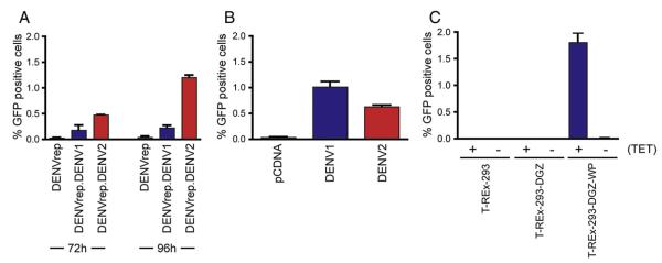
Production of DENV RVPs by complementation. (A) DENV RVPs were produced by transfection of HEK-293T cells with plasmids encoding a DENV sub-genomic replicon and the structural proteins of DENV1 (Western Pacific strain) or DENV2 (16681 strain). Supernatants were harvested at the indicated times and titered on Raji-DCSIGNR cells. Infection was measured as a function of the percentage of GFP expressing cells at 72 h post-infection by flow-cytometry. (B) DENV RVPs were produced by transfection of a T-REx-293 cell line (T-REx-293-DGZ) that stably propagates a DENV2 sub-genomic replicon. Supernatants were harvested 72 h post-transfection and analyzed as in panel A. (C) DENV RVPs were produced using an inducible cell line that stably propagates a DENV2 replicon and expresses DENV structural proteins in a tetracycline dependent fashion. Parental T-REx-293 cells, T-REx-293-DGZ, and T-REx-293-DGZ-WP cells were induced in fresh low glucose media in the presence (+) or absence (−) of 1 μg/ml tetracycline. Supernatants were harvested 72 h post-transfection and analyzed as in panel A.
To improve the yield of infectious DENV RVPs, two additional complementation strategies were evaluated. Flaviviruses replicate in the cytoplasm of infected cells (Lindenbach, 2007). However, the DENV replicon employed in these experiments are transcribed in the nucleus and must be exported intact to the cytoplasm, a process that could be limited by the utilization of splicing signals, or elements that inhibit RNA export, present in the native DENV2 genomic RNA sequence. Therefore, we produced a cell line (T-REx-293-DGZ) that stably propagates the DENV replicon RNA, and transfected it with a plasmid encoding the DENV structural proteins. DENV RVP titers achieved using this approach were similar to those obtained when transfecting the replicon plasmid directly (Fig. 1B). To address the possibility that the low titers of DENV RVPs reflected limitations in transfection efficiency, we also constructed cell lines that propagate the DENV replicon and can be induced to express DENV C-prM-E. Using cell lines expressing either the structural proteins of DENV1 (T-REx-293-DGZ-WP) or DENV2 (T-REx-293-DGZ-16681) strains, we were able to produce RVPs in a tetracycline-inducible fashion, albeit with modest infectious titers (~103 IU/ml) (Fig. 1C). Titration studies varying the concentration of tetracycline added to the cell line revealed that the titer of infectious RVPs was relatively constant over a broad range of induction conditions (Fig. S1). Since each complementation approach yielded similar low titers of DENV RVPs, we investigated whether the limitations in DENV RVP production, relative to our analogous approaches for WNV, may reflect other aspects of DENV virion production, assembly and release.
Impact of temperature on DENV RVP production kinetics and decay
Because protein stability and folding efficiency may be greater at lower temperatures, we investigated whether more efficient DENV RVPs production could be achieved at reduced temperatures. T-REx-293-DGZ-WP cells were induced with tetracycline, incubated at the indicated temperatures and sampled for the release of infectious RVPs over the course of several days (Fig. 2A). Cells incubated at lower temperatures produced infectious RVPs more slowly than those incubated at 37 °C, consistent with the slower kinetics of structural gene expression in cells at lower temperatures (data not shown). In contrast, production of DENV RVPs at 37 °C was relatively transient and achieved modest titers relative to the peak RVP titers achieved using lower temperatures (Fig. 2A). The titer of DENV RVPs in supernatants of cells at 37 °C decreased significantly (46.6±0.1 fold, n=6, p<0.0001) between 24 and 48 h (Fig. 2D), whereas titers at 28 °C and 30 °C increased during the same time frame ((16.4±0.4 fold, p=0.0004) and (3.4±0.3 fold, p=0.0012) for 28 °C and 30 °C, respectively). By comparison, while the kinetics of WNV RVP production were also affected by temperature in a similar fashion, WNV RVP infectious titers at both 24 and 48 h were similar (Figs. 2B and 2E). Furthermore, that the production of DENV RVPs by HEK-293T cells transfected with DENV structural genes and a WNV replicon was also transient at 37 °C (Figs. 2C and 2F) suggests the rapid decrease in DENV RVP titers released from T-REx-293-DGZ-WP cells does not reflect unique properties of the DENV replicon packaged by these particles nor the inducible cell line used to produce them. We noted that the kinetics of RVPs released from transfected cells are slightly delayed relative to the inducible cell line. This may reflect the time required for transcription of the WNV DNA “launched” replicon RNA and the initiation of replicon RNA replication. This was not due to an intrinsic difference in the replication rate between WNV and DENV replicons, as the former replicates faster and to higher levels than the DENV replicon (Fig. S2).
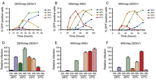
Temperature-dependent production of RVPs. (A) DENV1 RVPs were produced by induction of T-REx-293-DGZ-WP cells with tetracycline, followed by culture at the indicated temperatures. Culture supernatants were collected at the indicated times post-induction and titered using Raji-DCSIGNR cells. Infection was measured as a function of the percentage of GFP expressing cells at 72 h post-infection by flow-cytometry. (B) WNV RVPs were produced by transfection of HEK-293T cells with plasmids encoding a sub-genomic WNV replicon and WNV C-prM-E. Supernatants were collected at the indicated times post-transfection and titered as described above. (C) DENV1 RVPs that encapsidate a WNV genome were produced by transfection of HEK-293T cells with plasmids encoding a sub-genomic WNV replicon and DENV1 C-prM-E (Western Pacific strain). DENV RVPs were harvested at the indicated times and titered as described above. (D–F) To obtain a more quantitatively rigorous understanding of the temperature-dependent differences in DENV and WNV RVP production, RVPs were produced in replicate wells of a 6-well plate as described for A–C. RVP-containing supernatants were collected at 24 h and 48 h after induction and titered using Raji-DCSIGNR cells in triplicate. Infection is expressed relative to the titer achieved at 48 h post-infection at 37 °C. Error bars reflect the standard error of two independent production experiments.
To investigate the mechanisms that limit the duration and overall titer of DENV RVP production at 37 °C, we next measured the rate of RVP release from producer cells, and their decay in culture supernatants. The rate of virion release was estimated by measuring the titer of RVPs produced from cells during a four-hour window at 24 or 48 h post-induction or transfection (DENV and WNV, respectively). As suggested by our longitudinal and cross-sectional studies above (Figs. 2A and 2B), the rate of infectious DENV RVP production decreased significantly between days one and two for cells cultured at 37 °C (99.5± 0.1 fold, p=0.0032)(Fig. 3A), corresponding to the dramatic decrease in infectious DENV RVP titer present in culture supernatants by day two observed in longitudinal and cross-sectional sampling (Fig. 2A). By comparison, the rate of infectious WNV RVP release was only modestly reduced during the same interval. Infectious DENV RVP release from T-REx-293-DGZ-WP cells incubated at 28 °C and 30 °C increased significantly between day one and two (5.74±0.04 fold, p=0.0022, and 1.94±0.04 fold, p=0.0057, respectively). Analysis of the decay of the infectivity of DENV RVPs at each production temperature indicates that they are significantly less stable at 37 °C (T1/2=8.3 h, n=7) as compared to 28 °C (T1/2=34.2 h, n=6)(p<0.0001)(Fig. 4). WNV RVPs are significantly more stable than DENV RVPs at both 28 °C (T1/2=65.7 h, n=4) (p=0.0025) and 37 °C (T1/2=15.1 h, n=4) (p=0.0294).
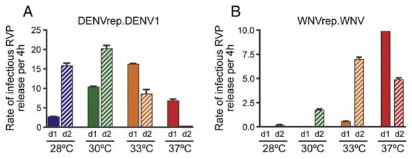
Temperature-dependent changes in the rate of DENV RVP production. The rate of infectious DENV and WNV RVP production at 24 and 48 h post-induction/transfection was investigated. DENV RVPs were produced by induction of T-REx-293-DGZ-WP cells (A) and WNV RVPs were produced by transfection of HEK-293T cells (B) as described in Fig. 2 and the Materials and methods. Culture supernatants were removed from duplicate wells at 22 h (d1) or 46 h (d2) post-induction and immediately replaced with conditioned media pre-equilibrated at the indicated temperature. Supernatant was collected 4 h later and titered in duplicate using Raji-DCSIGNR cells. Infection was measured as a function of the percent of GFP expressing cells at 72 h (A) or 48 h (B) post-infection by flow-cytometry. The data presented is representative of three independent experiments.
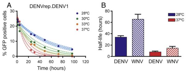
Stability of WNV and DENV RVPs. (A) DENV RVPs were produced using T-REx-293-DGZ-WP cells and aliquoted (500 μl) into wells of a 48-well plate. Plates were incubated at the indicated temperatures. At the indicated times, RVPs were removed from duplicate wells and frozen at −80 °C. The titer of RVPs from each time point was determined simultaneously by infecting Raji-DCSIGNR cells with serial two-fold dilutions of RVPs in complete RPMI-1640 medium. Infection was measured as a function of GFP expression at 72 h post-infection by flow-cytometry. Non-linear regression analysis was performed to generate one-phase exponential decay curves using GraphPad Prism version 4.0b. Dotted lines indicate the 95% confidence interval for the regression analysis. (B) The average half-life of WNV and DENV RVPs at 28 °C or 37 °C is displayed. Error bars indicate the standard error of multiple independent measurements of the stability of DENV and WNV at each temperature (n=6 and n=7 for DENV and WNV at 28 °C; n=7 and n=4 for DENV and WNV at 37 °C). Statistical comparisons were made using GraphPad Prism version 4.0b (unpaired t test).
Together, our data indicates that differences in the efficiency of WNV and DENV RVP production reflect both the dynamics of RVP production and differences in the stability of infectious RVPs at 37 °C. The titer of DENV RVPs produced at 37 °C is limited due to the transient release of virions by cells as measured functionally (Fig. 3A) and by the presence of RVP-associated replicon RNA in the supernatant (Fig. S3). Why DENV RVP producing cells stop releasing infectious virus particles is presently unclear, but does not appear to reflect overt cytotoxicity conferred by DENV protein expression. LDH-release from RVP producing cells increased only modestly with time at 37 °C, and was not dependent upon DENV structural gene expression. The level of replicon replication in 293T producer cells also decreased with time in culture at this temperature (~2-fold) (Fig. S4). As neither change in producer cell phenotype occurred with kinetics that parallel the rapid reduction of DENV RVP production observed above, it remains unclear whether these factors govern RVP production efficiency, or reflect changes that occur during prolonged culture of 293T cells at high density. Instead, the transient production of DENV RVPs at 37 °C revealed by longitudinal sampling (Fig. 2A) can be explained by the termination of infectious RVP release shortly after day one post-induction/transfection, followed by the decay of virions already released into the media with a half-life of approximately 8 h. Production at lower temperatures is more efficient because it increases the time during which producer cells release DENV RVPs, as well as decreases the rate of virion decay in the culture supernatant.
Infection of multiple cell lines with DENV RVPs
The expression of DCSIGN (CD209) and DCSIGNR (CD209L) on cell lines has been shown to significantly increase the efficiency of DENV infection, reflecting the role of these lectins in promoting more efficient and durable attachment of the virus to target cells (Navarro-Sanchez et al., 2003; Tassaneetrithep et al., 2003). Thus, to maximize the sensitivity of our RVP production optimization experiments we utilized a cell line that expresses DCSIGNR. We next explored whether DENV RVPs produced using the plasmid complementation approach described above were capable of infecting cell lines that do not express known attachment factors, including those commonly employed in studies of flaviviruses (BHK21 and Vero). DENV and WNV RVPs were produced by transfection of HEK-293Ts with plasmids encoding the structural proteins of either virus and a WNV replicon encoding the Renilla luciferase gene. The rationale for the selection of a WNV replicon was: i.) our studies described above did not suggest that the use of a heterologous replicon was associated with a significant reduction in RVP titer (compare Figs. 2B and C), ii.) to control for post-entry differences in replication between DENV and WNV, and iii.) the kinetics of reporter gene expression by WNV replicons is more rapid than that observed with DENV replicons of similar design (25 h vs 98 h for WNV and DENV replicons, respectively). Serial four-fold dilutions of RVPs were used to infect eleven cell lines of varying origin. Analysis of the signal obtained at the highest RVP dilution tested indicates varying degrees of permissiveness, as expected from published studies (Fig. 5) (Anderson, 2003; Clyde et al., 2006; Diamond et al., 2000). Signals greater than 1000-fold above background were obtained for both DENV and WNV RVPs on the most permissive lines, whereas infection of the Jurkat T cell line was not observed with either WNV or DENV RVPs. To get a more precise estimate of the relative infectivity of DENV and WNV RVPs on these lines, we next measured the genome content of each preparation and used the resulting data to normalize the level of infectivity between the two types of virus particles. To our surprise, we found that the ratio between infectivity and the relative numbers of replicon genomes in stocks of DENV RVPs was relatively low as compared to WNV RVPs, despite the similar infectious titers observed using several different cell lines (Fig. 5B). Interestingly, as a function of the relative number of genome copies, DENV RVPs were more infectious than WNV in certain cell lines (e.g. BHK) that do not express any known flavivirus attachment factor, suggesting that these particular strains of DENV (16681) and WNV (NY99) might utilize distinct attachment factors to infect certain cell lines (Fig. 5B).
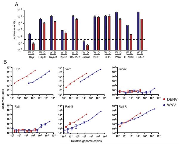
Infection of a panel of cell lines with DENV RVPs. (A) WNV and DENV RVPs were produced in HEK-293T cells transfected with a WNV sub-genomic replicon and plasmids encoding either WNV (blue) or DENV2 16881 (red) structural genes. Serial four-fold dilutions of RVPs were used to infect each cell line in quadruplicate as described in the Materials and methods section. Infection was scored as a function of Renilla luciferase activity 48 h post-infection. The signal obtained with undiluted RVPs is shown, with error bars indicating the standard error. (B) The infectivity of each population of RVPs was normalized as a function of the relative genomic RNA content of each preparation. Genomic RNA was measured using real-time qRT-PCR as described (Geiss et al., 2005; Houng et al., 2001).
Neutralization of DENV RVPs
We have employed WNV RVPs extensively to investigate the functional properties and mechanisms of action of neutralizing antibodies (Mehlhop et al., 2007; Nelson et al., 2008; Oliphant et al., 2007, 2006; Pierson et al., 2006, 2007). The key parameter that governs the precision and reproducibility of RVP-based measurements of neutralization is the ability to measure the outcome of antibody– virion interactions under conditions that satisfy the law of mass action (“antibody excess”) (Andrewes, 1933; Klasse and Sattentau, 2001; Pierson et al., 2006). Practically, this requires that RVPs are produced with a particle-to-infectious-particle ratio such that the concentration of antigen in RVP preparations is negligible with respect to the affinity of the antibodies investigated. This can be validated simply by studying the neutralization potency of antibodies using multiple concentrations of RVPs (Pierson et al., 2006). To validate the utility of our DENV RVP production approaches, we performed neutralization studies with three different concentrations of DENV1 RVPs produced in the T-REx-293-DGZ-WP cell line, and DENV1 immune sera obtained from experimentally-infected rhesus macaques (Whitehead et al., 2003). A similar study was also performed using DENV2 RVPs and DENV2 immune sera from experimentally-infected macaques (data not shown) (Blaney et al., 2004). Significant differences in neutralization titer were not observed with increasing dilution (Fig. 6A), nor were differences noted when DENV1 (Fig. 6B) or DENV2 (data not shown) RVPs produced by transfection or induction of our stable cell lines were studied in parallel. Overall, the variability of repeated assays of DENV1 (n=12) and DENV2 (n=4) immune sera using RVPs of the same serotype was approximately 15 and 26 percent of the average value, respectively (Fig. 6C). Together, these results highlight the utility of DENV for studying the functional properties of DENV neutralizing antibodies.

Measuring antibody-mediated neutralization using DENV RVPs. (A) To demonstrate that RVPs allow for the study of the functional properties of antibodies under conditions that satisfy the law of mass action (Andrewes, 1933; Klasse and Sattentau, 2001; Pierson et al., 2006), nine serial four-fold dilutions of DENV1-immune primate sera were incubated for 1 h at room temperature with serial dilutions of DENV1 RVPs. RVP-antibody complexes were then added to Raji-DCSIGNR cells in triplicate. Infection was measured as a function of GFP expression using flow-cytometry. Error bars display the standard error of triplicate infections. Non-linear regression analysis was used to calculate the EC50. (B) RVPs produced from T-REx-293-DGZ-WP cells and by plasmid transfection are neutralized equivalently. Dose response curves were generated using DENV1 or DENV2 (data not shown) immune sera as described above. (C) The neutralization titer of antibody present in the pooled DENV1 or DENV2-immune sera is shown. The error bar represents the average reciprocal dilution calculated from independent dose response curves composed of nine dilutions of sera performed in duplicate (DENV1, n=12 and DENV2, n=4, respectively). The error bar represents the standard error.
Concluding remarks
Pseudoinfectious reporter viruses that allow virus entry to be measured as a function of reporter gene expression have been employed extensively for many viruses, including flaviviruses. WNV RVPs produced by genetic complementation have provided an extremely quantitative and high-throughput approach for the study of antibody-mediated neutralization. While the direct application of these methods for the production of DENV RVPs was possible, by comparison to WNV, they were inefficient and of limited utility (Davis et al., 2006a, 2006b; Whitby et al., 2005). The identification of factors that limit the production of infectious DENV RVPs allowed the production of DENV RVPs with a sufficient titer to allow study of the functional properties of DENV antibodies (Mehlhop et al., 2007). In addition, the development of stable cell lines that can be induced to produce pseudoinfectious DENV RVPs represents a novel, low-cost approach that may be appropriately scaled to allow the study of the immune response of large numbers of infected or vaccinated individuals.
Materials and methods
Maintenance and production of cell lines
All cell lines were grown at 37 °C in the presence of 7% CO2. HEK-293T, HeLa, BHK21, HT1080, and Huh-7 cell lines were passaged in complete Dulbecco’s modified Eagle medium (DMEM)(complete DMEM: DMEM containing Glutamax and 25 mM Hepes, supplemented with 10% fetal bovine serum (FBS)(HyClone, Logan, UT) and 100 U/ ml penicillin–streptomycin (PS)). K562, Jurkat, and Raji cell lines were maintained in complete RPMI-1640 medium (complete RPMI) supplemented with 10% FBS and 100 U/ml penicillin–streptomycin. K562 and Raji lines that stably express DC-SIGN or DCSIGNR have been described elsewhere (Davis et al., 2006b; Pierson et al., 2007). Vero cells were cultured in complete Minimum Essential Medium (complete MEM) containing 10% FBS and 100 U/ml PS.
HEK-293T cell lines that stably propagate DENV replicons and can be induced to produce DENV RVPs were produced using Invitrogen’s T-REx-293 cell lines that constitutively express the Tet repressor. Parental T-REx-293 cell lines were propagated in complete DMEM supplemented with 5 μg/ml blasticidin S. A T-REx-293 cell line that stably propagates a DENV replicon was produced by infecting parental T-REx-293 cells with RVPs that encapsidate the DENV replicon, followed by selection in the presence of 5 μg/ml blasticidin S and 300 μg/ml zeocin (creating the T-REx-293-DGZ cell line). Inducible cell lines that release DENV RVPs were produced by transfection of the T-REx-293-DGZ cell line with plasmids that express the C-prM-E proteins of DENV strains Western Pacific (DENV1) and 16681 (DENV2). The resulting cell lines were selected and passaged in complete DMEM supplemented with 5 μg/ml blasticidin S, 300 μg/ml zeocin, and 500 μg/ml G418.
Plasmids
The WNV sub-genomic replicon and C-prM-E expression plasmid used throughout the study were described previously (Pierson et al., 2006). The DENV2 replicon was constructed as described below. DENV C-prM-E expression constructs (Western Pacific and 16681 strains) were constructed by topoisomerase mediated cloning into the pENTR/ D Gateway cloning entry vector. Inserts were sequenced and then transferred into pcDNA 6.2 and pT-REx-DEST30 destination vectors. The latter vector was used to construct the tetracycline-inducible cell lines described above.
Construction of a DNA-launched DENV2 replicon
A sub-genomic DENV2 replicon was constructed by modification of a molecular clone of the DENV2 strain 16681 using an approach described initially by Varnavski et al. (2000). Starting material for the work was the pD2/IC-30P molecular clone of the 16681 strain of DENV (Kinney et al., 1997). First, the majority of sequence encoding the structural genes of 16681 was deleted, and the T7 promoter of the pD2/IC-30P construct was replaced with the CMV immediate-early promoter/enhancer. To do this in the smallest number of steps, we performed overlapping PCR to fuse a fragment of a previously described sub-genomic WNV replicon that encodes the CMV promoter, WNV 5′ untranslated region, a small portion of the WNV capsid, and FMDV 2a autoprotease, and a MluI site with the DEN ORF (Pierson et al., 2006). The resulting PCR fragment was cleaved at BstX1 and MluI sites engineered into the primers and inserted into the pD2/IC-30P vector using unique SacI and KpnI sites. Importantly, the SacI site present in pD2/IC-30P was destroyed during this step, allowing the replacement of the WNV sequence in this cloning intermediate with sequence encoding the DEN 5′ UTR and 24 residues of DENV capsid. This was accomplished using a single PCR fragment and novel SacI and MluI sites engineered into the primers. The ribozyme of hepatitis delta virus (HDV) was then introduced into the 3′ end of the replicon using overlap extension PCR and introduced into the replicon via unique AvrII and ClaI sites present in the vector backbone. Subsequently, a GFP/Zeocin resistance reporter gene was inserted into the unique MluI site located just downstream of the FMDV2a autoprotease.
Production of reporter virus particles
RVPs were generated using two different approaches: i.) by transfection with plasmids encoding a sub-genomic replicon and structural genes (C-prM-E) as described (Pierson et al., 2006), or ii.) by induction of C-prM-E expression in 293T-Rex cell lines that stably propagate a DENV2 replicon. Production of RVPs by transfection was accomplished by introducing plasmids encoding a replicon and structural proteins (in a 1:2 ratio, by mass) into pre-plated HEK-293T cells using Lipofectamine 2000. RVPs were also produced by transfection of T-REx-293-DGZ cells with plasmids encoding DENV structural proteins. Four hours after transfection, culture media was removed and replaced with a low glucose formulation of DMEM containing 10% FBS and 100 U/ml PS and incubated at the indicated temperatures. RVP production using T-REx-293-DGZ-WP or T-REx-293-DGZ-16681 inducible cells lines was achieved following the addition of 1 μg/ml tetracycline. Using optimal production conditions for DENV RVPs, transfected cellswere incubated at 30 °C for approximately 72 h. RVP-containing supernatants were harvested, filtered using a 0.22 μm syringe filter and stored at −80 °C in complete low glucose DMEM.
RVP infections
The infectious titer of RVPs was typically determined by using Raji-DCSIGNR cells. Serial two-fold dilutions of RVPs were prepared using complete RPMI, and then added to Raji-DCSIGNR cells plated in 96-well plates at 5×10⁴ cells per well on the day of the assay. Infections were performed in a total volume of 200 μl. The percentage of cells infected at each dilution was scored as a function of GFP expression using flow-cytometry two days (when RVPs package a WNV replicon) or three days (when RVPs package a DENV replicon) post-infection. The rationale for performing serial dilutions of virus in each experiment reflects the lack of a linear relationship between virus dilution and the percentage of infected cells at all dilutions. In all cases, comparisons between RVP preparations were made at dilutions on the linear parts of the infection–dilution curve.
To compare the relative capacity of different cell lines to support E protein-mediated WNV or DENV entry, adherent cells were plated the day before the assay in 48-well plates ((BHK21, H1080, Huh-7 (20 k cells/well); Vero (30 k cells/well); HEK293T (40 k cells/well)). On the next day, media was removed and cells were infected with 100 μl of serial four-fold dilutions of RVPs. Cells were cultured at 37 °C overnight, and then media was replaced with 200 μl of a low glucose formulation of DMEM. In contrast, suspension cell lines (Raji, K562, and Jurkat) were plated in 96-well plates on the day of the assay (50 k cells/well) and infected with serial dilutions of RVPs (200 μl total volume). The relative infectivity was measured as a function of Renilla luciferase reporter gene expression at two days post-infection according to the manufacturers instructions.
Western blotting
Envelope protein expression in RVP-producing cells was detected by Western blot using the flavivirus pan-specific monoclonal antibody 4G2. Cells were lysed in 200 μl cell lysis buffer (1×PBS; 50 mM Tris. HCl; 150 mM NaCl; 2 mM EDTA; 1% Triton-X; protease inhibitor cocktail), spun briefly to remove cellular debris, and analyzed by BCA following the manufacturer’s protocol (BCA Protein Assay Kit—Pierce). Roughly 50 μg of total protein was analyzed by SDS-PAGE under non-reducing conditions and immunoblotted with 4G2 at 1 μg/ml.
Quantitative real-time PCR
The genomic RNA content of RVP populations was measured using a modification of a previously described protocol (Hanna et al., 2005). Briefly, RVP-containing supernatants were treated with 100 U recombinant DNAse I, followed by RNA isolation using the QiaAmp Viral RNA kit per the manufacturers instructions (Qiagen, Hilden, GE). Amplification of viral genomic RNA was accomplished using the Superscript III one-step RT-PCR system (Invitrogen, Carlsbad, CA) and primers specific for either the 3′ untranslated region of WNV lineage II replicon (Geiss et al., 2005) or dengue 2 replicon (Houng et al., 2001).
Neutralization of DV reporter virus particles
Neutralization of DENV RVPs was performed as described previously for WNV RVPs (Nelson et al., 2008; Pierson et al., 2006, 2007). Experiments using RVPs encapsidating a WNV replicon were analyzed on day two, whereas studies employing a DENV replicon were analyzed on day three. Antibody potency was calculated from dose–response data by non-linear regression analysis using GraphPad Prism v4.0b as described (Pierson et al., 2007).
Supplementary Material
Fig S1
Fig S2
Fig S3
Fig S4
Sup. legends
Acknowledgments
This work was supported by the Intramural Research Program of the NIH, National Institutes of Allergy and Infectious Diseases (NIAID) and by the Pediatric Dengue Vaccine Initiative (PDVI). We are grateful to Qing Xu for technical support and Dr. Bridget Puffer for providing the DENV C-prM-E expression vectors used throughout this study, for excellent technical assistance, and productive discussions. We thank Drs. Christopher Buck, Subhajit Poddar, and Michael Diamond for useful discussions and their comments on the manuscript.
Footnotes
Appendix A. Supplementary data Supplementary data associated with this article can be found, in the online version, at 10.1016/j.virol.2008.08.021.
References
- Alvarez M, Rodriguez-Roche R, Bernardo L, Vazquez S, Morier L, Gonzalez D, Castro O, Kouri G, Halstead SB, Guzman MG. Dengue hemorrhagic fever caused by sequential dengue 1–3 virus infections over a long time interval: Havana epidemic, 2001–2002. Am. J. Trop. Med. Hyg. 2006;75(6):1113–1117. [Abstract] [Google Scholar]
- Anderson R. Manipulation of cell surface macromolecules by flaviviruses. Adv. Virus Res. 2003;59:229–274. [Europe PMC free article] [Abstract] [Google Scholar]
- Andrewes C.H.a.E., W.J. Observations on anti-phage sera. I. ‘The percentage law’ Br. J. Exp. Pathol. 1933;14:367–376. [Google Scholar]
- Blaney JE, Jr., Hanson CT, Hanley KA, Murphy BR, Whitehead SS. Vaccine candidates derived from a novel infectious cDNA clone of an American genotype dengue virus type 2. BMC. Infect Dis. 2004;4:39. [Europe PMC free article] [Abstract] [Google Scholar]
- Clyde K, Kyle JL, Harris E. Recent advances in deciphering viral and host determinants of dengue virus replication and pathogenesis. J. Virol. 2006;80(23):11418–11431. [Europe PMC free article] [Abstract] [Google Scholar]
- Davis CW, Mattei LM, Nguyen HY, Ansarah-Sobrinho C, Doms RW, Pierson TC. The Location of asparagine-linked glycans on West Nile virions controls their interactions with CD209 (dendritic cell-specific ICAM-3 grabbing nonintegrin) J. Biol. Chem. 2006a;281(48):37183–37194. [Abstract] [Google Scholar]
- Davis CW, Nguyen HY, Hanna SL, Sanchez MD, Doms RW, Pierson TC. West Nile virus discriminates between DC-SIGN and DC-SIGNR for cellular attachment and infection. J. Virol. 2006b;80(3):1290–1301. [Europe PMC free article] [Abstract] [Google Scholar]
- Diamond MS, Edgil D, Roberts TG, Lu B, Harris E. Infection of human cells by dengue virus is modulated by different cell types and viral strains. J. Virol. 2000;74(17):7814–7823. [Europe PMC free article] [Abstract] [Google Scholar]
- Endy TP, Nisalak A, Chunsuttitwat S, Vaughn DW, Green S, Ennis FA, Rothman AL, Libraty DH. Relationship of preexisting dengue virus (DV) neutralizing antibody levels to viremia and severity of disease in a prospective cohort study of DV infection in Thailand. J. Infect Dis. 2004;189(6):990–1000. [Abstract] [Google Scholar]
- Fayzulin R, Scholle F, Petrakova O, Frolov I, Mason PW. Evaluation of replicative capacity and genetic stability of West Nile virus replicons using highly efficient packaging cell lines. Virology. 2006;351(1):196–209. [Abstract] [Google Scholar]
- Gehrke R, Ecker M, Aberle SW, Allison SL, Heinz FX, Mandl CW. Incorporation of tick-borne encephalitis virus replicons into virus-like particles by a packaging cell line. J. Virol. 2003;77(16):8924–8933. [Europe PMC free article] [Abstract] [Google Scholar]
- Geiss BJ, Pierson TC, Diamond MS. Actively replicating West Nile virus is resistant to cytoplasmic delivery of siRNA. Virol. J. 2005;2:53. [Europe PMC free article] [Abstract] [Google Scholar]
- Goto A, Yoshii K, Obara M, Ueki T, Mizutani T, Kariwa H, Takashima I. Role of the N-linked glycans of the prM and E envelope proteins in tick-borne encephalitis virus particle secretion. Vaccine. 2005;23(23):3043–3052. [Abstract] [Google Scholar]
- Gubler DJ, Meltzer M. Impact of dengue/dengue hemorrhagic fever on the developing world. Adv. Virus Res. 1999;53:35–70. [Abstract] [Google Scholar]
- Gubler D, K.G., Markoff L. Flaviviruses. In: H.P., Knipe DM, Griffin DE, Lamb RA, Martin MA, editors. Fields Virology, 1. 5th 2 vols. Lippincott Wlliams & Wilkins; Philadelphia: 2007. pp. 1153–1252. [Google Scholar]
- Halstead SB. Neutralization and antibody-dependent enhancement of dengue viruses. Adv. Virus Res. 2003;60:421–467. [Abstract] [Google Scholar]
- Hanna SL, Pierson TC, Sanchez MD, Ahmed AA, Murtadha MM, Doms RW. N-linked glycosylation of West Nile virus envelope proteins influences particle assembly and infectivity. J. Virol. 2005;79(21):13262–13274. [Europe PMC free article] [Abstract] [Google Scholar]
- Harvey TJ, Liu WJ, Wang XJ, Linedale R, Jacobs M, Davidson A, Le TT, Anraku I, Suhrbier A, Shi PY, Khromykh AA. Tetracycline-inducible packaging cell line for production of flavivirus replicon particles. J. Virol. 2004;78(1):531–538. [Europe PMC free article] [Abstract] [Google Scholar]
- Hayasaka D, Yoshii K, Ueki T, Iwasaki T, Takashima I. Sub-genomic replicons of Tick-borne encephalitis virus. Arch. Virol. 2004;149(6):1245–1256. [Abstract] [Google Scholar]
- Houng HS, Chung-Ming Chen R, Vaughn DW, Kanesa-thasan N. Development of a fluorogenic RT-PCR system for quantitative identification of dengue virus serotypes 1–4 using conserved and serotype-specific 3′ noncoding sequences. J. Virol. Methods. 2001;95(1–2):19–32. [Abstract] [Google Scholar]
- Jones CT, Patkar CG, Kuhn RJ. Construction and applications of yellow fever virus replicons. Virology. 2005;331(2):247–259. [Abstract] [Google Scholar]
- Khromykh AA, Westaway EG. Subgenomic replicons of the flavivirus Kunjin: construction and applications. J. Virol. 1997;71(2):1497–1505. [Europe PMC free article] [Abstract] [Google Scholar]
- Khromykh AA, Kenney MT, Westaway EG. trans-Complementation of flavivirus RNA polymerase gene NS5 by using Kunjin virus replicon-expressing BHK cells. J. Virol. 1998a;72(9):7270–7279. [Europe PMC free article] [Abstract] [Google Scholar]
- Khromykh AA, Varnavski AN, Westaway EG. Encapsidation of the flavivirus kunjin replicon RNA by using a complementation system providing Kunjin virus structural proteins in trans. J. Virol. 1998b;72(7):5967–5977. [Europe PMC free article] [Abstract] [Google Scholar]
- Kinney RM, Butrapet S, Chang GJ, Tsuchiya KR, Roehrig JT, Bhamarapravati N, Gubler DJ. Construction of infectious cDNA clones for dengue 2 virus: strain 16681 and its attenuated vaccine derivative, strain PDK-53. Virology. 1997;230(2):300–308. [Abstract] [Google Scholar]
- Klasse PJ, Sattentau QJ. Mechanisms of virus neutralization by antibody. Curr. Top Microbiol. Immunol. 2001;260:87–108. [Abstract] [Google Scholar]
- Lai CY, Hu HP, King CC, Wang WK. Incorporation of dengue virus replicon into virus-like particles by a cell line stably expressing precursor membrane and envelope proteins of dengue virus type 2. J. Biomed. Sci. 2008;15(1):15–27. [Abstract] [Google Scholar]
- Lindenbach BD, Thiel HJ, Rice CM. Flaviviridae: the viruses and their replication. In: H.P.M., D.M., Knipe, Griffin DE, Lamb RA, Martin MA, editors. Fields Virology, 1. 5th 2 vols. Lippincott Williams & Wilkins; Philadelphia: 2007. pp. 1101–1152. [Google Scholar]
- Mackenzie JS, Gubler DJ, Petersen LR. Emerging flaviviruses: the spread and resurgence of Japanese encephalitis, West Nile and dengue viruses. Nat. Med. 2004;10(12 Suppl):S98–109. [Abstract] [Google Scholar]
- Mehlhop E, Ansarah-Sobrinho C, Johnson S, Engle M, Fremont DH, Pierson TC, Diamond MS. Complement protein C1q inhibits antibody-dependent enhancement of flavivirus infection in an IgG subclass-specific manner. Cell Host Microbe. 2007;2(6):417–426. [Europe PMC free article] [Abstract] [Google Scholar]
- Molenkamp R, Kooi EA, Lucassen MA, Greve S, Thijssen JC, Spaan WJ, Bredenbeek PJ. Yellow fever virus replicons as an expression system for hepatitis C virus structural proteins. J. Virol. 2003;77(2):1644–1648. [Europe PMC free article] [Abstract] [Google Scholar]
- Monath TP. Dengue and yellow fever-challenges for the development and use of vaccines. N. Engl. J. Med. 2007;357(22):2222–2225. [Abstract] [Google Scholar]
- Mongkolsapaya J, Dejnirattisai W, Xu XN, Vasanawathana S, Tangthawornchaikul N, Chairunsri A, Sawasdivorn S, Duangchinda T, Dong T, Rowland-Jones S, Yenchitsomanus PT, McMichael A, Malasit P, Screaton G. Original antigenic sin and apoptosis in the pathogenesis of dengue hemorrhagic fever. Nat. Med. 2003;9(7):921–927. [Abstract] [Google Scholar]
- Navarro-Sanchez E, Altmeyer R, Amara A, Schwartz O, Fieschi F, Virelizier JL, Arenzana-Seisdedos F, Despres P. Dendritic-cell-specific ICAM3-grabbing non-integrin is essential for the productive infection of human dendritic cells by mosquito-cell-derived dengue viruses. EMBO. Rep. 2003;4(7):723–728. [Europe PMC free article] [Abstract] [Google Scholar]
- Nelson S, Jost CA, Xu Q, Ess J, Martin JE, Oliphant T, Whitehead SS, Durbin AP, Graham BS, Diamond MS, Pierson TC. Maturation of West Nile virus modulates sensitivity to antibody-mediated neutralization. PLOS Pathogens. 2008;4:e1000060. [Europe PMC free article] [Abstract] [Google Scholar]
- Oliphant T, Nybakken GE, Engle M, Xu Q, Nelson CA, Sukupolvi-Petty S, Marri A, Lachmi BE, Olshevsky U, Fremont DH, Pierson TC, Diamond MS. Antibody recognition and neutralization determinants on domains I and II of West Nile Virus envelope protein. J. Virol. 2006;80(24):12149–12159. [Europe PMC free article] [Abstract] [Google Scholar]
- Oliphant T, Nybakken GE, Austin SK, Xu Q, Bramson J, Loeb M, Throsby M, Fremont DH, Pierson TC, Diamond MS. Induction of epitope-specific neutralizing antibodies against West Nile virus. J. Virol. 2007;81(21):11828–11839. [Europe PMC free article] [Abstract] [Google Scholar]
- Pang X, Zhang M, Dayton AI. Development of Dengue virus type 2 replicons capable of prolonged expression in host cells. B.M.C. Microbiol. 2001;1:18. [Europe PMC free article] [Abstract] [Google Scholar]
- Pierson TC, Diamond MS, Ahmed AA, Valentine LE, Davis CW, Samuel MA, Hanna SL, Puffer BA, Doms RW. An infectious West Nile virus that expresses a GFP reporter gene. Virology. 2005;334(1):28–40. [Abstract] [Google Scholar]
- Pierson TC, Sanchez MD, Puffer BA, Ahmed AA, Geiss BJ, Valentine LE, Altamura LA, Diamond MS, Doms RW. A rapid and quantitative assay for measuring antibody-mediated neutralization of West Nile virus infection. Virology. 2006;346(1):53–65. [Abstract] [Google Scholar]
- Pierson TC, Xu Q, Nelson S, Oliphant T, Nybakken GE, Fremont DH, Diamond MS. The stoichiometry of antibody-mediated neutralization and enhancement of West Nile virus infection. Cell Host Microbe. 2007;1(2):135–145. [Europe PMC free article] [Abstract] [Google Scholar]
- Rothman AL. Dengue: defining protective versus pathologic immunity. J. Clin. Invest. 2004;113(7):946–951. [Europe PMC free article] [Abstract] [Google Scholar]
- Sanchez MD, Pierson TC, McAllister D, Hanna SL, Puffer BA, Valentine LE, Murtadha MM, Hoxie JA, Doms RW. Characterization of neutralizing antibodies to West Nile virus. Virology. 2005;336(1):70–82. [Abstract] [Google Scholar]
- Sanchez MD, Pierson TC, Degrace MM, Mattei LM, Hanna SL, Del Piero F, Doms RW. The neutralizing antibody response against West Nile virus in naturally infected horses. Virology. 2007;359(2):336–348. [Abstract] [Google Scholar]
- Scholle F, Girard YA, Zhao Q, Higgs S, Mason PW. trans-Packaged West Nile virus-like particles: infectious properties in vitro and in infected mosquito vectors. J. Virol. 2004;78(21):11605–11614. [Europe PMC free article] [Abstract] [Google Scholar]
- Shi PY, Tilgner M, Lo MK. Construction and characterization of subgenomic replicons of New York strain of West Nile virus. Virology. 2002;296(2):219–233. [Abstract] [Google Scholar]
- Stephens HA, Klaythong R, Sirikong M, Vaughn DW, Green S, Kalayanarooj S, Endy TP, Libraty DH, Nisalak A, Innis BL, Rothman AL, Ennis FA, Chandanayingyong D. HLA-A and -B allele associations with secondary dengue virus infections correlate with disease severity and the infecting viral serotype in ethnic Thais. Tissue Antigens. 2002;60(4):309–318. [Abstract] [Google Scholar]
- Tassaneetrithep B, Burgess TH, Granelli-Piperno A, Trumpfheller C, Finke J, Sun W, Eller MA, Pattanapanyasat K, Sarasombath S, Birx DL, Steinman RM, Schlesinger S, Marovich MA. DC-SIGN (CD209) mediates dengue virus infection of human dendritic cells. J. Exp. Med. 2003;197(7):823–829. [Europe PMC free article] [Abstract] [Google Scholar]
- Varnavski AN, Young PR, Khromykh AA. Stable high-level expression of heterologous genes in vitro and in vivo by noncytopathic DNA-based Kunjin virus replicon vectors. J. Virol. 2000;74(9):4394–4403. [Europe PMC free article] [Abstract] [Google Scholar]
- Vaughn DW, Green S, Kalayanarooj S, Innis BL, Nimmannitya S, Suntayakorn S, Endy TP, Raengsakulrach B, Rothman AL, Ennis FA, Nisalak A. Dengue viremia titer, antibody response pattern, and virus serotype correlate with disease severity. J. Infect Dis. 2000;181(1):2–9. [Abstract] [Google Scholar]
- Whitby K, Pierson TC, Geiss B, Lane K, Engle M, Zhou Y, Doms RW, Diamond MS. Castanospermine, a potent inhibitor of dengue virus infection in vitro and in vivo. J. Virol. 2005;79(14):8698–8706. [Europe PMC free article] [Abstract] [Google Scholar]
- Whitehead SS, Falgout B, Hanley KA, Blaney JE, Jr., Markoff L, Murphy BR. A live, attenuated dengue virus type 1 vaccine candidate with a 30-nucleotide deletion in the 3′ untranslated region is highly attenuated and immunogenic in monkeys. J. Virol. 2003;77(2):1653–1657. [Europe PMC free article] [Abstract] [Google Scholar]
- Whitehead SS, Blaney JE, Durbin AP, Murphy BR. Prospects for a dengue virus vaccine. Nat. Rev. Microbiol. 2007;5(7):518–528. [Abstract] [Google Scholar]
- WHO . Dengue Haemorrhagic Fever: Diagnosis, Treatment, Prevention and Control. Geneva: 1997. [Google Scholar]
- Yoshii K, Hayasaka D, Goto A, Kawakami K, Kariwa H, Takashima I. Packaging the replicon RNA of the Far-Eastern subtype of tick-borne encephalitis virus into single-round infectious particles: development of a heterologous gene delivery system. Vaccine. 2005;23(30):3946–3956. [Abstract] [Google Scholar]
- Yoshii K, Goto A, Kawakami K, Kariwa H, Takashima I. Construction and application of chimeric virus-like particles of tick-borne encephalitis virus and mosquito-borne Japanese encephalitis virus. J. Gen. Virol. 2008;89(Pt 1):200–211. [Abstract] [Google Scholar]
Full text links
Read article at publisher's site: https://doi.org/10.1016/j.virol.2008.08.021
Read article for free, from open access legal sources, via Unpaywall:
https://europepmc.org/articles/pmc3428711?pdf=render
Citations & impact
Impact metrics
Article citations
B cell receptor dependent enhancement of dengue virus infection.
PLoS Pathog, 20(10):e1012683, 31 Oct 2024
Cited by: 0 articles | PMID: 39480886 | PMCID: PMC11556684
SARS-CoV-2 Neutralization Assays Used in Clinical Trials: A Narrative Review.
Vaccines (Basel), 12(5):554, 18 May 2024
Cited by: 2 articles | PMID: 38793805 | PMCID: PMC11125816
Review Free full text in Europe PMC
The Dynamic Landscape of Capsid Proteins and Viral RNA Interactions in Flavivirus Genome Packaging and Virus Assembly.
Pathogens, 13(2):120, 28 Jan 2024
Cited by: 1 article | PMID: 38392858 | PMCID: PMC10893219
Review Free full text in Europe PMC
Defining the impact of flavivirus envelope protein glycosylation site mutations on sensitivity to broadly neutralizing antibodies.
mBio, 15(2):e0304823, 09 Jan 2024
Cited by: 1 article | PMID: 38193697 | PMCID: PMC10865826
The effect of single mutations in Zika virus envelope on escape from broadly neutralizing antibodies.
J Virol, 97(11):e0141423, 09 Nov 2023
Cited by: 0 articles | PMID: 37943046 | PMCID: PMC10688354
Go to all (87) article citations
Data
Data behind the article
This data has been text mined from the article, or deposited into data resources.
BioStudies: supplemental material and supporting data
Similar Articles
To arrive at the top five similar articles we use a word-weighted algorithm to compare words from the Title and Abstract of each citation.
Pseudo-infectious reporter virus particles for measuring antibody-mediated neutralization and enhancement of dengue virus infection.
Methods Mol Biol, 1138:75-97, 01 Jan 2014
Cited by: 25 articles | PMID: 24696332
Dengue reporter virus particles for measuring neutralizing antibodies against each of the four dengue serotypes.
PLoS One, 6(11):e27252, 09 Nov 2011
Cited by: 49 articles | PMID: 22096543 | PMCID: PMC3212561
Zika Virus Is Not Uniquely Stable at Physiological Temperatures Compared to Other Flaviviruses.
mBio, 7(5):e01396-16, 06 Sep 2016
Cited by: 32 articles | PMID: 27601578 | PMCID: PMC5013301
Using a Virion Assembly-Defective Dengue Virus as a Vaccine Approach.
J Virol, 92(21):e01002-18, 12 Oct 2018
Cited by: 12 articles | PMID: 30111567 | PMCID: PMC6189489
Funding
Funders who supported this work.
Intramural NIH HHS (2)
Grant ID: ZIA AI000957
Grant ID: ZIA AI000957-05


