Abstract
Context
Tissue eosinophilia in oral squamous cell carcinoma has been well - recognized. Studies have reported both favorable and unfavorable prognoses associated with tissue eosinophils in oral squamous cell carcinoma. However, the role of eosinophils in the development of tumor is still unclear.Aims
The present study was an attempt to elucidate the potential role of tissue eosinophils in oral leukoplakia, a potentially malignant lesion.Settings and design
To count eosinophils in tissues of normal subjects and oral leukoplakia cases. To compare tissue eosinophil count (TEC) between normal and oral leukoplakia cases. To compare TEC between dysplastic and non-dysplastic cases of oral leukoplakia and to correlate with degree of epithelial dysplasia.Materials and methods
A total of 85 cases (59 cases of oral leukoplakia and 26 normal oral tissues) constituted the study material. Tissue eosinophils were counted in 10 different high- power fields.Statistical analysis used
Non-parametric tests (Mann-Whitney U-test, Kruskal-Wallis test, Mann-Whitney post hoc analysis and Spearman's correlation statistics).Results
Mean eosinophil count (MEC) in oral leukoplakia cases was significantly more when compared to normal subjects. MEC in dysplastic cases of oral leukoplakia was significantly more when compared to those without epithelial dysplasia (Mann-Whitney U-test). Furthermore, MEC was directly proportional to the degree of epithelial dysplasia (Spearman's correlation statistics).Conclusions
TEC may be used as an adjunct to predict the malignant transformation of dysplastic cases of oral leukoplakia. Eosinophilic infiltration in oral dysplastic cases should prompt a thorough evaluation for invasiveness, especially when features of invasion are absent or suspected in smaller biopsy specimens. Use of TEC as a prognostic indicator demands larger sample size and mandates long-term follow-up.Free full text

Role of tissue eosinophils in oral Leukoplakia: A pilot study
Abstract
Context:
Tissue eosinophilia in oral squamous cell carcinoma has been well - recognized. Studies have reported both favorable and unfavorable prognoses associated with tissue eosinophils in oral squamous cell carcinoma. However, the role of eosinophils in the development of tumor is still unclear.
Aims:
The present study was an attempt to elucidate the potential role of tissue eosinophils in oral leukoplakia, a potentially malignant lesion.
Settings and Design:
To count eosinophils in tissues of normal subjects and oral leukoplakia cases. To compare tissue eosinophil count (TEC) between normal and oral leukoplakia cases. To compare TEC between dysplastic and non-dysplastic cases of oral leukoplakia and to correlate with degree of epithelial dysplasia.
Materials and Methods:
A total of 85 cases (59 cases of oral leukoplakia and 26 normal oral tissues) constituted the study material. Tissue eosinophils were counted in 10 different high- power fields.
Statistical Analysis Used:
Non-parametric tests (Mann-Whitney U-test, Kruskal-Wallis test, Mann-Whitney post hoc analysis and Spearman's correlation statistics).
Results:
Mean eosinophil count (MEC) in oral leukoplakia cases was significantly more when compared to normal subjects. MEC in dysplastic cases of oral leukoplakia was significantly more when compared to those without epithelial dysplasia (Mann-Whitney U-test). Furthermore, MEC was directly proportional to the degree of epithelial dysplasia (Spearman's correlation statistics).
Conclusions:
TEC may be used as an adjunct to predict the malignant transformation of dysplastic cases of oral leukoplakia. Eosinophilic infiltration in oral dysplastic cases should prompt a thorough evaluation for invasiveness, especially when features of invasion are absent or suspected in smaller biopsy specimens. Use of TEC as a prognostic indicator demands larger sample size and mandates long-term follow-up.
INTRODUCTION
Wharton Jones (1846) first described the eosinophils as “coarse granular cells” and later Paul Ehrlich in 1880 as “eosinophils”.[1] Eosinophils show abundant cytoplasm with coarse reflective granules.[2] They are distinguished by their tinctorial properties exhibiting bright red staining with acid aniline dyes.[3] Eosinophils appear to be pleiotropic and play a significant role in health and disease. These multifunctional leukocytes are engaged in initiation and propagation of a variety of inflammatory responses comprising parasitic, helminth, viral and bacterial infections; allergic diseases; tissue injury and also are modulators of innate and adaptive immunity.[4]
Tissue eosinophilia has been recognized in many neoplasms including oral squamous cell carcinoma.[5] The inflammatory cells in tumor stroma are a result of host response to tumor cells. Similar inflammatory infiltrate may be seen in potentially malignant lesions such as leukoplakia, though not many studies are done on this.[6,7] Many oral carcinomas are preceded by these potentially malignant lesions/conditions. These potentially malignant lesions show simple hyperkeratosis to different degrees of epithelial dysplasia (mild, moderate and severe); and can be associated with risk of oral carcinoma.
A diagnosis of leukoplakia (a potentially malignant lesion) is made when a white lesion at clinical examination cannot be clearly diagnosed as any other disease of oral mucosa, except for a lesion associated with the use of tobacco. The diagnosis should be supported by microscopic findings to establish the presence or absence of epithelial dysplasia and to exclude malignancy or other mucosal lesions.[8,9]
Review of English literature revealed very few studies on tissue eosinophilia in oral leukoplakia, a potentially malignant lesion. Hence, the present study aimed at elucidating the role of tissue eosinophils in oral leukoplakia. Moreover, tissue eosinophilia was correlated with the degree of epithelial dysplasia in dysplastic cases of oral leukoplakia, thereby checking whether tissue eosinophilia could be used as an adjunct to predict malignant transformation.
MATERIALS AND METHODS
Tissue blocks belonging to cases diagnosed as oral leukoplakia (and confirmed by microscopic findings) from the archives of the Department of Oral and Maxillofacial Pathology, constituted the study sample.
Tissue blocks of normal oral samples, obtained from the patients during minor oral surgical procedures (from the Departments of Oral and Maxillofacial Surgery and Periodontics) were included as control group. The cases for tissue eosinophil count (TEC) were selected at random and they were continuous biopsies.
A total of 85 cases (26 normal oral mucosal samples and 59 of oral leukoplakia) constituted the study sample.
Cases diagnosed as oral leukoplakia (including 50 cases of homogenous leukoplakia, eight cases of speckled leukoplakia and one case of proliferative verrucous leukoplakia) were included in the study. Fifty – two were male patients and seven were females; age group was from the second to seventh decade; 49 cases had presented lesion on buccal mucosa, five on commissural area, four on labial mucosa and one case on lateral border of the tongue. Fifty-eight out of 59 cases were associated with tobacco habit and one case was not associated, for which a provisional diagnosis of idiopathic leukoplakia was given.
Cases with any other lesion other than oral leukoplakia; patients with systemic diseases and allergic conditions; oral mucosal sample with inflammation and periodontal disease were excluded from the study.
All the procedures were done under strict aseptic precautions. Written informed consent was obtained from the patients. The confidentiality of case details procured for the study purpose was maintained. Clearance from the institutional ethical board was obtained.
Tissue sections of 4 – 5 μm thick were prepared from formalin fixed paraffin embedded tissue blocks, using semiautomatic soft-tissue microtome and were transferred onto clean glass slides coated with suitable adhesive. The sections were stained with H and E stains.
Eosinophils were counted in 10 random high-power fields (using modified Zig Zag pattern) without overlap and recorded as the number of eosinophils/10 high power fields [Figures [Figures11–4]. Cells with bilobed nucleus and intensely red cytoplasmic granules were accepted as eosinophils. Red blood cells with superimposed mononuclear and polymorphonuclear leukocytes were excluded. Eosinophils in lymphovascular channels were not included.
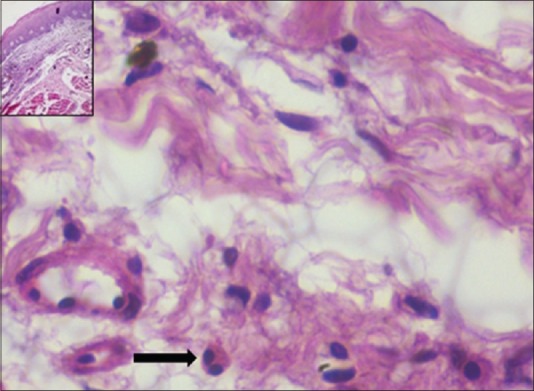
The section shows tissue eosinophil in a case of hyperkeratotic lesion (H&E stain, x400). Inset: Scanner view of the lesion (H&E stain, x40)
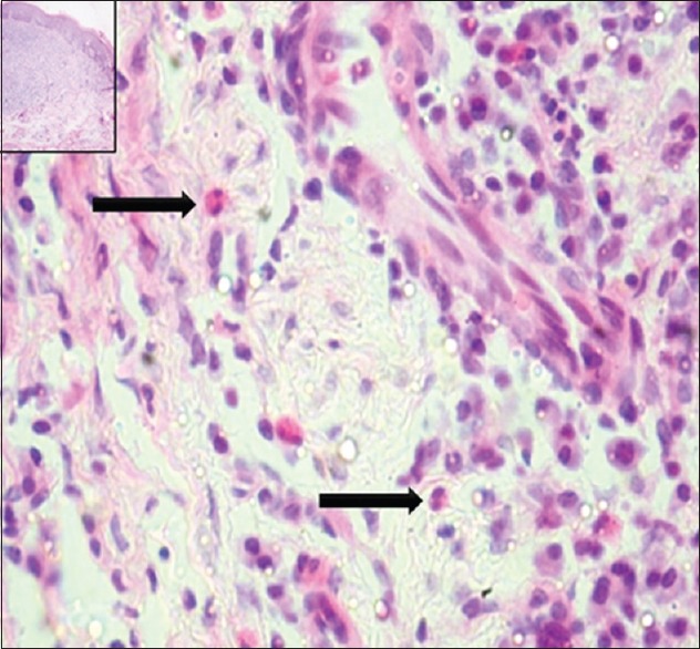
The section shows tissue eosinophil in a case of severe epithelial dysplasia (H&E stain, x400). Inset: Scanner view of the lesion (H&E stain, x40)
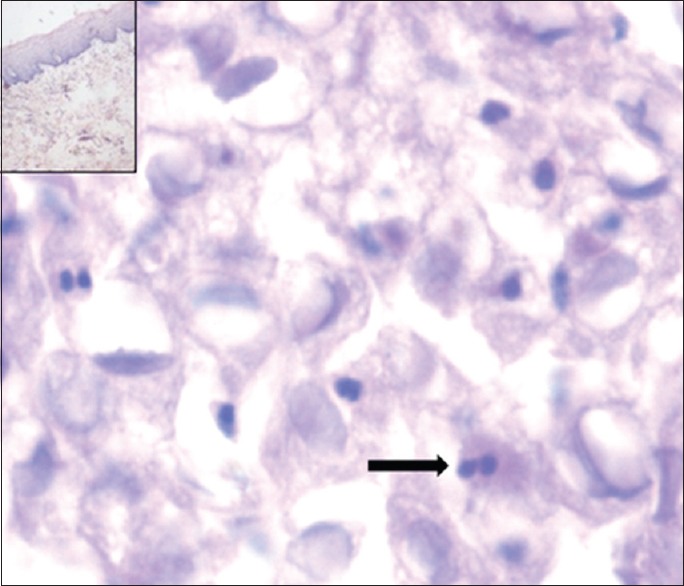
The section shows tissue eosinophil in a case of mild epithelial dysplasia (H&E stain, x400). Inset: Scanner view of the lesion (H&E stain, x40)
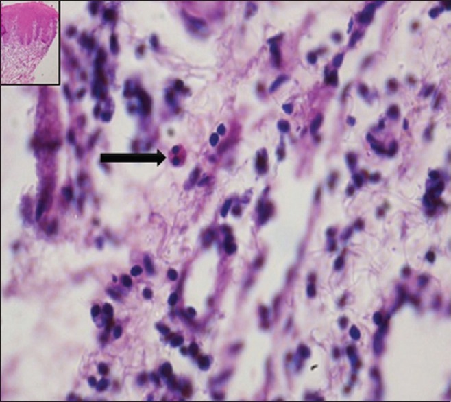
The section shows tissue eosinophil in a case of moderate epithelial dysplasia (H&E stain, x400). Inset: Scanner view of the lesion (H&E stain, x40)
Burkhart and Maerkar grading system was used earlier as some of the cases were received with a provisional diagnosis of oral leukoplakia with superimposed candidiasis; however, these cases did not reveal the presence of candidal hyphae. For the purpose of the present study, the selected cases were graded again using the WHO system.[8,9,10]
Grading of epithelial dysplasia was done by four observers and a common opinion given by three observers was taken into consideration.
The data were entered in the master chart for further interpretation and evaluation.
As the sample size was not uniform among the study groups, non-parametric tests (Mann-Whitney U-test, Kruskal - Wallis test and Spearman's correlation statistics) were used for analysis of the data.
RESULTS
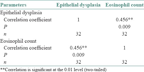
A total of 85 cases (59 cases of oral leukoplakia and 26 of normal subjects) were evaluated for TEC. The TEC was found to be significantly more in oral leukoplakia cases when compared to normal tissues [Table 1]. On comparing, raised level of TEC was observed in dysplastic cases of oral leukoplakia when compared to non-dysplastic cases [Table 2]. Mean eosinophil count (MEC) was observed to be higher in severe grade of epithelial dysplasia when the count was compared among mild, moderate and severe grades of epithelial dysplasia [Table 3]. In addition, the eosinophil count was directly proportional to the degree of epithelial dysplasia [Table 4].
Table 1
Comparison of eosinophil count between oral leukoplakia cases and controls using Mann–Whitney U-test

Table 2
Comparison of eosinophil count between nondysplastic and dysplastic cases using Mann–Whitney U-test

Table 3
Comparison of mean eosinophil count among different dysplastic lesions using Kruskal–Wallis test followed by Mann–Whitney U-test post-hoc analysis

Table 4
Spearman's correlation statistics to correlate the relationship of epithelial dysplasia and eosinophil count
DISCUSSION
The development of invasive cancer is multifactorial, associated with genetic alterations coupled with significant changes in host stromal, inflammatory/immune cells and endothelial cells. Host's immune response to neoplastic process is expressed as peritumoral and intratumoral inflammatory infiltrates.[11] The recruitment and activation of eosinophils toward the microenvironment of tumor involves a complex mechanism, mediated by inflammatory cytokines and chemokines, chiefly related to Th 2 response. Interleukin-4 (IL-4) and IL-13 are potent inducers of eotaxin chemokines that would explain the eosinophilia associated with Th2 responses. Activation of eosinophil involves histamine and eosinophilic chemotactic factor A in mast cells, neutrophil peptides, eosinophil stimulator and promoter substances in lymphocytes, eotaxin and C5a complement.[2] Even though eosinophils are frequently seen in human cancer, their functional role in malignancy still remains an ambiguity. The reported studies reveal favorable as well as unfavorable prognoses of squamous cell carcinoma associated with tissue eosinophilia. Some studies have shown no influence on patients’ outcome reflecting controversy attached with tumor-associated tissue eosinophilia.[11]
Hence, the present study was designed to investigate the role of tissue eosinophils in oral precancer.
Oral leukoplakia, a potentially malignant lesion, was defined by the WHO as a white patch or plaque that cannot be characterized clinically or histologically as any other disease.[12]
Oral leukoplakia is considered to be a clinicopathologic concept reflecting the biology of cellular atypia and epithelial dysplasia.[13] The reported prevalence of leukoplakia is 0.4%-0.7%. The incidence is higher in people who are habitual smokers or drinkers.[14]
The malignant transformation of oral leukoplakia has been reported to be 17.5%. The development of squamous carcinoma was seen in hyperkeratotic epithelial region with an average time of 8.1 years. Non-smoking, type of clinical lesion, and duration of the lesion have been advocated as additional attributes for malignant transformation of oral leukoplakia and more aggressive management has been suggested.[15]
A follow - up study on a hospital-based population comprising 166 patients with oral leukoplakia showed 2.9% annual malignant transformation rate. The median follow-up period in the study was 29 months. Factors associated with higher risk of malignant transformation were females, the absence of smoking habits in women and a non-homogenous clinical appearance.[16]
Figures on incidence and prevalence rates of oral leukoplakia as well as malignant transformation do vary according to the population studied and is dependent on the sample size.
Very few studies have been conducted on tissue eosinophilia in precancer. Some studies have found higher eosinophil counts in invasive tumors when compared to in situ neoplastic lesions. This suggests elevated eosinophil count as a histopathological marker associated with stromal invasion.[17,18]
Search in Google search engine with keywords such as “eosinophils in oral leukoplakia” in different combinations revealed only one reported study by Jain et al. They have assessed tissue eosinophilia using image analysis on Congo red stained sections of oral epithelial dysplasia and oral squamous cell carcinoma (metastatic and non-metastatic groups). In their study, MEC among mild, moderate and severe epithelial dysplasia groups in oral leukoplakia category was statistically insignificant.[7] In contrast, the present study revealed statistically significant difference in MEC in different grades of epithelial dysplasia of oral leukoplakia cases. In addition, MEC was directly proportional to the degree of epithelial dysplasia. Not all cases of severe epithelial dysplasia showed raised eosinophil count. Statistical analysis has shown that there are 60% chances to see higher eosinophil count with increase in severity of epithelial dysplasia [Tables [Tables33 and and4].4]. In addition, MEC was found to be more in cases of oral leukoplakia when compared to normal tissues [Table 1] and MEC was observed to be more again in dysplastic cases of oral leukoplakia on comparing it with non-dysplastic cases [Table 2].
Another interesting finding observed during the study was the presence of significant number of mast cells, especially in those cases which showed raised eosinophil count. It is well-known that eosinophilic chemotactic factor A and histamine of mast cells are responsible for activation of eosinophils. Therefore, mast cells may be responsible for the recruitment of tissue eosinophils.
The present study was an initial attempt to assess the TEC in oral leukoplakia cases. All the clinical variants of oral leukoplakia were included for the study and majority of the cases were found to be associated with the history of tobacco habit.
Scope for further research
TEC may be evaluated in different types of oral leukoplakia (especially between homogenous and non-homogenous variants) and further may be correlated with degree of epithelial dysplasia
Special stains such as Congo red and carbol chromotrope may be used to check the eosinophil counts with accuracy, especially when the morphology of eosinophils in hematoxylin and eosin stained sections becomes difficult to appreciate in suspicious cases
Age, sex, race and habit- matched control studies may be carried out on larger population to enhance our knowledge on the potential role of eosinophils in the development of oral neoplasms
Needless to say that long- term follow-up of the study subjects is mandatory to know the malignant transformation rate and the association of tissue eosinophilia as a prognostic indicator.
CONCLUSION
The present study was an attempt to assess the role of tissue eosinophils in oral leukoplakia. MEC was found to be more in oral leukoplakia cases when compared to normal tissues. The eosinophil count was more in dysplastic cases of oral leukoplakia on comparison with non-dysplastic cases. In addition, the TEC was directly proportional to the degree of epithelial dysplasia in cases of oral leukoplakia. Hence, the present study recommends the quantitative assessment of tissue eosinophils to be a part of the routine histopathological evaluation of oral leukoplakia cases. Higher eosinophil counts in dysplastic cases should prompt a thorough evaluation for invasiveness.
Financial support and sponsorship
Nil.
Conflicts of interest
There are no conflicts of interest.
REFERENCES
Articles from Journal of Oral and Maxillofacial Pathology : JOMFP are provided here courtesy of Wolters Kluwer -- Medknow Publications
Full text links
Read article at publisher's site: https://doi.org/10.4103/0973-029x.174647
Read article for free, from open access legal sources, via Unpaywall:
https://europepmc.org/articles/pmc4774279
Citations & impact
Impact metrics
Citations of article over time
Smart citations by scite.ai
Explore citation contexts and check if this article has been
supported or disputed.
https://scite.ai/reports/10.4103/0973-029x.174647
Article citations
Eosinophils in Oral Disease: A Narrative Review.
Int J Mol Sci, 25(8):4373, 16 Apr 2024
Cited by: 2 articles | PMID: 38673958 | PMCID: PMC11050291
Review Free full text in Europe PMC
The Immune Cells in the Development of Oral Squamous Cell Carcinoma.
Cancers (Basel), 15(15):3779, 26 Jul 2023
Cited by: 6 articles | PMID: 37568595 | PMCID: PMC10417065
Review Free full text in Europe PMC
Precancerous Lesions of the Head and Neck Region and Their Stromal Aberrations: Piecemeal Data.
Cancers (Basel), 15(8):2192, 07 Apr 2023
Cited by: 0 articles | PMID: 37190121 | PMCID: PMC10137082
Review Free full text in Europe PMC
Genetic Changes Driving Immunosuppressive Microenvironments in Oral Premalignancy.
Front Immunol, 13:840923, 27 Jan 2022
Cited by: 10 articles | PMID: 35154165 | PMCID: PMC8829003
Review Free full text in Europe PMC
Tumour-associated tissue eosinophilia (TATE) in oral squamous cell carcinoma: a comprehensive review.
Histol Histopathol, 36(2):113-122, 28 Sep 2020
Cited by: 7 articles | PMID: 32985680
Review
Go to all (7) article citations
Lay summaries
Plain language description
Similar Articles
To arrive at the top five similar articles we use a word-weighted algorithm to compare words from the Title and Abstract of each citation.
Eosinophils: An imperative histopathological prognostic indicator for oral squamous cell carcinoma.
J Oral Maxillofac Pathol, 23(2):307, 01 May 2019
Cited by: 7 articles | PMID: 31516251 | PMCID: PMC6714271
Evaluation of CD44 and TGF-B Expression in Oral Carcinogenesis.
J Dent (Shiraz), 22(1):33-40, 01 Mar 2021
Cited by: 8 articles | PMID: 33681421 | PMCID: PMC7921764
Accumulation of the p53 tumor-suppressor gene product in oral leukoplakia.
Otolaryngol Head Neck Surg, 111(6):758-763, 01 Dec 1994
Cited by: 15 articles | PMID: 7527509
Oral leukoplakia: a clinicopathological review.
Oral Oncol, 33(5):291-301, 01 Sep 1997
Cited by: 138 articles | PMID: 9415326
Review


