Abstract
Free full text

Fluorescence probe measurement of the intralysosomal pH in living cells and the perturbation of pH by various agents.
Abstract
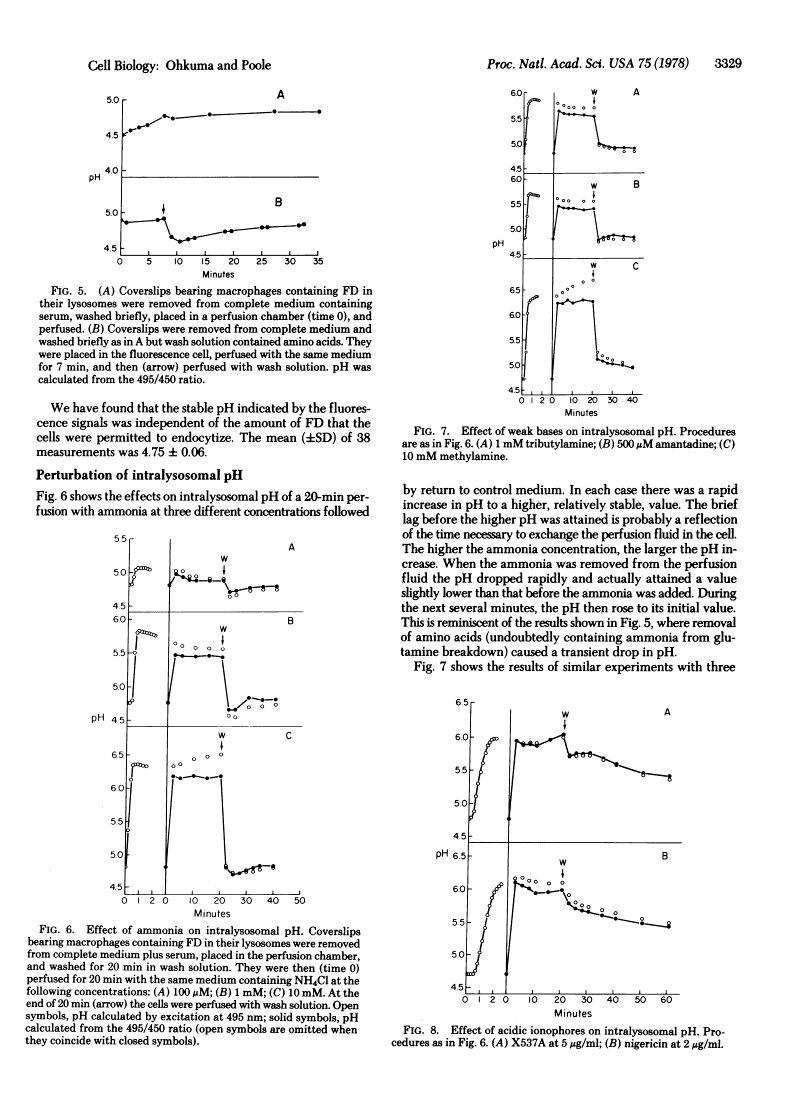
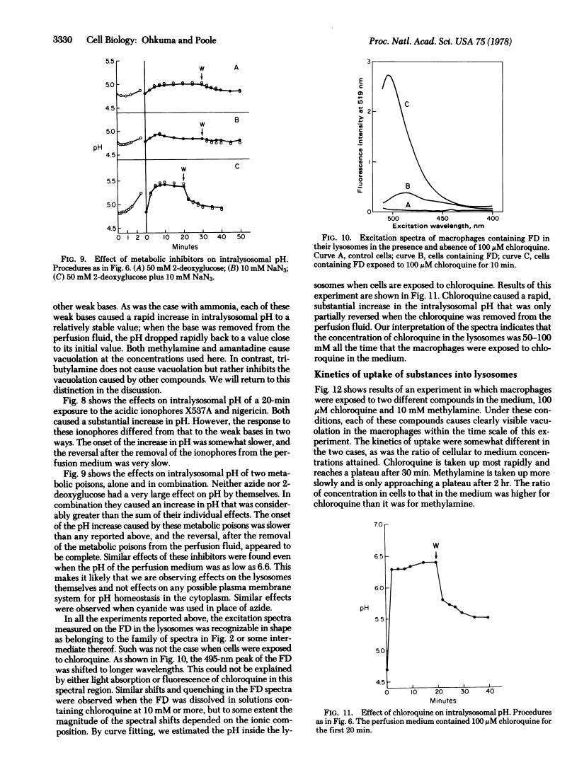
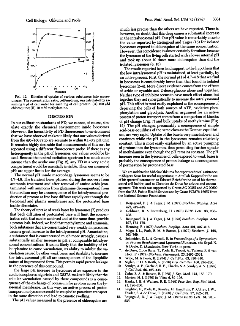
A quantitative method is described for the measurement of intralysosomal pH in living cells. Fluorescein isothiocyanate-labeled dextran (FD) is endocytized and accumulates in lysosomes where it remains without apparent degradation. The fluorescence spectrum of this compound changes with pH in the range 4-7 and is not seriously affected by FD concentration, ionic strength, or protein concentration. Living cells on coverslips are mounted in a spectrofluorometer cell and can be perfused with various media. The normal pH inside macrophage lysosomes seems to be 4.7-4.8, although it can drop transiently as low as 4.5. Exposure of the cells to various weak bases and to acidic potassium ionophores causes the pH to increase. The changes in pH are much more rapid than is the intralysosomal accumulation of the weak bases. Inhibitors of glycolysis (2-deoxyglucose) and of oxidative phosphorylation (cyanide or azide) added together, but not separately, cause the intralysosomal pH to increase. These results provide evidence for the existence of an active proton accumulation mechanism in the lysosomal membrane and support the theory of lysosomal accumulation of weak bases by proton trapping.
Full text
Full text is available as a scanned copy of the original print version. Get a printable copy (PDF file) of the complete article (890K), or click on a page image below to browse page by page. Links to PubMed are also available for Selected References.
Selected References
These references are in PubMed. This may not be the complete list of references from this article.
- Reijngoud DJ, Tager JM. The permeability properties of the lysosomal membrane. Biochim Biophys Acta. 1977 Nov 14;472(3-4):419–449. [Abstract] [Google Scholar]
- Goldman R, Rottenberg H. Ion distribution in lysosomal suspensions. FEBS Lett. 1973 Jul 1;33(2):233–238. [Abstract] [Google Scholar]
- Riejngoud DJ, Tager JM. Measurement of intralysosomal pH. Biochim Biophys Acta. 1973 Jan 24;297(1):174–178. [Abstract] [Google Scholar]
- Henning R. pH gradient across the lysosomal membrane generated by selective cation permeability and Donnan equilibrium. Biochim Biophys Acta. 1975 Aug 20;401(2):307–316. [Abstract] [Google Scholar]
- Mego JL, Farb RM, Barnes J. An adenosine triphosphate-dependent stabilization of proteolytic activity in heterolysosomes. Evidence for a proton pump. Biochem J. 1972 Jul;128(4):763–769. [Europe PMC free article] [Abstract] [Google Scholar]
- de Duve C, de Barsy T, Poole B, Trouet A, Tulkens P, Van Hoof F. Commentary. Lysosomotropic agents. Biochem Pharmacol. 1974 Sep 15;23(18):2495–2531. [Abstract] [Google Scholar]
- Wibo M, Poole B. Protein degradation in cultured cells. II. The uptake of chloroquine by rat fibroblasts and the inhibition of cellular protein degradation and cathepsin B1. J Cell Biol. 1974 Nov;63(2 Pt 1):430–440. [Europe PMC free article] [Abstract] [Google Scholar]
- Seglen PO, Reith A. Ammonia inhibition of protein degradation in isolated rat hepatocytes. Quantitative ultrastructural alterations in the lysosomal system. Exp Cell Res. 1976 Jul;100(2):276–280. [Abstract] [Google Scholar]
- Somlyo AP, Garfield RE, Chacko S, Somlyo AV. Golgi organelle response to the antibiotic X537A. J Cell Biol. 1975 Aug;66(2):425–443. [Europe PMC free article] [Abstract] [Google Scholar]
- COHN ZA, BENSON B. THE DIFFERENTIATION OF MONONUCLEAR PHAGOCYTES. MORPHOLOGY, CYTOCHEMISTRY, AND BIOCHEMISTRY. J Exp Med. 1965 Jan 1;121:153–170. [Europe PMC free article] [Abstract] [Google Scholar]
- Morton HJ. A survey of commercially available tissue culture media. In Vitro. 1970 Sep-Oct;6(2):89–108. [Abstract] [Google Scholar]
- Leighton F, Poole B, Beaufay H, Baudhuin P, Coffey JW, Fowler S, De Duve C. The large-scale separation of peroxisomes, mitochondria, and lysosomes from the livers of rats injected with triton WR-1339. Improved isolation procedures, automated analysis, biochemical and morphological properties of fractions. J Cell Biol. 1968 May;37(2):482–513. [Europe PMC free article] [Abstract] [Google Scholar]
- Reijngoud DJ, Tager JM. Chloroquine accumulation in isolated rat liver lysosomes. FEBS Lett. 1976 Apr 15;64(1):231–235. [Abstract] [Google Scholar]
Associated Data
Articles from Proceedings of the National Academy of Sciences of the United States of America are provided here courtesy of National Academy of Sciences
Full text links
Read article at publisher's site: https://doi.org/10.1073/pnas.75.7.3327
Read article for free, from open access legal sources, via Unpaywall:
http://www.pnas.org/content/75/7/3327.full.pdf
Citations & impact
Impact metrics
Citations of article over time
Alternative metrics
Smart citations by scite.ai
Explore citation contexts and check if this article has been
supported or disputed.
https://scite.ai/reports/10.1073/pnas.75.7.3327
Article citations
Streamlined analysis of drug targets by proteome integral solubility alteration indicates organ-specific engagement.
Nat Commun, 15(1):8923, 16 Oct 2024
Cited by: 0 articles | PMID: 39414818 | PMCID: PMC11484808
Ammonia-induced lysosomal and mitochondrial damage causes cell death of effector CD8<sup>+</sup> T cells.
Nat Cell Biol, 26(11):1892-1902, 11 Sep 2024
Cited by: 0 articles | PMID: 39261719
Elongation of Very Long-Chain Fatty Acids (ELOVL) in Atopic Dermatitis and the Cutaneous Adverse Effect AGEP of Drugs.
Int J Mol Sci, 25(17):9344, 28 Aug 2024
Cited by: 0 articles | PMID: 39273293 | PMCID: PMC11395647
Review Free full text in Europe PMC
Protocol for the application of single-cell damage in murine intestinal organoid models.
STAR Protoc, 5(3):103153, 30 Jul 2024
Cited by: 0 articles | PMID: 39088328 | PMCID: PMC11342180
Macropinocytosis Is the Principal Uptake Mechanism of Antigen-Presenting Cells for Allergen-Specific Virus-like Nanoparticles.
Vaccines (Basel), 12(7):797, 18 Jul 2024
Cited by: 0 articles | PMID: 39066435 | PMCID: PMC11281386
Go to all (1,076) article citations
Other citations
Similar Articles
To arrive at the top five similar articles we use a word-weighted algorithm to compare words from the Title and Abstract of each citation.
Effect of weak bases on the intralysosomal pH in mouse peritoneal macrophages.
J Cell Biol, 90(3):665-669, 01 Sep 1981
Cited by: 444 articles | PMID: 6169733 | PMCID: PMC2111912
Fluorescein conjugates as indicators of subcellular pH. A critical evaluation.
Exp Cell Res, 150(1):29-35, 01 Jan 1984
Cited by: 59 articles | PMID: 6198189
Intralysosomal accumulation of polyanions. II. Polyanion internalization and its influence on lysosomal pH and membrane fluidity.
J Cell Biol, 93(3):875-882, 01 Jun 1982
Cited by: 33 articles | PMID: 6181075 | PMCID: PMC2112131
[Segregation function of the cell and its molecular mechanisms].
Tsitologiia, 28(4):387-402, 01 Apr 1986
Cited by: 0 articles | PMID: 2872743
Review


