Abstract
Free full text

This article has been retracted. Retraction in:
Modulation of miR-10a-mediated TGF-β1/Smads signaling affects atrial fibrillation-induced cardiac fibrosis and cardiac fibroblast proliferation
Abstract
Atrial fibrillation (AF) rat models and rat cardiac fibroblasts (CFs) with overexpressed or inhibited miR-10a were used to investigate the possible role of miR-10a-mediated transforming growth factor-β (TGF-β1)/Smads signaling in cardiac fibrosis and fibroblast proliferation in rats with AF. Gene ontology and pathway enrichment analyses were used to identify the possible function of miR-10a in cardiac fibrosis. The results showed that overexpressed miR-10a significantly prolonged the duration of AF, further elevated the collagen volume fraction (CVF), and increased the viability of CFs in AF rats; these findings were in contrast with the findings for rats with inhibition of miR-10a (all P<0.05). Moreover, miR-10a overexpression could promote miR-10a, collagen-I, collagen III, α-SMA, and TGF-β1 protein expression and increase the levels of hydroxyproline but reduced Smad7 protein expression in atrial tissues and CFs in AF rats. Not surprisingly, inhibiting miR-10a led to completely contrasting results (all P<0.05). Moreover, TGF-β1 treatment could reverse the inhibitory effect of miR-10a down-regulation on cardiac fibrosis in CFs. Bioinformatics analysis and luciferase reporter assay results demonstrated that miR-10a bound directly to the 3′-UTR of BCL6, which is involved in cell growth and proliferation. Thus, our study indicate that down-regulation of miR-10a may inhibit collagen formation, reduce atrial structure remodeling, and decrease proliferation of CFs, eventually suppressing cardiac fibrosis in AF rats via inhibition of the TGF-β1/Smads signaling pathway.
Introduction
Atrial fibrillation (AF), widely considered the most common cardiac rhythm disorder, significantly increases the risk of morbidity and mortality from shock and heart failure and has rapidly become a major medical problem worldwide [1–3]. The pathogenesis of AF is extremely complex and is reportedly associated with various factors, such as atrial structural remodeling, electrical remodeling, inflammation, autonomic nervous system dysfunction, and oxidative stress, amongst other factors [4–7]. Additionally, there is evidence that cardiac fibroblasts (CFs) can differentiate into myofibroblasts via proliferation and activation during cardiac remodeling, causing extracellular matrix (ECM) accumulation and inducing cardiac fibrosis that ultimately leads to cardiac remodeling [8,9]. The formation of cardiac fibrosis therefore plays a key role in the incidence and development of cardiac structural remodeling, which may also constitute an important component of AF pathogenesis [10].
MiRNAs are small noncoding RNA molecules consisting of 22–25 nts. They can regulate target genes at the post-transcriptional level by degrading their target mRNAs or inhibiting their translation [11,12]. MiRNA (miR)-10a is a member of the miR-10 family, which has been shown to play a direct role in the proliferation and differentiation of several cell types [13]. In recent years, miR-10a has been shown to be closely related to the occurrence and progression of cardiovascular-related diseases, such as coronary artery disease (CAD) [14]. Moreover, miR-10a has been suggested as a biomarker for heart failure prognosis, since it is abnormally expressed in rats with heart failure [15]. It can also promote the expression of angiogenic factors to improve angiogenesis and cardiac function in mice after myocardial infarction [16]. Of note, miR-10a can promote the progression of hepatic fibrosis through modulating the TGFβ1/Smads signaling pathway [17]. However, data regarding the effects of miR-10a on cardiac fibrosis, particularly in CFs, remain limited. Transforming growth factor-β (TGF-β) belongs to the TGF-β superfamily of cytokines and plays a crucial role in numerous developmental and pathological processes, such as inflammation, fibrosis, angiogenesis, and tumor development [18,19]. According to previous studies, TGF-β1 can also participate in the proliferation, transformation, and migration of CFs via the TGF-β/Smads signal transduction pathway, thereby promoting cardiac fibrosis [20,21]. Given these prior findings, we hypothesized that miR-10a could play a vital role in cardiac fibrosis in AF patients. Therefore, by constructing AF rat models and obtaining CFs via isolation, we investigated whether miR-10a could mediate TGF-β1/Smads signaling to regulate cardiac fibrosis in AF, thus providing a novel theoretical foundation for the clinical treatment of AF.
Materials and methods
Ethics statement
All animal experiments were performed according to local protocols concerning the care and use of laboratory animals and in strict obedience with the Guide for the Care and Use of Laboratory Animals published by the National Institutes of Health in the U.S.A. (number 85-23, revised 1996) [22].
Establishment and grouping of AF rat models
Forty healthy male Sprague–Dawley rats, each weighing 400–450 g, were used to construct AF rat models as previously described [23]. Rats in experimental groups were administered a calcium chloride (10 mg/ml)-acetylcholine (66 μg/ml) mixture (SelleckBio, U.S.A.) via tail vein injection (1 ml/kg) once per day for seven consecutive days. Rats in the control group were injected with equal volumes of normal saline. Electrocardiogram (ECG) measurements were recorded, and P-wave absence and the appearance of typical f-waves were indicators of the successful establishment of AF rat models [24]. Next, rats were randomly divided into four groups of ten animals each: Control (normal rats), Model (AF rat models injected with the same volume of a vehicle control), agomiR-10a (AF rat models injected with agomiR-10a), and antagomiR-10a (AF rat models injected with antagomiR-10a). More specifically, 5 μl of antagomiR-10a/agomiR-10a (7.5 nmol) was injected into the left atrial wall at five different injection points with a Hamilton microsyringe (33-gauge needle) immediately after operation. The antagomiR-10a and agomiR-10a were synthesized by Ribobio Co., Ltd. (Guangzhou, China). Rats in the Control group were injected with equal volumes of normal saline, and rats in the Model group were injected with equal volumes of miR-10a negative vehicle control. The injection was performed once per day for three consecutive days. Then, the biosignal collecting and processing system Med Lab-U/4C501H (Nanjing Medease Science and Technology Co., Ltd) was used to detect the duration of AF, which was defined as the duration from the appearance to the termination of AF. Later, rats were killed by cervical dislocation, and the thoracic cavity was opened to remove the heart. After rinsing the heart in precooled normal saline (4°C) to remove the blood, the atria were separated out, followed by removing the connective tissues, fat and blood vessels in the atria, and rinsing the atria with precooled D-Hanks solution three times. Atrial tissue samples were used to prepare paraffin-embedded sections for HE staining and Masson’s trichrome staining, and the remaining atrial tissues were cryopreserved in EP tubes in a freezer at −80°C.
Isolation and culture of CFs
SD rats were killed by cervical dislocation and immersed in 75% alcohol for 10 min before removing the atria, cutting the atria into pieces with sterile eye scissors, and centrifuging the tissues at 1000 rpm for 5 min in a centrifuge. Next, the supernatant was discarded, and tissue samples were transferred into a conical flask, digested with 2.5 g/l trypsin for 30 min at 37°C, washed twice with PBS buffer, and centrifuged for 5 min at 1000 rpm. After removing the supernatant, cells were washed three times with DMEM/F12 culture fluid (Sigma, U.S.A.) supplemented with 1 ml of 10% FBS (Gibco, Australia). CFs were isolated by removing cardiac myocytes using the differential adhesion method, suspended in 20% FBS-containing culture fluid, inoculated into cell culture flasks, and incubated in an incubator (37°C and 5% CO2). When cells were subcultured for the second or third generation, immunohistochemical fibronectin staining was performed for the identification of CFs. After that, CFs at the logarithmic growth phase were removed and seeded into 96-well plates (100 μl/well, Orange Scientificx, Belgium). Cells were assigned into five groups: Control (CFs without any treatment), miR-10a mimics (CFs transfected with miR-10a mimics), miR-10a inhibitors (CFs transfected with miR-10a inhibitors), miR-10a NC (CFs transfected with miR-10a negative control), and miR-10a inhibitors+TGF-β1 (CFs transfected with miR-10a inhibitors and cultured for 24 h in culture medium containing 10 ng/ml TGF-β1 and 10% FBS). Opti-MEM culture medium containing miR-10a mimics/inhibitors or miR-10a negative control was used to transfect cells. Final concentrations of the miR-10a mimics/inhibitors or miR-10a negative control in each well during transfection were 20 nmol/l. Cell transfection was performed with Lipofectamine 2000 (Invitrogen, U.S.A.).
HE staining and Masson’s trichrome staining
Atrial tissues were collected and fixed in Davidson’s solution for 24 h before routine dehydration, hyalinization, wax-dipping, and paraffin embedment. Next, an ultrathin semiautomatic microtome (Shandon325, U.K.) was used to prepare ten serial sections (each 3 μm in thickness), which were baked at 50°C for 1 h. Then, HE staining was performed to observe the pathological changes of cardiac tissues under a microscope (Leica DM LB2, Germany). Masson’s trichrome staining was used for the detection of collagen fibers in tissues. Briefly, after routine staining with Weigert’s iron Hematoxylin–Biebrich scarlet–Aniline Blue (Beyotime Institute of Biotechnology), five visual fields were randomly selected and imaged from the stained tissues under a high-power lens, and Image ProPlus 6.0 software was used to analyze the pathological changes. The collagen volume fraction (CVF) of cardiac tissues was calculated as follows: CVF = collagen area/total area under observation × 100%.
Hydroxyproline level determination
According to the manufacturer’s instructions for the hydroxyproline assay kit (Solarbio, U.S.A.), atrial tissues and the supernatants of CF solutions were isolated to determine the optical density (OD value) of the samples at a wavelength of 550 nm in a microplate reader, and the level of hydroxyproline was calculated accordingly.
CCK-8 assay
CFs of rats from each group were inoculated on to 96-well plates (Orange Scientific, Belgium) at 104 cells/well in 100 μl culture medium, and five replicates and five blank wells for each experimental group. Next, cells were incubated in 5% CO2 at 37°C. The plates were removed at 24 and 48 h for the addition of CCK-8 solution at 10 μl/well (Solarbio, Beijing, China), followed by another 2 h of incubation. OD values at a wavelength of 450 nm were determined using a Microplate reader (MRX MRX-HD, U.S.A.), and the viability of CFs was calculated according to the following formula: Cell viability rate = (OD value of experimental group/OD value of blank control group) × 100%.
qRT-PCR
Total RNA was extracted from tissues and cells with an RNA extraction kit (Omega, U.S.A.), determined for purity and concentration with a UV spectrophotometer (UV-1800, Japan), and tested for RNA integrity via agarose gel electrophoresis. Primer 5.0 software was used to design the primer sequences of miR-10a, a-SMA, collagen-I and collagen III, which were synthesized by Sangon Biotech (Shanghai) Co., Ltd. (Table 1). Next, the cDNA was synthesized from total RNA using the Primescript™ RT reagent kit (Takara, Japan), and qRT-PCR was performed with a SYBR® premix ExTaq™ kit (Takara, Japan). Using U6 as an internal reference gene for miR-10a and GAPDH for other genes, the 2−ΔΔCt method was used to analyze the relative expression of target genes.
Table 1
| Gene | Primer sequences |
|---|---|
| miR-10a | F: 5′-CATTCGGATCCCAAGAACAGAC-3′ |
| R: 5′-GGGAGAATTCGGGGAGAGTTC-3′ | |
| α-SMA | F: 5′-TGGCCACTGCTGCTTCCTCTTCTT-3′ |
| R: 5′-GGGGCCAGCTTCGTCATACTCCT-3′ | |
| Collagen-I | F: 5′-CCAGCGGTGGTTATGACTTCA-3′ |
| R: 5′-TGCTGGCTCAGGCTCTTGA-3′ | |
| Collagen III | F: 5′-GCGGCTTTTCACCATATTACG-3′ |
| R: 5′-CGTTAACAGACTTGAGTGAAG-3′ | |
| U6 | F: 5′-GCGGTCCGTGAA-3′ |
| R: 5′-GTGTGCGGTCCGAGGT-3′ | |
| GAPDH | F: 5′-GAGGCTCTCTTCCAGCCTT-3′ |
| R: 5′-AGGGTGTAAAATCAGCTCA-3′ |
Western blot analysis
Cardiac tissues were isolated from rats, then lysis buffer was added (100–200 μl for 20 mg tissues), and a glass homogenizer was used for homogenization, followed by centrifugation at 12000 rpm for 15 min. The supernatant was collected for SDS/PAGE, and electronic transfer was employed to transfer isolated proteins to the nitrocellulose filter, which was placed in 5% skim milk-PBS buffer for blocking for 1 h. Subsequently, primary antibodies were added for overnight incubation at 4°C, including anti-TGF-β1 antibody (ab92486), anti-Smad7 antibody (ab55493), and anti-GAPDH antibody (ab8245, Abcam). Subsequently, the filter was rinsed with PBS buffer three times and horseradish peroxidase (HRP)–conjugated secondary antibodies (CwBio Technology Co., Ltd.) were added for 1 h of incubation at room temperature. Finally, the membranes were rinsed with PBS buffer three times and visualized using the chemiluminescence method. Relative expression levels of proteins are presented as the gray value ratio of the target band to a reference band, with GAPDH as the internal reference.
Bioinformatics analysis of miR-10a
miRanda (union of http://microRNA.org and miRBase), PicTar, and TargetScan algorithms were employed for the prediction of miR-10a target genes. Functional analysis of the predicted targets genes was performed using the Database for Annotation, Visualization and Integrated Discovery (DAVID) online tool (https://david.ncifcrf.gov/home.jsp). From this database, GO enrichment and KEGG pathway analysis were used. The GO enrichment analysis included biological processes (BPs), cell component (CC), and molecular function (MF).
Luciferase assay
Given the strong connection between miR-10a expression and CF proliferation investigated in the present study, we focussed on genes related to cell growth proliferation. BCL6, TFAP2C, and NR5A2 (all involved in the regulation of cell proliferation) were selected from the genes listed in Table 2 for further analysis via reporter assay. The luciferase reporter assay was performed as previously described [25].
Table 2
| Category | Term | P-value | Genes | FDR |
|---|---|---|---|---|
| GOTERM_BP_DIRECT | GO:0021766~hippocampus development | 0.003 | PAFAH1B1, ID4, NR4A3 | 4.079 |
| GOTERM_BP_DIRECT | GO:0045944~positive regulation of transcription from RNA polymerase II promoter | 0.007 | NCOA6, ID4, NR5A2, HOXD10, GTF2H1, ARNT | 8.668 |
| GOTERM_BP_DIRECT | GO:0030036~actin cytoskeleton organization | 0.014 | FHL3, PAFAH1B1, BCL6 | 16.517 |
| GOTERM_CC_DIRECT | GO:0005634~nucleus | 0.033 | ACTG1, ZMYND11, FHL3, ID4, BCL6, SLC25A1, TFAP2C, NR4A3, FXR2, HOXD10, BTBD11 | 29.152 |
| GOTERM_BP_DIRECT | GO:0030099~myeloid cell differentiation | 0.036 | NCOA6, BCL6 | 37.201 |
| GOTERM_BP_DIRECT | GO:0007405~neuroblast proliferation | 0.036 | PAFAH1B1, ID4 | 37.201 |
| GOTERM_BP_DIRECT | GO:0042127~regulation of cell proliferation | 0.044 | BCL6, TFAP2C, NR5A2 | 43.938 |
| GOTERM_CC_DIRECT | GO:0090575~RNA polymerase II transcription factor complex | 0.049 | NR5A2, ARNT | 40.211 |
Statistical analysis
Data analysis was conducted using statistical software SPSS 22.0. Measurement data were presented as means ± S.D., and intergroup comparison was performed with the Student’s t test. Differences amongst multiple groups were compared with one-way ANOVA. P<0.05 indicated significant differences.
Results
Expression of miR-10a and fibrosis-related genes in rats and CFs
As shown in Figure 1, rats in the Model group had higher expression levels of miR-10a, collagen-I, collagen III, and α-SMA than rats in the Control group (all P<0.05). Additionally, the expression levels of miR-10a, collagen-I, collagen III, and α-SMA were dramatically higher in rats from the agomiR-10a group but lower in rats from the antagomiR-10a group compared with the Model rats (all P<0.05). Regarding the CFs, these molecules were dramatically increased in the miR-10a mimics group, whereas they were significantly reduced in the miR-10a inhibitors the group in comparison with Control group (all P<0.05). However, compared with the miR-10a inhibitors group, the miR-10a inhibitors + TGF-β1 group did not show any notable differences in miR-10a expression (P>0.05), although they had significantly increased expression levels of collagen-I, collagen III, and α-SMA (all P<0.05).
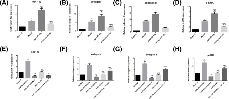
(A–D) Expression levels of miR-10a (A), collagen-I (B), collagen III (C), and α-SMA (D) as determined by qRT-PCR in rats; *, P<0.05 compared with the Control group; #, P<0.05 compared with the Model group; %, P<0.05 compared with the agomiR-10a group. (E–H) Expression level of miR-10a (E), collagen-I (F), collagen III (G), and α-SMA (H) as determined by qRT-PCR in CFs; *, P<0.05 compared with the Control group; #, P<0.05 compared with the miR-10a mimics group; %, P<0.05 compared with the miR-10a inhibitors group.
AF rat model establishment and AF duration comparison
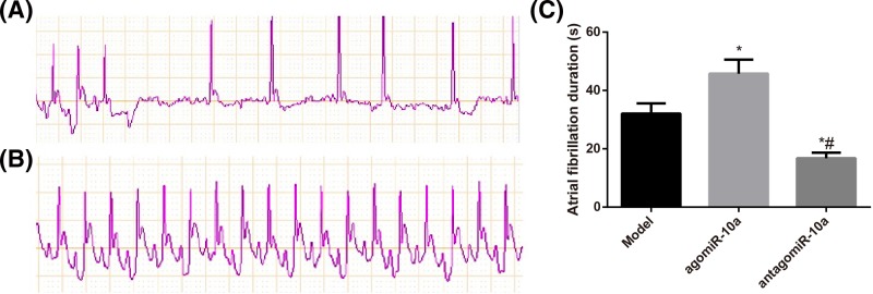
Each day after tail vein injection of the acetylcholine–calcium chloride mixture, the typical AF ECG pattern was observed. All AF model rats showed marked AF seizures, with the appearance of f-waves instead of p-waves and different RR durations. In contrast, rats in the Control group showed sinus rhythms with p-waves and similar RR durations. Additionally, the agomiR-10a group had a prolonged duration of AF, whereas the antagomiR-10a group showed deceased AF durations compared with Model AF rats (Figure 2, all P<0.05).
Morphological changes in cardiac tissues
According to the evaluation of HE staining and Masson’s trichrome staining (Figure 3A), cardiac cells in the Control group showed normal morphologies. The arrangement of cardiac cells in the Model group was loose and irregular, and the muscle fibers were elongated and thickened with indistinct transverse striation and local tissue fibrosis, as well as cardiac cell hypertrophy and increased collagenous fibers. Additionally, rats in the agomiR-10a group had more severe tissue fibrosis, while those in the antagomiR-10a group showed a tight arrangement, thinner and shorter muscle fibers, visible transverse striations, and decreased collagenous fibers. On the other hand, compared with the Control group, CVF was increased significantly in rats in the Model group (P<0.05). CVF was remarkably higher in rats from the agomiR-10a group but significantly lower in rats from the antagomiR-10a group compared with Model group (all P<0.05, Figure 3B).

(A) Representative microscopic images of cardiac tissues shown by HE staining and Masson’s trichrome staining. Fibrosis is indicated in blue by Masson’s trichrome. (B) Comparison of the CVF amongst different groups; *, P<0.05 compared with the Control group; #, P<0.05 compared with the Model group; %, P<0.05 compared with the agomiR-10a group.
Hydroxyproline level in rats and CFs
As shown in Figure 4, the Model rats had markedly increased hydroxyproline levels compared with the Control group (P<0.05). At the same time, compared with the Model group, the hydroxyproline level increased substantially in the agomiR-10a group and decreased significantly in the antagomiR-10a group (all P<0.05). Regarding CFs, these cells had dramatically elevated hydroxyproline levels in the miR-10a mimics group and reduced hydroxyproline levels in the miR-10a inhibitors group compared with Controls (all P<0.05). Moreover, these levels were significantly increased in the miR-10a inhibitors + TGF-β1 group compared with the miR-10a inhibitors group (P<0.05).
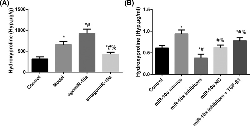
Hydroxyproline levels in cardiac tissues of rats (A) and supernatants of CF solutions (B). (A) *, P<0.05 compared with the Control group; #, P<0.05 compared with the Model group; %, P<0.05 compared with the agomiR-10a group. (B) *, P<0.05 compared with the Control group; #, P<0.05 compared with the miR-10a mimics group; %, P<0.05 compared with the miR-10a inhibitors group.
Effect of miR-10a on the viability of CFs
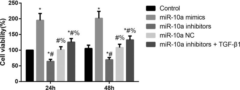
As shown in Figure 5, compared with CFs in the Control group, the cell viability was significantly increased in the miR-10a mimics group but was dramatically decreased in the miR-10a inhibitors group (both P<0.05). Additionally, the miR-10a inhibitors + TGF-β1 group showed significantly greater CF viability than the miR-10a inhibitors group (P<0.05).
Expression of TGF-β1/Smads pathway-related proteins in rats and CFs
TGF-β1 protein levels were up-regulated while Smad7 protein levels were down-regulated in atrial tissues from the Model group compared with rats in the Control group (both P<0.05, Figure 6). The agomiR-10a group showed markedly elevated TGF-β1 protein levels and substantially decreased Smad7 protein levels compared with the Model group, whereas the antagomiR-10a group showed significant changes in the opposite direction (all P<0.05). On the other hand, the CFs in the miR-10a mimics group had significantly increased TGF-β1 levels but decreased Smad7 levels, whereas those in the miR-10a inhibitors group changed in the opposite direction regarding these two proteins compared with the Control group (all P<0.05). Meanwhile, the CFs in the miR-10a inhibitors+TGF-β1 group showed elevated TGF-β1 protein levels and markedly lower Smad7 protein levels than those in the miR-10a inhibitors group (both P<0.05).
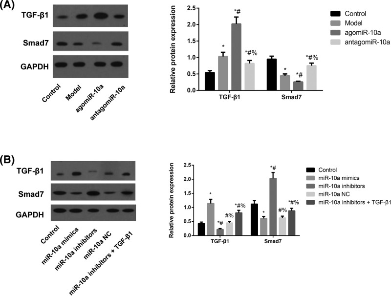
The expressions of TGF-β1/Smads pathway-related proteins in atrial tissues of rats (A) and CFs (B). (A) Expression levels of TGF-β1 and Smad7 proteins in atrial tissues of rats as detected by Western blotting analysis and normalized to GAPDH. *, P<0.05 compared with the Control group; #, P<0.05 compared with the Model group; %, P<0.05 compared with the agomiR-10a group. (B) Expression levels of TGF-β1 and Smad7 proteins amongst different groups of CFs as detected by Western blotting analysis and normalized to GAPDH. *, P<0.05 compared with the Control group; #, P<0.05 compared with the miR-10a mimics group; %, P<0.05 compared with the miR-10a inhibitors group.
Prediction and functional analysis of miR-10a targets
To further investigate the functional roles of miR-10a, we sought to predict the possible targets of miR-10a and analyze their involvement in possible BPs. Our results show that a total of 44 genes (ACTG1, DOCK11, MAPRE1, SDC1, ANKRD12, DVL3, NARG1, SFRS1, ARNT, ELAVL2, NCOA6, SLC25A1, ASXL1, FHL3, NCOR2, SLC38A2, BCL6, FXR2, NR4A3, SMAP1, BCL2L2, GRM3, NR5A2, SON, BTBD11, GTF2H1, PAFAH1B1, SVOP, BTBD14B, HNRPK, POPDC2, TFAP2C, CNNM4, HOXD10, PURB, USF2, CTDSPL, ID4, RAP2A, WNK3, DAZAP1, IL1RAPL1, RB1CC1, and ZMYND11) were simultaneously predicted by all three algorithms (miRanda, PicTar, and TargetScan), and these are subsequently referred to as stringently predicted targets.
GO enrichment and KEGG pathway analysis
According to the results of DAVID-based GO analysis (Table 2), genes were significantly enriched that were involved in the regulation of hippocampus development and positive regulation of transcription during BP; the highest enrichment of CC was in regulation of the nucleus. Additional KEGG pathway analysis showed that genes were enriched in the Wnt signaling pathway, proteoglycans in cancer and the Notch signaling pathway, amongst others, although these enrichments were not statistically significant (all P>0.05).
miR-10a targets BCL6
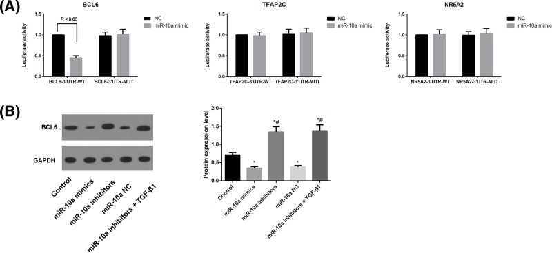
To elucidate the tumorigenic role of miR-10a in CFs, we further verified several putative targets of miR-10a. As shown in Figure 7A, luciferase assays indicated that miR-10a mimic treatment significantly reduced the luciferase activity of the BCL6-3′UTR (P<0.05) compared with the negative control (NC). Mutation of the predicted binding site on the 3′-UTRs abolished this inhibitory effect of the miR-10a mimic. In contrast, miR-10a mimic treatment had no effect on luciferase activity of the TFAP2C-3′UTR and NR5A2-3′UTR. We further determined the expression of BCL6 in different transfection groups of CFs by Western blot analysis. The results showed that the expression of BCL6 was reduced in the miR-10a mimics group, whereas it was significantly increased in the miR-10a inhibitors and miR-10a inhibitors+TGF-β1 groups in comparison with the Control group (all P<0.05, Figure 7B). Therefore, we have demonstrated that miR-10a can target BCL6 by binding to its 3′-UTR in CFs.
Discussion
AF rat models were successfully established in our study; we found that AF duration was prolonged with agomiR-10a treatment but was significantly shortened via inhibition of miR-10a with antagomiR-10a, suggesting that miR-10a overexpression could at least partially aggravate AF whereas suppression of miR-10a expression could exert therapeutic effects. Through HE staining and Masson’s trichrome staining, we observed that miR-10a overexpression aggravated atrial fibrosis and enhanced CVF, whereas miR-10a inhibition had the opposite effect. It is well-established that atrial fibrosis is the most typical manifestation of atrial structural remodeling in AF patients [26], which can block the continuity of cardiomyocyte bundles to interfere with local conduction and induce re-entrant arrhythmias. Thus, inhibiting fibrosis could effectively reduce the incidence and shorten the duration of AF [27,28]. Similar to our findings, Huang et al. [29] also observed that antagomir-21 treatment could appreciably reduce the duration and incidence of AF, as well as further down-regulate the expression of α-SMA, collagen-1, and collagen-3 to ameliorate atrial fibrosis. Moreover, the AF duration and recurrence rate of AF in patients has shown a positive correlation with the CVF in atrial tissues [30]. In our study, we measured the levels of hydroxyproline in rats, which represents levels of collagen [31], and observed that miR-10a down-regulation led to a reduction in hydroxyproline levels, suggesting that suppressing miR-10a may repress cardiac fibrosis and block atrial structural remodeling during AF to shorten AF duration, inhibit collagen formation, and ameliorate AF.
More specifically, the major manifestation of atrial fibrosis is the reconstruction of ECM, and the main components of ECM in the atrium are pro-proteins, the majority of which are collagen-I and collagen III [32,33]. The synthesis and secretion of collagens mainly appears in fibroblasts. When CFs are affected by abnormal stimulation, a large number of different collagens are produced to disrupt the balance between collagen synthesis and degradation, leading to the excessive collagen accumulation in the ECM and eventually inducing atrial fibrosis [26,30]. In addition to serving as a marker protein expressed by fibroblasts, α-SMA also participates in the formation of ECM and directly influences the appearance of atrial fibrosis [34,35]. Therefore, we also determined the expression levels of α-SMA, collagen-I, and collagen III in rats with different treatments. Overexpressed miR-10a significantly up-regulated the expression levels of α-SMA, collagen-I, and collagen III, while these expression levels were reduced through down-regulation of miR-10a, suggesting that inhibiting miR-10a is somehow able to reduce collagen levels to ameliorate atrial fibrosis.
Wang et al. [36] discovered that miR-27b could block the Smad-2/3 pathway to reduce the incidence rate and shorten the duration of AF to inhibit atrial fibrosis, along with reduced expression levels of α-SMA, collagen-I, and collagen III. To investigate these effects in our work, CFs were isolated and treated with miR-10a mimics or inhibitors. Consequently, overexpressed miR-10a could substantially enhance the proliferation of CFs, with elevated expression levels of α-SMA, collagen-I, and collagen III and increased hydroxyproline levels, whereas down-regulation of miR-10a caused by miR-10a inhibitors led to completely opposite effects. As reported by Zhou et al. [17], miR-10a can promote the proliferation of hepatic fibroblasts and accelerate hepatic fibrosis by activating the TGF-βl/Smads signaling pathway. Therefore, the TGF-βl/Smads pathway was measured in this study. Overexpression of miR-10a could up-regulate TGF-βl but down-regulate Smad7 expression in AF rats and CFs, whereas inhibiting miR-10a resulted in an opposite trend, which suggested that miR-10a may activate the TGF-βl/Smads signaling pathway to promote cardiac fibrosis. As noted, TGF-β1 can act on CFs to induce massive synthesis of fibronectins and increase the expressions of collagen I and III in cardiac tissues, thus disrupting the balance of collagen ratios and reducing the number of matrix metalloproteinases (MMPs), which eventually causes a reduction in ECM degradation and the formation of cardiac fibrosis [37–39]. The key transduction molecule in the TGF-β signaling pathway is the cytoplasmic protein Smads [40]. The TGF-β complex can bind to R-Smads and Co-Smads to form heteromers and thereby exerts its biological effect by regulating the transcription of target genes [41,42]. Smad7 can block the phosphorylation of R-Smads and inhibit the oligomerization of activated R-Smads with Co-Smads, thereby negatively regulating the TGF-β signaling transduction pathway [43,44]. In a study by He et al. [45], Smad7 down-regulation could promote TGF-β1-mediated cardiac fibrosis, which is one of the major mechanisms of cardiac fibrosis. Liu et al. also reported that osthol treatment could alleviate TGF-β1-induced cardiac fibrosis with increased Smad7 expression and reduced collagen I/III ratios and α-SMA and ECM deposition [46]. Importantly, He et al. [47] found that miR-21 could down-regulate Smad 7 to enhance TGF-β1/Smad signaling and promote AF-induced cardiac fibrosis. As miRNAs function post-transcriptionally during BPs; therefore, definitive identification of miRNA target genes is essential. For this reason, we used bioinformatics methods to predict miR-10a target genes, and then analyzed the possible mechanisms of its role in cardiac fibrosis. BCL6, TFAP2C, NR5A2—all involved in regulation of cell proliferation—were further analyzed by luciferase assays and the results indicated that miR-10a could target BCL6 by binding directly to its 3′-UTR. Coincidentally, it has been reported that a high expression level of BCL6 induces ells to be refractory to the TGF-β antiproliferative response, whereas knockdown of BCL6 expression partially restores this TGF-β response [48]. These findings strongly suggest that miR-10a is closely correlated with cardiac fibrosis. It may potentially function through interactions with its target gene BCL6, which is involved in the TGF-βl/Smads pathway. Future verification studies are needed to verify the BCL6 and TGF-βl/Smads pathway effects of miR-10a in cardiac fibrosis. However, we also found in the present study that TGF-β1 can reverse the inhibitory effects of miR-10a inhibitors in the reduction in cardiac fibrosis, further suggesting that the regulatory role of miR-10a is realized by modulation of TGF-βl/Smads signal transduction pathway.
In summary, we found that down-regulating miR-10a could reduce the proliferation of CFs, inhibit collagen formation, and decrease atrial structural remodeling by blocking the TGF-β1/Smads signaling pathway, thereby suppressing AF-induced cardiac fibrosis.
Acknowledgments
We thank the reviewers for their useful and informative comments regarding the present study.
Abbreviations
| AF | atrial fibrillation |
| BCL6 | B cell lymphomas 6 |
| BP | biological process |
| CC | cell component |
| CCK-8 | cholecystokinin-8 |
| CF | cardiac fibroblast |
| CVF | collagen volume fraction |
| DAVID | Database for Annotation, Visualization and Integrated Discovery |
| DMEM/F12 | Dulbecco’s modified Eage medium: nutrient mixture F-12 |
| ECG | electrocardiogram |
| ECM | extracellular matrix |
| EP | Eppendorf |
| GAPDH | glyceraldehyde-3-phosphate dehydrogenase |
| GO | Gene Ontology |
| HE | Haematoxilin-Eosin |
| HRP | horseradish peroxidase |
| KEGG | Kyoto Encyclopedia of Genes and Genomes |
| NR5A2 | nuclear receptor subfamily 5, group A, member 2 |
| OD | optical density |
| qRT-PCR | Quantitative reverse transcriptase real-time polymerase chain reaction |
| SD | Sprague-Dawley |
| Smads | drosophila mothers against decapentaplegic proteins |
| TFAP2C | Transcription factor activator protein-2γ |
| TGF-β | transforming growth factor-β |
| Wnt | Wingless/int-1 class |
| α-SMA | Alpha smooth muscle actin |
Competing interests
The authors declare that there are no competing interests associated with the manuscript.
Author contribution
P.-F.L. designed the work. R.-H.H. conceived and supervised the work. S.-B.S. performed experiments. R.L. performed the statistical analysis. Q.-T.W. and G.-T.R. interpreted the results, and B.Y. drafted the study.
References
Articles from Bioscience Reports are provided here courtesy of Portland Press Ltd
Full text links
Read article at publisher's site: https://doi.org/10.1042/bsr20181931
Read article for free, from open access legal sources, via Unpaywall:
https://portlandpress.com/bioscirep/article-pdf/39/2/BSR20181931/844842/bsr-2018-1931.pdf
Citations & impact
Impact metrics
Citations of article over time
Article citations
Genetically predicted inflammatory proteins and the risk of atrial fibrillation: a bidirectional Mendelian randomization study.
Front Cardiovasc Med, 11:1375750, 26 Jun 2024
Cited by: 0 articles | PMID: 38988665 | PMCID: PMC11234858
Research progress of non-coding RNA in atrial fibrillation.
Front Cardiovasc Med, 10:1210762, 14 Jul 2023
Cited by: 0 articles | PMID: 37522088 | PMCID: PMC10379658
Review Free full text in Europe PMC
Experimental study on the effect of chlorhexidine gluconate (CG)-induced atrial fibrillation on renal water and sodium metabolism.
Sci Rep, 13(1):4016, 10 Mar 2023
Cited by: 0 articles | PMID: 36899056 | PMCID: PMC10006165
Noncoding RNAs and Cardiac Fibrosis.
Rev Cardiovasc Med, 24(2):63, 14 Feb 2023
Cited by: 1 article | PMID: 39077397 | PMCID: PMC11273127
Review Free full text in Europe PMC
Mechanism of action of non-coding RNAs and traditional Chinese medicine in myocardial fibrosis: Focus on the TGF-β/Smad signaling pathway.
Front Pharmacol, 14:1092148, 09 Feb 2023
Cited by: 8 articles | PMID: 36843918 | PMCID: PMC9947662
Review Free full text in Europe PMC
Go to all (25) article citations
Data
Data behind the article
This data has been text mined from the article, or deposited into data resources.
BioStudies: supplemental material and supporting data
Quick GO (Showing 8 of 8)
- (1 citation) GO - GO0005634
- (1 citation) GO - GO0007405
- (1 citation) GO - GO0090575
- (1 citation) GO - GO0030099
- (1 citation) GO - GO0021766
- (1 citation) GO - GO0045944
- (1 citation) GO - GO0030036
- (1 citation) GO - GO0042127
Show less
Similar Articles
To arrive at the top five similar articles we use a word-weighted algorithm to compare words from the Title and Abstract of each citation.
Apigenin suppresses TGF-β1-induced cardiac fibroblast differentiation and collagen synthesis through the downregulation of HIF-1α expression by miR-122-5p.
Phytomedicine, 83:153481, 27 Jan 2021
Cited by: 9 articles | PMID: 33607460
Rapid atrial pacing induces myocardial fibrosis by down-regulating Smad7 via microRNA-21 in rabbit.
Heart Vessels, 31(10):1696-1708, 11 Mar 2016
Cited by: 41 articles | PMID: 26968995 | PMCID: PMC5043001
Resveratrol Inhibits the TGF-β1-Induced Proliferation of Cardiac Fibroblasts and Collagen Secretion by Downregulating miR-17 in Rat.
Biomed Res Int, 2018:8730593, 17 Dec 2018
Cited by: 26 articles | PMID: 30648109 | PMCID: PMC6311767
Computational model predicts paracrine and intracellular drivers of fibroblast phenotype after myocardial infarction.
Matrix Biol, 91-92:136-151, 21 Mar 2020
Cited by: 26 articles | PMID: 32209358 | PMCID: PMC7434705
Review Free full text in Europe PMC

