Abstract
Background
West Nile virus (WNV) is a flavivirus that is recognized as one of the common causes of arboviral neurological disease in the world. WNV infections usually manifest with constitutional symptoms such as fever, fatigue, myalgia, rash, arthralgia, and headache. Neuroinvasive WNV infections are characterized by signs and symptoms suggestive of meningitis, encephalitis, meningoencephalitis, and acute flaccid paralysis. In addition, many patients with neuroinvasive WNV infection develop a wide range of movement disorders. This article aims to comprehensively review the spectrum and natural course of the movement disorders observed in patients with neuroinvasive WNV infections.Methods
A literature search was performed in March 2019 (in PubMed and EMBASE) to identify articles for this review.Results
Movement disorders observed in the context of WNV infections include tremor, opsoclonus-myoclonus, parkinsonism, myoclonus, ataxia, and chorea. Most often, these movement disorders resolve within a few weeks to months with an indolent course. The commonly observed tremor phenotypes include action tremor of the upper extremities (bilateral > unilateral). Tremor in patients with West Nile meningitis subsides earlier than that in patients with West Nile encephalitis/acute flaccid paralysis. Opsoclonus-myoclonus in WNV infections responds well to intravenous immunoglobulins/plasmapheresis/corticosteroids. Parkinsonism has been reported to be mild in nature and usually lasts for a few weeks to months in the majority of the patients.Conclusion
A wide spectrum of movement disorders is observed in neuroinvasive WNV infections. Longitudinal studies are warranted to obtain better insights into the natural course of these movement disorders.Free full text

Spectrum of Movement Disorders in Patients With Neuroinvasive West Nile Virus Infection
ABSTRACT
Background
West Nile virus (WNV) is a flavivirus that is recognized as one of the common causes of arboviral neurological disease in the world. WNV infections usually manifest with constitutional symptoms such as fever, fatigue, myalgia, rash, arthralgia, and headache. Neuroinvasive WNV infections are characterized by signs and symptoms suggestive of meningitis, encephalitis, meningoencephalitis, and acute flaccid paralysis. In addition, many patients with neuroinvasive WNV infection develop a wide range of movement disorders. This article aims to comprehensively review the spectrum and natural course of the movement disorders observed in patients with neuroinvasive WNV infections.
Methods
A literature search was performed in March 2019 (in PubMed and EMBASE) to identify articles for this review.
Results
Movement disorders observed in the context of WNV infections include tremor, opsoclonus–myoclonus, parkinsonism, myoclonus, ataxia, and chorea. Most often, these movement disorders resolve within a few weeks to months with an indolent course. The commonly observed tremor phenotypes include action tremor of the upper extremities (bilateral > unilateral). Tremor in patients with West Nile meningitis subsides earlier than that in patients with West Nile encephalitis/acute flaccid paralysis. Opsoclonus–myoclonus in WNV infections responds well to intravenous immunoglobulins/plasmapheresis/corticosteroids. Parkinsonism has been reported to be mild in nature and usually lasts for a few weeks to months in the majority of the patients.
Conclusion
A wide spectrum of movement disorders is observed in neuroinvasive WNV infections. Longitudinal studies are warranted to obtain better insights into the natural course of these movement disorders.
West Nile (WN) virus (WNV) is a mosquito‐borne, single‐stranded RNA flavivirus, which has become a substantial public health concern throughout the world. This neurotropic virus was first isolated from the blood of a febrile patient in the West Nile district of northern Uganda in 1940.1 Serologically, WNV is a member of the Japanese encephalitis serocomplex.2 Although WNV is a major public health concern in the United States, it is also among the most broadly distributed arboviruses in the world. Its seroprevalence is remarkable in the general population of North Africa, Eastern Mediterranean, Southern Europe, and India. The mosquito vectors for WNV are broadly distributed throughout the world, and the geographic range of WNV transmission and disease has been expanding during the past few decades.3 The incubation period of the clinical illness usually ranges from 2 to 14 days, and approximately 25% of the people infected with WNV develop the WN fever.4 It is not fully understood why only a few of the infected people become symptomatic. Previous studies have documented an association of high viral load and female gender with the development of WN fever.4 The symptoms of WN fever are usually sudden in onset and include low‐grade fever, chills, headache, malaise, myalgia, eye pain, morbilliform or maculo‐papular rashes, and vomiting.2 Less than 1% of WNV infections result in the neuroinvasive disease process; however, the prevalence of neuroinvasive infections is higher in elderly patients, alcoholics, and transplant recipients.2, 5 Neuroinvasive WNV infections commonly manifest with meningitis (with typical meningeal symptoms such as neck stiffness, photophobia, or headaches), encephalitis (altered mental status, confusion, lethargy, or seizures), or meningo‐encephalitis. One of the characteristic symptoms of neuroinvasive WNV infection is areflexic or hyporeflexic acute flaccid paralysis (AFP) in the absence of any sensory involvement.6, 7 Although WNV infection is self‐limiting, some of the symptoms, especially that of the neuroinvasive diseases, might persist for a long time, resulting in considerable functional disability.8 A longitudinal study on a large cohort of patients with a history of WNV infection in Houston, TX, revealed that 40% of the patients who had clinical disease continued to experience WNV‐associated morbidity up to 8 years after the infection, and importantly, the proportion was even higher (up to 80%) for those who initially presented with encephalitis.9 In a recently published study, the same group of authors reported the persistence of abnormal neurologic examination (reduced strength, abnormal reflexes, and tremors) and cognitive dysfunction (impaired immediate and delayed memory).10 These data underscore the chronicity of the neurological symptoms associated with WNV infection.
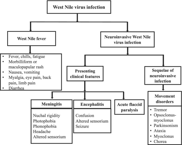
In addition to meningitis, encephalitis, and AFP, many patients with neuroinvasive WNV infection develop a wide range of movement disorders that are often seen as the sequelae of the WNV infection (Fig. (Fig.11).11 The frequent emergence of some of the movement disorders in neuroinvasive WNV infection is likely secondary to the specific neurotropism of WNV for the extrapyramidal structures that include the deep gray matter nuclei, especially the substantia nigra, thalami, and the cerebellum.12, 13, 14 It is important to be familiar with the movement disorders associated with the WNV infection as certain movement disorders such as tremor might persist for a long time (in 10%) after the infection and might worsen the activities of daily living.10 Although movement disorders comprise a major part of the neuroinvasive WNV infections, there is a paucity of articles in the current literature that are dedicated understanding the spectrum, pattern, and the natural course of movement disorders in this infectious disease. Through this systematic review, we discuss the published literature on movement disorders in the background of neuroinvasive WNV infections to obtain insights into their spectrum and natural course.
Methodology
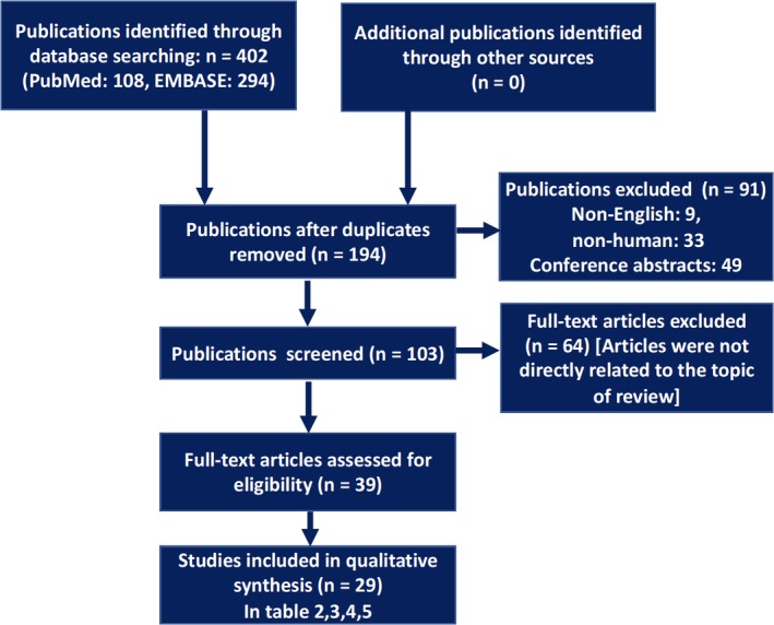
In April 2019, 2 of the authors (A.L. and S.O.M.) used 2 different databases (PubMed and EMBASE) to search for the pertinent literature using the terms “West Nile” and with the following additional search terms: “Movement Disorders,” “Hyperkinetic,” “Hypokinetic,” “Parkinsonism,” “Tremor,” “Ataxia,” “Dystonia,” “Myoclonus,” “Opsoclonus,” “Chorea,” “Athetosis,” “Ballismus,” and “Tics” (Table (Table1).1). With these terms, we covered the search for all the possible hyperkinetic and hypokinetic movement disorders observed in the clinical practice. This search yielded 402 articles (Table (Table1,1, Fig. Fig.2).2). A total of 29 articles (summarized in Tables Tables2,2, ,3,3, ,4,4, ,5)5) were shortlisted for the final review after excluding the articles that were not published in English, nonhuman studies, conference abstracts, articles that were unrelated to this review (general reviews on WNV/not discussed movement disorders), and duplicates. A checklist of the preferred reporting items for systematic reviews and meta‐analyses was used to organize the literature search.15
Table 1
Results of search for articles from PubMed and EMBASE using various key words and their combinations
| Keywords and Combinations | Number of Publications (PubMed) | Number of Publications (EMBASE) |
|---|---|---|
| “West Nile” AND “Movement disorders” | 10 | 17 |
| “West Nile” AND “Hyperkinetic” | 1 | 2 |
| “West Nile” AND “Hypokinetic” | 0 | 0 |
| “West Nile” AND “Parkinsonism” | 7 | 30 |
| “West Nile” AND “Tremor” | 14 | 76 |
| “West Nile” AND “Ataxia” | 42 | 88 |
| “West Nile” AND “Dystonia” | 1 | 7 |
| “West Nile” AND “Myoclonus” | 18 | 42 |
| “West Nile” AND “Opsoclonus” | 12 | 21 |
| “West Nile” AND “Chorea” | 2 | 9 |
| “West Nile” AND “Athetosis” | 0 | 0 |
| “West Nile” AND “Ballismus” | 0 | 0 |
| “West Nile” AND “Tics” | 1 | 2 |
| Total number of articles obtained | 108 | 294 |
Table 2
Studies that have reported tremor in patients with West Nile virus infection
| Authors, Year, Reference | Number of Patients | Percent With Tremor | Type of Tremor |
|---|---|---|---|
| Sejvar et al. 200320 | 16 | 15 (94) | Static or postural in all patients, intention tremor in 2 patients. |
| Carson et al. 200617 | 49 | 10 (20) | Upper extremity intention tremor in 9 patients Head tremor in one patient. |
| Téllez‐Zenteno et al. 201319 | 57 | 10 (17.5) | Postural and kinetic tremor of the upper extremities (details were not provided) |
| Hart et al. 201418 | 55 | At presentation: 19 (35%) | Not described clearly During follow up: 11 (20%) |
| Murray et al. 201810 | 117 | 12 (~10%) | Rest tremors (predominantly in upper extremities) |
| Kleinschmidt‐DeMasters et al. 200412 | 11 (transplant recipients) | 4 (36.3) | Isolated postural and kinetic tremor, 1; with myoclonus, 1; with myoclonus and parkinsonism, 2 |
Table 3
Studies reporting opsoclonus‐myoclonus syndrome in West Nile virus infection
| Authors, Year, Reference | Age/Gender | Additional Involuntary Movements | Paraneoplastic Panel | Natural Course |
|---|---|---|---|---|
| Sayao et al. 200430 | 39/female | Gait ataxia, bilateral UE intention tremor | Details NA | Spontaneous improvement after 2 weeks |
| Khosla et al. 200526 | 62/male | Limb ataxia | Negative | Spontaneous improvement after 2 months |
| Alshekhlee et al. 200622 | 53/male | Action tremor of both UE | Negative | 3 months: mild improvement 8 months: complete remission |
| Afzal et al. 201421 | 43/female | Limb ataxia | Negative | Excellent response to IVIg, symptoms were minimal during follow‐up |
| Cooper and Said 201424 | 48/female | Nil | Negative | Complete recovery after 6 months |
| Bîrluţiu and Bîrluţiu 201423 | 57/female | Nil | Negative | Death after 4 weeks (OMS persisted) |
| Hébert et al. 201725 | 63/female | Tremor of jaw and bilateral UE | Negative | Complete remission after 8 weeks (IVIg therapy) |
| Tan et al. 201728 | 47/male | Generalized tremor | Negative | Complete remission after 4‐weeks (IV steroid) |
| 47/female | Gait ataxia | Negative | Complete remission after 4 weeks (IV steroid) | |
| Zaltzman et al. 201729 | 61/female | Truncal ataxia head and UE tremor | Negative | Complete remission after 3 months (IV steroid) |
| 55/male | Truncal ataxia UE action tremor | Negative | Complete remission in a few months | |
| Radu et al. 201827 | 75/female | Nil | Negative | Complete improvement in 6 months |
UE, upper extremities; IV, intravenous; IVIg, intravenous immunoglobulins; OMS, opsoclonus‐myoclonus syndrome; NA: Not available.
Table 4
Studies that have reported parkinsonism in patients with West Nile virus infection
| Author, Year, Reference | Subjects (age/gender) | Other Involuntary Movements | MRI/CT | Outcome |
|---|---|---|---|---|
| Sejvar et al. 200320 | 11 in a cohort of 16 (WNM, 5; WNE, 8; AFP, 3) | Myoclonus and tremor | Imaging abnormality Correlated with parkinsonism in 2 patients | Parkinsonism persisted in 5/11 patients |
| Robinson et al. 200341 | 71/female | Nil | Normal | Recovered within 3 weeks |
| 81/female | Intention tremor, myoclonus | Normal | Resolution of symptoms by sixth day | |
| Pepperell et al. 200342 | 2 in a cohort of 64 hospitalized patients | Details NA | NA | NA |
| Burton et al. 200440 | 72/male | Nil | Normal (CT) | Resolution of symptoms in weeks |
| Kleinschmidt‐DeMasters et al. 200412 | 56/male (s/p: liver transplantation) | Postural and intentional tremor, myoclonus | Signal changes in left hippocampus | Died after 6 months |
| 61/male (s/p: lung transplantation) | Postural and intentional tremor, myoclonus | White‐matter changes in subcortical area | Resolution of parkinsonism after few months |
MRI, magnetic resonance imaging; CT, computed tomography; WNM, West Nile meningitis; WNE, West Nile encephalitis; AFP, acute flaccid paralysis; s/p, status post.
Table 5
Other movement disorders reported in patients with West Nile virus infection
| Author, Year, Reference | Subject (age/gender) | Predominant Involuntary Movements | Other Involuntary Movements | Outcomes |
|---|---|---|---|---|
| Kanagarajan et al. 200345 | 66/female | Truncal and gait ataxia | Intention tremor of both upper limbs | Symptoms resolved within few weeks |
| DeBiasi et al. 200544 | 12/male | Gait ataxia | Nil | Complete recovery within 3 weeks |
| Moon et al. 200547 | 2/female | Acute gait ataxia | Nil | Mild improvement during 9 days of hospitalization |
| Popescu et al. 200848 | 43/male | Gait ataxia | Nil | Successful recovery with corticosteroids and IVIg |
| Kooli et al. 201746 | 36/male | Acute onset of ataxia | Bilateral postural tremor, nystagmus | Favorable outcome with symptomatic management |
| Cunha et al. 201249 | 60/male | Generalized chorea | Nil | Improvement over several weeks |
| Josekutty et al. 201350 | 44/male | Myoclonus | Nil | No improvement |
| Maharaj et al. 201751 | 60/male | Late onset myoclonus | Nil | Improvement during 1 week |
Results
Tremor in Patients With WNV Infection
Tremor has been documented by several published studies on patients with neuroinvasive WNV infections (Table (Table2).2). Moreover, a population‐based study aimed at exploring the clinical factors associated with WNV infection (by comparing WNV cases with a control group of symptomatic patients in whom no WNV infection was established) revealed that tremor is 1 of the features that is highly predictive of acute WNV infection,16 thus highlighting the fact that tremor is an important clinical feature of WNV infection. A total of 6 published studies have described the presence of tremor in patients with neuroinvasive WNV infection6, 10, 12, 17, 18, 19 (Table (Table2).2). Although action tremor (postural and/or kinetic) of the upper extremities is the most commonly reported phenomenology, rest tremor of the upper extremities10 and head tremor17 were reported in a few patients. In addition, myoclonus and parkinsonism coexisted with tremor in 1 of the case series published by Kleinschmidt‐DeMasters and colleagues.12 Although these studies provided important information about the prevalence and phenomenology of tremor in neuroinvasive WNV infection, there was a paucity of data on the natural course of tremor and its neural corelates.
Sejvar and colleagues described the clinical features of 16 patients with WNV infection 8 months after the diagnosis and documented the presence of movement disorders in 15 patients; tremor being the most common phenomenology (tremor, 94%; parkinsonism, 69%; myoclonus, 31%).20 Although all of the patients with WN encephalitis (n = 8) and AFP (n = 3) in this series developed tremor, 4 of 5 patients with WN meningitis developed tremor. Tremor emerged after day 5 of the illness in 60% (9/15) of the patients. During the follow‐up evaluation after 8 months, none of the patients with WN meningitis or AFP had any neurological sequalae; however, 5 of the 8 patients with WN encephalitis continued to have postural and/or kinetic tremor. This perhaps suggests a more severe and widespread pathology in patients with WN encephalitis when compared with that in patients with WN meningitis. Another case series reported the presence of tremor in 35% patients at the time of presentation (only 1 patient had isolated tremor), and during follow‐up evaluation after 90 days, tremor persisted in 20% of the patients.18 However, details about the phenomenology of tremor, pattern of neuroinvasive infection (encephalitis or meningitis or AFP), and its association with the natural course of tremor was not described in this study. In a longitudinal study during 13 months, Carson and colleagues17 reported the presence of tremor in 10 patients (intention tremor, 9; head tremor, 1) in a cohort of 49 patients with WNV infection. At the follow‐up evaluation after 13 months, 6 of those 10 patients continued to have tremor.17 In a recently published study, Murray and colleagues 9reported the presence of tremor in 10% of the patients (12/117), and of those patients 12 had tremor, 11 had upper extremity tremor (bilateral, 7; unilateral, 4), and 2 had facial tremor (bilateral, 1; unilateral, 1). Details regarding the onset and natural course of tremor were not described. Tremor may coexist with other movement disorders such as myoclonus and parkinsonism in patients with neuroinvasive WNV infection. Kleinschmidt‐DeMasters and colleagues12 examined 11 transplant recipients who were infected by WNV and reported the presence of tremor in 4 patients. Of those 4 patients, 1 had isolated tremors, 2 had a combination of tremor and myoclonus, and 1 had a combination of tremor, parkinsonism, and myoclonus.
Opsoclonus–Myoclonus in WNV Infections
There are several published reports on opsoclonus–myoclonus‐ataxia syndrome (OMAS) in patients with WNV infection.21, 22, 23, 24, 25, 26, 27, 28, 29, 30 Of the 12 cases of OMAS reported so far, 9 were associated with other involuntary movements that include action tremor, truncal ataxia, and jaw tremor (Table (Table3).3). Interestingly, the majority (8 of 12) of the reported cases were women and were >
> 40 years of age. Of the 12 cases, 11 had a benign outcome with complete remission of the opsoclonus and myoclonus within months, whereas only 1 patient died 4 weeks after the onset of symptoms, and opsoclonus–myoclonus syndrome persisted till death.23 It is important to note that in the majority of the aforementioned reports (Table (Table3),3), patients with OMAS were treated with intravenous immunoglobulins (IVIg) or intravenous steroids. Hence, it is unclear if the resolution of OMAS in the background of WNV infections represents the natural course of the infection or it is the effect of IVIg/steroids. Additional studies comparing the outcomes of patients on IVIg/steroids and those without IVIg/steroids might provide a clear answer to this question.
A female preponderance of OMAS and its resolution after treatment with corticosteroids or IVIg perhaps provide a clue toward the potential mechanism of genesis of this movement disorder in neuroinvasive WNV infection. Although neurotropism of WNV for extrapyramidal structures grossly explains the common occurrence of movements disorders in WNV infections, the emergence of OMAS possibly has an underlying postinfectious or parainfectious autoimmune process. Molecular mimicry could be playing a crucial role in this context. The capability of WNV to trigger immune cross‐reactions through molecular mimicry has been involved in several postinfectious myasthenic syndromes.31, 32 In addition, several case reports have described postinfectious, immune‐mediated neurologic events associated with flaviviral infections.33, 34
Parkinsonism in Neuroinvasive WNV Infection
Parkinsonism is one of the commonly reported movement disorders in viral encephalitis, most notably secondary to infections by influenza virus,35 Coxsackie virus,36 Powassan virus,37 Japanese encephalitis,38 and human immune deficiency virus (HIV).39 There are few case series/case reports that have documented the presence of parkinsonism in patients with WNV infection (Table (Table44).12, 20, 40, 41, 42 In all of these reports, parkinsonism was mild, and it subsided within a few weeks after the infection.
Sejvar and colleagues,20 in a study focused on the assessment of the outcomes of WNV infection, documented parkinsonism in 11 of 16 patients (69%) with anti‐WNV antibodies. Although parkinsonism persisted in 5 of those 11 patients, symptoms were benign and did not interfere with activities of daily life. The authors reported that in 2 patients the magnetic resonance imaging (MRI) abnormalities could be correlated with the parkinsonian features.
It is presumed that direct infection of the basal ganglia by WNV possibly results in parkinsonian symptoms, and autopsy studies are warranted to get more insight into the pathogenesis and natural course of parkinsonism in WNV‐infected patients. Although several studies have reported extensive pathological changes in the substantia nigra in patients with neuroinvasive WNV infections, not all of the patients with abnormalities in the substantia nigra were reported to have parkinsonian signs. For instance, Bosanko and colleagues14 described a patient with severe‐involvement substantia nigra (patchy marked loss of neurons and pigment incontinence in the pars compacta) that correlated with presence of parkinsonian symptoms, whereas 1 of the cases described by Schafernak and Bigio43 did not have parkinsonism even though autopsy revealed multifocal necrosis and macrophage influx in the substantia nigra and red nuclei. This differential outcome of WNV infection in the background of substantia nigra involvement raises the question of why all the patients with substantia nigra involvement do not develop parkinsonism, and it underscores the requirement of additional autopsy‐based studies to understand this complex phenomenon.
Other Movement Disorders in Patients With WNV Infection
It is evident from the articles described previously that tremor, opsoclonus–myoclonus, and parkinsonism are the common movement disorders seen in patients with neuroinvasive WNV infection. In addition, there are 5 case reports that have described ataxia as sequalae of neuroinvasive WN infection (Table (Table55).9, 44, 45, 46, 47, 48 Ataxia resolved within 1 to 3 weeks in all of the aforementioned case reports. In 2 of those 5 case reports,45, 46 tremor (postural and intentional) coexisted with ataxia, whereas the remaining cases reported the presence of isolated gait ataxia.9, 44, 47, 48 These 2 case reports46, 47 with acute gait ataxia along with tremor of upper limbs indicate probable cerebellar involvement. However, neuroimaging in these 2 cases of ataxia did not reveal any structural abnormality that could explain the onset of ataxia. Popescu and colleagues48 described a patient with symptoms similar to that of Bickerstaff's encephalitis (fever, ophthalmoplegia, ataxia, hyperreflexia, and disturbance of consciousness) who was subsequently found to be positive for the WNV IgM and IgG. This patient had good improvement of all the symptoms, including ataxia after treatment with corticosteroids and IVIg.
Cunhna and colleagues49 published the only case of acute generalized chorea reported so far in a patient infected with WNV. In this report on a 60‐year‐old man, chorea involving all of the extremities and trunk was the only involuntary movement, and the movements stopped in a few weeks (Table (Table5).5). Josekutty and colleagues50 reported myoclonus along with meningoencephalitic syndrome, spastic quadriparesis, and hyperreflexia in a 44‐year old man. He was subsequently diagnosed as being infected with both HIV and WNV with MRI pictures characteristic of viral encephalitic illness (patchy enhancement in the thalami bilaterally with extension to cerebral peduncles). Recently, Maharaj and colleagues51 reported an interesting case where a 60‐year‐old male patient with WNV infection developed late‐onset myoclonus of the left arm, face, and, neck 8 weeks after the diagnosis of WNV infection. Considering the fact that a majority of the movement disorders resolve within a few weeks, the onset of myoclonus 8 weeks after the diagnosis is unusual, and it further expands the spectrum as well as the pattern of movement disorders in WNV infections.
Discussion
In this article, we comprehensively review the spectrum of movement disorders in patients with neuroinvasive WNV infection. Of all the movement disorders, the most commonly reported phenomenologies include tremor, opsoclonus–myoclonus, and parkinsonism. In patients having tremor, action tremor of the upper extremities (bilateral > unilateral) are the most commonly reported tremor phenotype. In addition, the majority of the patients who predominantly had opsoclonus–myoclonus also had action tremor of both upper extremities. Parkinsonism is not as common as tremor or opsoclonus–myoclonus, and in most of the reported cases, it is transient and mild enough not to interfere in the activities of daily life. In addition to these 3 movement disorders (tremor, opsoclonus–myoclonus, and parkinsonism), there are reports of acute gait ataxia, myoclonus, and generalized chorea that expand the spectrum of movement disorders in neuroinvasive WNV infections. Although most of the case reports have suggested resolution of opsoclonus–myoclonus within a few weeks after the treatment with corticosteroids and IVIg, information on treatment is limited in patients with other forms of movement disorders. From the limited information available from the longitudinal studies, it appears that the movement disorders (especially tremor) persist for a longer duration in patients with WN encephalitis when compared with those with WN meningitis at presentation. Additional longitudinal studies are warranted to confirm this information as it has important prognostic implications.
Although the emergence of movement disorders is common in neuroinvasive WNV infections, the pathogenesis remains elusive at present. Moreover, most of the published studies have not found unambiguous neuroimaging correlates for the aforementioned movement disorders. As described previously, in addition to the neurotropism of WNV, molecular mimicry during the parainfectious or postinfectious phases could be playing a crucial role in the genesis of movement disorders, especially opsoclonus–myoclonus. Although it appears that there is a preponderance of female gender for the emergence of opsoclonus–myoclonus, a similar pattern is not evident for other phenomenologies. Although there is no clear reason why some patients develop tremor and some develop parkinsonism or opsoclonus–myoclonus, it is possibly linked to the differences in the pathogenesis.
Considering the steady increase in the number of outbreaks of WVN infections worldwide, research on various aspects related to movement disorders would have significant implications. Longitudinal studies with detailed documentation of the movement disorders looking into the potentially associated factors such as age, gender, type of WNV infection (encephalitis or meningitis or AFP), and outcome of the symptoms would provide important insights into the natural course of movement disorders in WNV infections. Functional neuroimaging studies involving functional MRI or positron emission tomography may improve the understanding of the neural correlates of movement disorders in the setting of WNV infections.
Considering the common occurrence of movement disorders in WNV infections, it is important for healthcare providers across specialties to be familiar with the phenomenology of these movement disorders. This has important implications in the early clinical diagnosis of neuroinvasive WNV infections. The combined presence of AFP and movement disorders would certainly help to tilt the diagnosis toward neuroinvasive WNV infection because AFP is uncommon in other encephalitidies that present with movement disorders (eg, Japanese encephalitis),52 and movement disorders are uncommon in other conditions that present with AFP (eg, poliomyelitis, Guillain‐Barré syndrome).53 Although one would test for WNV in presentations of AFP with or without the presence of a movement disorder, familiarity with the aforementioned phenomenologies would help in taking clinical decisions in resource‐limited settings where diagnostic tests for WNV might not be available. In addition, knowledge about the natural course of the common movement disorders in neuroinvasive WNV infection would help in providing appropriate counseling regarding the outcome of the illness.
Author Roles
(1) Research Project: A. Conception, B. Organization, C. Execution; (2) Statistical Analysis: A. Design, B. Execution, C. Review and Critique (3) Manuscript: A. Writing of the First Draft, B. Review and Critique.
A.L.: 1A, 1B, 1C, 2A, 2B, 3A
A.K.: 1A, 1B, 2A, 2C, 3A
S.O.M.: 1A, 1B, 2A, 2B, 3B
Disclosures
Ethical Compliance Statement
The authors confirm that they have read the Journal's position on issues involved in ethical publication and affirm that this work is consistent with those guidelines. The authors confirm that the approval of an institutional review board and patient consent was not required for this work.
Funding Sources and Conflict of Interest
No specific funding was received for this work. The authors declare that there are no conflicts of interest relevant to this work.
Financial Disclosures for the previous 12 months
The authors declare that there are no additional disclosures to report.
References
Articles from Movement Disorders Clinical Practice are provided here courtesy of Wiley
Full text links
Read article at publisher's site: https://doi.org/10.1002/mdc3.12806
Read article for free, from open access legal sources, via Unpaywall:
https://onlinelibrary.wiley.com/doi/pdfdirect/10.1002/mdc3.12806
Citations & impact
Impact metrics
Citations of article over time
Alternative metrics
Smart citations by scite.ai
Explore citation contexts and check if this article has been
supported or disputed.
https://scite.ai/reports/10.1002/mdc3.12806
Article citations
Clinical and Diagnostic Features of West Nile Virus Neuroinvasive Disease in New York City.
Pathogens, 13(5):382, 03 May 2024
Cited by: 1 article | PMID: 38787234 | PMCID: PMC11123700
Infections in the Etiology of Parkinson's Disease and Synucleinopathies: A Renewed Perspective, Mechanistic Insights, and Therapeutic Implications.
J Parkinsons Dis, 14(7):1301-1329, 01 Jan 2024
Cited by: 0 articles | PMID: 39331109 | PMCID: PMC11492057
Review Free full text in Europe PMC
Overview of management of infection-related movement disorders with focus on specific-infections.
Clin Park Relat Disord, 10:100233, 11 Jan 2024
Cited by: 1 article | PMID: 38304096 | PMCID: PMC10831291
Neurological and neuromuscular manifestations in patients with West Nile neuroinvasive disease, Belgrade area, Serbia, season 2022.
Neurol Sci, 45(2):719-726, 22 Aug 2023
Cited by: 0 articles | PMID: 37606743
Acute Parkinsonism with West Nile Virus Infection.
Ann Indian Acad Neurol, 26(5):801-803, 01 Sep 2023
Cited by: 2 articles | PMID: 38022432 | PMCID: PMC10666871
Go to all (16) article citations
Similar Articles
To arrive at the top five similar articles we use a word-weighted algorithm to compare words from the Title and Abstract of each citation.
Neurologic manifestations and outcome of West Nile virus infection.
JAMA, 290(4):511-515, 01 Jul 2003
Cited by: 325 articles | PMID: 12876094
Neuroinvasive West Nile virus infection in solid organ transplant recipients.
Transpl Infect Dis, 25(1):e14004, 10 Jan 2023
Cited by: 4 articles | PMID: 36573623
Review
Surveillance for human West Nile virus disease - United States, 1999-2008.
MMWR Surveill Summ, 59(2):1-17, 01 Apr 2010
Cited by: 103 articles | PMID: 20360671
West Nile virus neuroinvasive disease associated with rituximab therapy.
J Neurovirol, 26(4):611-614, 29 May 2020
Cited by: 5 articles | PMID: 32472356
 1
1


