Abstract
Free full text

The importance of sonographic evaluation of muscle depth and thickness prior to the ‘tiny percutaneous needle biopsy’
Abstract
Biopsy of human skeletal muscle tissue is a widely used method in many research studies, where ‘the tiny percutaneous needle biopsy’ (TPNB) is one of the relatively simplest and safest procedures currently available. By using and contrasting ultrasound images of vastus lateralis of young and elderly subjects, this work highlights further the safety aspects of TPNB and stresses the importance of prior ultrasound evaluation of muscle depth and thickness in order to prevent wrong muscle group or tissue sampling in subsequent laboratory analyses.
Ethical Publication Statement
We confirm that we have read the Journal’s position on issues involved in ethical publication and affirm that this report is consistent with those guidelines. The study was approved by the Ethics Committee of “G. d’Annunzio” University (protocol n. 16/2019) and was in accordance to the Declaration of Helsinki.
Needle biopsy of human skeletal muscle tissue is a widely used method in many research studies in order to investigate the cellular and molecular aspects of skeletal muscle under different conditions including ageing,1 exercise,2 or disease states.3 Particularly useful in this respect is the ‘tiny percutaneous needle biopsy’ (TPNB) that without compromising the quality of the myofiber samples has proved to be a less invasive, easier and a safer procedure than the traditional conchotome,4 or the needle aspiration biopsy (see for more details Pietrangelo et al.).5,6 However, to identify and interpret the cellular and molecular aspects of the muscle tissue of interest at best, good quality muscle biopsy specimens ought to be obtained before any subsequent laboratory analyses.
Fig 1.
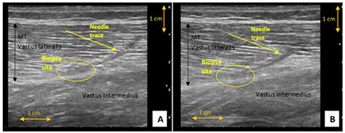
Ultrasound images of vastus lateralis, of a 32 years-old male subjects without any acute or chronic muscular disease (BMI: 23.1 Kg/m2, estimated Fat-Free Mass from bioimpedance: 80%); A- first day after biopsy, B - fourth day after biopsy; images from “Kanchenjunga Exploration & Physiology” research project
Fig. 2.
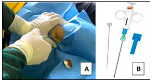
Tiny percutaneous needle biopsy from vastus lateralis muscle; A - Sterile procedure during muscle biopsy at a one-third distal distance between the greater trochanter and patellar top border performed by a trained medical doctor; B - Semi-automatic needle used for the biopsy (PRECISA® model 1310 - HS Hospital Service SpA, Latina, Italy)
Sonographic guidance for the prior assessment of muscle thickness (MT) or site of sampling has been already recommended previously; however, it is still too rarely performed.7,8 This work intends to further highlight the importance of ultrasound evaluation of muscle depth (MD) and MT prior to muscle biopsy collection and reports on the potential risks of obtaining different or inadequate muscle biopsy samples. . The ultrasound (US) images reported in this paper were acquired using longitudinal B-mode US scanning of the VL at the distal one-third of the length between the greater trochanter and the patellar top border, acquired with a 5-cm, 3-11 MHz linear array probe (Mylab Gamma, Esaote Biomedica, Genova, Italy). MT of VL was marked by either a black or white arrow (depending on the echogenicity of muscles) as a distance from superficial to deep aponeurosis. The aponeuroses appear as brightly, whitish-silvery colour, echogenic linear structures on the US images. A vertical yellow arrow marks MD.
Figure 1 shows two US images of VL of 32 years old male taken at 1st and 4th day after TPNB. A clear needle trace and the site of biopsy is visible in both images. As the figure shows, there is no evidence of oedema in both images, and the needle trace seems to be visibly reduced on the day 4th (panel B) compared to the day1st (panel A), suggesting a rapid and successful muscle wound healing process. TPNB is classed as a minor surgical procedure, the acute muscle damage is minimal, usually resulting only in slight soreness or discomfort lasting max. 1-3 days. Anyhow, healthcare providers should recommend restricting any intense physical activity for 24-to-48 h post-biopsy. This makes the TPNB as one of the safest and the least invasive muscle biopsy technique currently available.
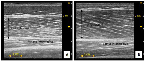
By using longitudinal ultrasound images of the vastus lateralis (VL) muscle obtained from three male subjects who participated in a recent research project “Kanchenjunga Exploration & Physiology”, where one of the research methods included muscle biopsy from (VL) using the TPNB technique (Fig 2), we could demonstrate the importance of performing a sonographic evaluation of MD and MT before taking muscle biopsy samples. Instead, Fig shows two US images of VL of the other two male subjects who participated in the study, detailing different muscle morphology. As may be observed in these images, each subject exhibited different MT (~2 cm vs. nearly 3 cm), which resulted in the biopsy specimens being taken from slightly different sites. The biopsy specimens from the first subject (48 years old) were taken at the border of the deep aponeurosis (panel A), whereas the biopsy samples from the second subject (25 years old) were taken from the middle of the VL muscle (panel B). Such findings are of laboratory importance as by taking the aponeurosis specimens together with the muscle any subsequent laboratory analyses could be affected, likely leading to some ‘skewed’ conclusions. In the previous subject, the biopsy samples were also taken near the deep aponeurosis (see Fig 1). It is, therefore, fundamental that prior US evaluation of MT and MD is performed. Fig 3 details also the biopsy site (orange circles in the images) after 7 weeks of TPNB with no visible needle traces, suggesting successful regeneration of muscle tissue without the formation of a connective tissue scar.
Fig. 3.
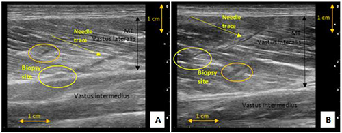
Ultrasound images of vastus lateralis; the orange circle shows a biopsy site after 7 weeks (pre-Nepal); A - 48 years-old male subjects without any acute or chronic muscular disease (BMI: 28.0 Kg/m2, estimated Fat- Free Mass from bioimpedance: 77%); B - 25 years-old male subjects without any acute or chronic muscular disease (BMI: 23.1 Kg/m2, estimated Fat-Free Mass from bioimpedance: 79%); images from “Kanchenjunga Exploration & Physiology” research project
Fig. 4.
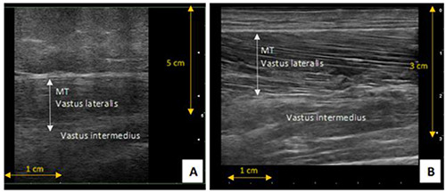
Ultrasound images of vastus lateralis of sarcopenic elderly subjects; A - 83 years old sarcopenic obese female; B - 84 years old sarcopenic male
Our findings are particularly important when considering the studies involving elderly individuals. Currently, many researchers are interested in identifying the exact cellular and molecular mechanisms behind sarcopenia using muscle biopsy samples in their laboratory investigations.9-11
With this respect, TPNB seems to be the most suitable muscle biopsy technique for the elderly. However, one should also recognise the fact that the elderly population is very heterogeneous, ranging from healthy active individuals to obese or sarcopenic; thus, it is of crucial importance to evaluate their muscles beforehand. For illustration, we present a selection of US images, detailing the heterogeneity in muscle morphology in the elderly population.
Fig 4 shows US images of VL of two sarcopenic and Fig 5 of two healthy active elderly individuals. As seen from the images, the MD and MT are very different in all the elderly individuals. In terms of the sarcopenic ones, the female’s VL is about 4-5 cm in-depth, under a prominent ~3cm layer of subcutaneous tissue (Fig 4, A), whereas the sarcopenic male (panel B) has virtually no subcutaneous tissue and his VL is no more than 2cm in-depth. A similar trend, however not so prominent, is visible in the healthy active female and male individuals, where the female has a small layer of subcutaneous tissue and smaller MT (Fig 5, A) than the male (panel B). Thus, extra caution must be taken before obtaining muscle biopsy samples from this population in order to prevent the wrong muscle group or tissue sampling. In fact, US has already been proposed as a useful tool for clinicians as a non-invasive, inexpensive and objective measurement of muscle mass and architecture in geriatrics,12,13 and it can also serve as a highly useful prior mean in the assistance to optimal muscle needle biopsies These observations seem to warrant the use of the TPNB as one of the safest and minimally invasive biopsy techniques currently available. In addition, prior use of MD and MT ultrasound evaluation seems a highly useful strategy to obtain good quality muscle biopsies by minimally invasive surgical approach, avoiding the risk of harvesting incorrect specimens.
Fig 5.
Ultrasound images of vastus lateralis at a one-third distal distance between great trochanter and patellar top border of healthy elderly subjects; A) 66 years old female; B) 68 years old male
List of acronyms
| TPNB | Tiny Percutaneous Needle Biopsy |
| MT | Muscle Thickness |
| MD | Muscle Depth |
| VL | Vastus Lateralis |
| US | ultrasound |
Funding Statement
Funding: The study was funded by: the “G. d’Annunzio” University grants to TP and VV; the “Departments of Excellence 2018–2022” initiative of the Italian Ministry of Education, University and Research to the Department of Neuroscience, Imaging and Clinical Sciences of “G. d’Annunzio” University, Italy; the project Q41 from the Charles University, Czech Republic to MS.
References
Articles from European Journal of Translational Myology are provided here courtesy of PAGEPress
Full text links
Read article at publisher's site: https://doi.org/10.4081/ejtm.2019.8851
Read article for free, from open access legal sources, via Unpaywall:
https://pagepressjournals.org/index.php/bam/article/download/8851/8552
Citations & impact
Impact metrics
Article citations
Microbiopsy Sampling for Examining Age-Related Differences in Skeletal Muscle Fiber Morphology and Composition.
Front Physiol, 12:756626, 10 Jan 2022
Cited by: 12 articles | PMID: 35082686 | PMCID: PMC8784837
Similar Articles
To arrive at the top five similar articles we use a word-weighted algorithm to compare words from the Title and Abstract of each citation.
Tiny percutaneous needle biopsy: An efficient method for studying cellular and molecular aspects of skeletal muscle in humans.
Int J Mol Med, 27(3):361-367, 14 Dec 2010
Cited by: 21 articles | PMID: 21165550
A method for the ultrastructural preservation of tiny percutaneous needle biopsy material from skeletal muscle.
Int J Mol Med, 32(4):965-970, 23 Jul 2013
Cited by: 5 articles | PMID: 23900509 | PMCID: PMC3812242
The reliability of muscle biopsies taken from vastus lateralis.
J Sci Med Sport, 2(4):333-340, 01 Dec 1999
Cited by: 10 articles | PMID: 10710011
The skeletal muscle microbiopsy method in exercise and sports science research: A narrative and methodological review.
Scand J Med Sci Sports, 32(11):1550-1568, 24 Aug 2022
Cited by: 1 article | PMID: 35904526
Review


