Abstract
Free full text

IFNγ is Critical for CAR T Cell Mediated Myeloid Activation and Induction of Endogenous Immunity
Abstract
Chimeric antigen receptor (CAR) T cells mediate potent antigen-specific antitumor activity, however, their indirect effects on the endogenous immune system is not well characterized. Remarkably, we demonstrate that CAR T cell treatment of mouse syngeneic glioblastoma activates intratumoral myeloid cells and induces endogenous T cell memory responses coupled with feed-forward propagation of CAR T responses. IFNγ production by CAR T cells and IFNγ-responsiveness of host immune cells is critical for tumor immune landscape remodeling to promote a more activated and less suppressive tumor microenvironment. The clinical relevance of these observations is supported by studies showing that human IL13Rα2-CAR T cells activate patient-derived endogenous T cells and monocyte/macrophages through IFNγ-signaling, as well as induce the generation of tumor-specific T cell responses in a responding patient with GBM. These studies establish that CAR T therapy has the potential to shape the tumor microenvironment, creating a context permissible for eliciting endogenous antitumor immunity.
INTRODUCTION
Glioblastoma (GBM) is among the deadliest cancers with very limited therapeutic options (1,2). Despite aggressive standard-of-care therapies, tumor recurrence is almost inevitable and uniformly lethal, with most patients not surviving beyond two years from diagnosis. While advances in immunotherapy have resulted in significant therapeutic improvements for many cancers, outcomes for patients in GBM have been disappointing (3). The complex myeloid-rich tumor microenvironment (TME) and heterogeneous nature of GBM remain barriers to the therapeutic success of immunotherapeutic approaches, including checkpoint blockade, vaccine therapy and chimeric antigen receptor (CAR) T cell therapy. A better understanding of the interplay between immunotherapy and the TME is required to develop more effective therapies for GBM and other solid tumors.
CAR T cell therapy is emerging as a promising strategy to treat cancer and may offer new therapeutic options for individuals diagnosed with GBM and other refractory brain tumors (4). Current efforts are focusing on expanding CAR T cell targets to address tumor heterogeneity (5–7), armored-CAR T cells introducing cytokines to improve CAR T cell persistence and potency (8–10), and optimizing route of delivery to improve trafficking (11). These preclinical studies have primarily utilized immunocompromised xenograft models to optimize antigen-dependent effector activity. However, to adequately address parameters that are critical for the therapeutic success of CAR T cell therapy preclinical models that take into consideration the host immune system are required. Recent studies in immunocompetent models of GBM establish a dynamic interplay between the endogenous immune system and CAR T cell responses, with phenotypic changes in the immune subsets observed post therapy (12,13). Nevertheless, detailed phenotypic analysis of these changes, as well as a mechanistic understanding of whether alterations in the TME may be a consequence of antigen-dependent tumor lysis or indirectly through CAR T cell-mediated cytokine production have not been well-elucidated.
Proinflammatory cytokines secreted by CAR T cells, such as IFNγ, may play an important role in activation and programming of the immune infiltrates in GBM TME. IFNγ can activate macrophage (14) and microglia (15), recruit and activate cytotoxic T cells, polarize CD4+ T cells into Th1 effector cells and impair tumor-promoting regulatory T cell (Treg) development and function (16–18). IFNs can additionally act as a key signal 3 to facilitate the activation and priming of tumor reactive T cells (19). Therefore, investigating the potential indirect effects of CAR T cell therapy on innate immune activation and adaptive immunity is critical for further optimization of this therapy for solid tumors.
While CAR T cell therapy in GBM and other solid tumors have not elicited the remarkable responses observed in hematological malignancies, clinical studies have demonstrated early evidence of safety and bioactivity in selected patients (20–24), although objective clinical responses have been limited. Our lead CAR program for GBM targets the IL13Ra2, a tumor associated antigen that is over-expressed by the majority of GBM (25). We previously optimized an IL13 (E12Y)-ligand 4-1BB/CD137-based CAR for clinical translation (6) and have initiated a phase I trial to evaluate the safety and feasibility of locorgenionally delivered IL13Ra2-CAR T cells for recurrent GBM. While this trial is ongoing, one patient of particular interest, who presented with recurrent multifocal GBM, remarkably achieved a complete response (CR) following locoregional delivery of IL13Rα2-CAR T cells, despite heterogeneous IL13Rα2 tumor expression. This case study demonstrates that CAR T cell therapy has the potential to elicit profound antitumor activity against one of the most difficult to treat solid tumors. Lessons learned from this clinical experience as well as other early clinical findings for CAR T cells in GBM suggest that in addition to targeting antigen positive cells, CAR T cells may also alter the TME with local changes in inflammatory cytokines, alterations in tumor immune infiltrates and clonal T cell populations (20–22). However, very little is understood regarding the therapeutic dependencies between CAR T cell therapy and the endogenous immune system.
Herein, we evaluated changes in the TME post CAR T cell therapy in both murine immune competent mouse models and patient-derived immune populations, focusing on our clinically validated IL13Rα2-CAR T cell platform, we show that CAR T cells not only target antigen positive tumors, but also promote the activation of host immune cells in the TME through a mechanism involving IFNγ signaling. Our findings highlight that CAR T therapy changes GBM immune landscape and induces activation of host immune cells which in turn can enhance CAR T therapy. Defining the immunological changes potentiated by CAR T cell therapy, along with specific immunological signatures required for effective antitumor responses, is critical for the successful advancement of CAR T cell therapy for solid tumors.
RESULTS
CAR T cells induce endogenous antitumor immune responses in GBM patient unique responder
Motivated by the unique responder in our IL13Ra2-CAR clinical trial, in which locoregionally delivered CAR T cells mediated a complete response in a setting of progressive multifocal GBM, we set out to investigate whether CAR T cells may have stimulated endogenous immunity that could have cooperated with CAR T therapy to elicit this profound therapeutic response (20). To determine whether this clinical response involved the induction of host immunity, T cells were isolated from the patient’s blood before CAR T cell treatment (Pre-CAR T) and during the response to CAR T therapy (Post-CAR T) (Fig. 1A). Isolated CD3+ T cells, confirmed to be CAR negative (Supplementary Fig. S1), were stimulated and expanded in the presence of the patient’s irradiated autologous tumor cell line. Endogenous CAR-negative T cells isolated during response (Post-CAR T) displayed enhanced tumor reactivity as indicated by increased intracellular IFNγ (3.9 fold increase) and greater tumor-dependent proliferation as compared to T cells isolated prior to the initiation of CAR T cell therapy (Pre-CAR T) (Fig. 1B–C). Importantly, subsequent co-culture assays with the autologous patient GBM tumor line or an irrelevant tumor line, established that T cells isolated during response to CAR T therapy exhibited enhanced tumor-specific killing compared to pre-treatment T cells (Fig. 1D). Tumor-specific proliferation and killing by endogenous T cells isolated from the blood during therapeutic response was independent of IL13Rα2 expression, as the ex vivo expanded autologous GBM line did not express the IL13Rα2 antigen (Fig. 1E). The same observation was not detected in a GBM patient that responded poorly to CAR T therapy (Supplementary Fig. S2A–D). These findings support the premise that the induction of endogenous immunity is important for productive CAR T cell therapy and is highly patient dependent, likely influenced by unique aspects of each tumor and the tumor microenvironment (TME). Furthermore, these observations provided the rationale to mechanistically investigate the potential of CAR T cells to promote the generation of tumor-specific T cell responses as well as evaluate the importance of host immune cells in a successful CAR T therapy.
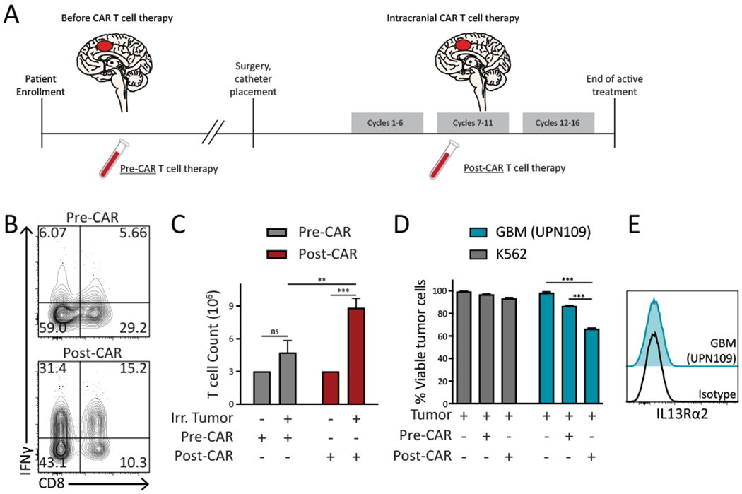
A, Treatment schema of a unique responder to IL13Rα2-CAR T therapy. T cells (CD3+) were isolated from peripheral blood prior to the initiation of therapy (Pre-CAR T) and during therapeutic response (Post-CAR). B, Representative flow cytometry showing intracellular IFNγ levels in patient T cells obtained before therapy (Pre-CAR) and during response (Post-CAR). T cells were cocultured with irradiated autologous tumor (UPN109) followed by a 4 hour stimulation. C, T cell count after 14-day co-culture with autologous irradiated (Irr.) patient tumor cell line. D, In vitro killing by patient T cells against autologous (UPN109) or nonspecific tumor line (K562) at 1:1, E:T ratio. E, Representative flow cytometry demonstrates the IL13Rα2 expression of the patient autologous (UPN109) tumor line. Data are presented as means ± s.e.m. (C and D) and were analyzed by two-tailed, unpaired Student’s t-test. *p < 0.05, **p < 0.01, ***p < 0.001 and ****p < 0.0001 for indicated comparison.
Murine IL13Rα2-CAR T cells mediate potent antitumor activity in immune competent models of GBM
To gain a deeper understanding of the interplay between CAR T cells, host immune responses and the GBM microenvironment, as well as interrogate the potential mechanisms involved (6,20), we established immunocompetent mouse models of our clinical IL13Rα2-CAR T cell platform. A mouse counterpart to our human IL13Rα2-targeted CAR was constructed(6), and composed of the IL-13(E12Y) tumor-targeting domain, murine CD8 hinge (mCD8h), murine CD8 transmembrane domain (mCD8tm), murine 4-1BB costimulatory domain (m4-1BB) and murine CD3 zeta (mCD3ζ). A T2A skip sequence separates the CAR from a truncated murine CD19 (mCD19t) used for cell tracking (Supplementary Fig. S3A). The engineering process resulted in a 70-85% transduction efficiency as assessed by the frequency of CD19t+ cells (Supplementary Fig. S3B). Phenotypic analyses indicated that, similar to human CAR T cells(6), the murine IL13Rα2-targeted CAR T cells (mIL13BBζ CAR T cells) contained comparable numbers of CD4+ and CD8+ T cell subsets with mixed early memory (CD62L+) and effector (CD62L−) T cell populations on day 4 of ex vivo expansion (Supplementary Fig. S3C and D). We designed a therapeutic setting based on K-Luc and GL261-Luc, two syngeneic, immunocompetent murine glioma models. K-Luc, a firefly luciferase engineered subline of KR158 (26), was used as it recapitulates the highly invasive features of GBM (Supplementary Fig. S3E). This tumor line is derived from a spontaneous glioma arising from Nf1, Trp53 mutant mice, and is poorly immunogenic as indicated by its unresponsiveness to anti-PD-1 checkpoint therapy (27). As a second model, we also used GL261 engineered to express ffLuc (GL261-luc), a non-invasive “bulky” glioma (Supplementary Fig. S3F), which was generated by chemical induction, contains high numbers of mutations, and in contrast to K-Luc, is responsive to anti-PD-1 immunotherapy (28,29). Both tumor lines were engineered to express murine IL13Rα2 (mIL13Rα2) (Supplementary Fig. S3E and S3F). mIL13BBζ CAR T cells specifically killed mIL13Rα2-engineered K-Luc and GL261-Luc cells, which was associated with production of inflammatory cytokines IFNγ and TNFα (Supplementary Fig. S3E and S3F), and were not responsive to IL13Rα2-negative parental tumor lines in vitro (Supplementary Fig. S3G).
We next evaluated CAR T cell antitumor activity against orthotopically engrafted glioma tumors in C57BL/6 immunocompetent mice. In IL13Rα2+ K-Luc tumor model, single intratumoral administration of mIL13BBζ CAR T cells seven days after tumor injection mediated potent in vivo antitumor activity and conferred a significant survival benefit (Fig. 2A–D). Mock T cell or PBS injected (untreated) tumor bearing mice did not exhibit reduced tumor progression (Supplementary Fig. S4A) and local treatment of CAR or Mock T cells did not result in toxicity associated weight loss (Supplementary Fig. S4B). To evaluate the impact of an intact immune system on CAR T cell-mediated antitumor responses, we compared anti-K-Luc activity in C57BL/6 mice to tumors engrafted in immunocompromised NOD-scid IL2Rgnull (NSG) models, which lack adaptive immune subsets. In a smaller tumor model (4-day old), with a less-developed TME, antitumor activity in C57BL/6 and NSG was equivalent, indicating CAR T cell functionality is comparable in both mouse strains and independent of an intact immune system (Supplementary Fig. S5A–C). However, surprisingly, the in vivo response to CAR T cell therapy was superior in immunocompetent C57BL/6 as compared to immunodeficient NSG mice (p<0.001) when the TME exhibited an established immune microenvironment (7-day engrafted tumor) (Supplementary Fig. S5D and S5E). This observation raised the possibility that tumor infiltrating immune cells may enhance the antitumor effects of CAR T cell therapy in GBM, and we sought to further investigate this observation in greater detail.
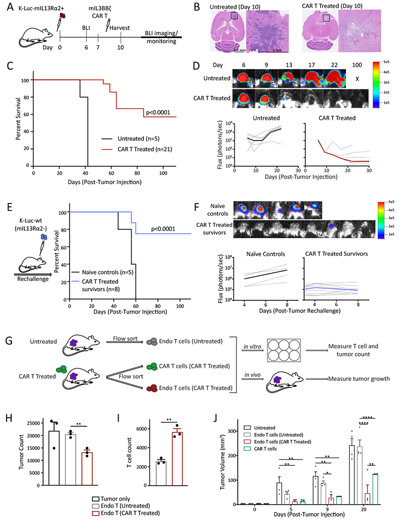
A, Schematic of in vivo experimental design. B, Representative images of hematoxylin and eosin (H&E) staining show invasive K-Luc in untreated brains and tumor elimination in CAR T treated brains. C, Survival curve of mice bearing K-Luc-mIL13Rα2+ tumors in untreated and CAR T treated groups. D, Representative bioluminescent (BLI) images (top) and flux values (bottom) show tumor growth in untreated and CAR T-treated groups. Individual mice are represented with dotted lines, while median flux is represented by the thick line. E, Survival of mice cured by CAR T therapy and rechallenged with IL13Rα2 negative K-Luc tumors. F, Representative bioluminescent (BLI) images from day 8 post-rechallenge tumor injection (top) and flux values (bottom) show tumor growth in naïve controls and survivors of CAR T therapy groups. Individual mice are represented with dotted lines, while median flux is shown by the thick line. G, Overview of experimental design. H, In vitro killing and I, expansion of endogenous T cells isolated from untreated or CAR T-treated mice against K-Luc-mIL13Rα2+ tumor cells (E:T, 10:1). J, Assessment of in vivo killing capacity of isolated CAR T cells and endogenous T cells from untreated or CAR T-treated cells in tumor (K-Luc-mIL13Rα2+) bearing mice. Data are representative of at least two independent experiments. Data are presented as means ± s.e.m. (H, I and J) and were analyzed by two-tailed, unpaired Student’s t-test. Differences between survival curves (C and E) were analyzed by log-rank (Mantel–Cox) test. *p < 0.05, **p < 0.01, ***p < 0.001 and ****p < 0.0001 for indicated comparisons.
CAR T therapy can promote immunological memory and the generation of tumor-specific T cells
To evaluate whether CAR T cells have the potential to induce endogenous antitumor immunity, cured mice following CAR T cell treatment were challenged with IL13Rα2-negative parental tumors. Indeed, in a more established TME (7 day engraftment before CAR T therapy), cured mice in the immunocompetent C57BL/6 model successfully rejected tumor rechallenge with IL13Rα2-negative K-Luc (Fig. 2E and and2F)2F) and GL261-Luc (Supplementary Fig. S6A and B) parental tumor cells, demonstrating that CAR T cells can promote immunological memory in two independent tumor models with differential responsiveness to anti-PD1 immunotherapy (27,29). The capacity of CAR T cells to induce endogenous immunity against IL13Rα2-negative tumor cells again required a more established TME, as mice cured in the small tumor model (4-day engraftment before CAR T therapy) were not capable of mounting antitumor responses following rechallenge with parental tumors (Supplementary Fig. S7A and B). Indeed, flow cytometry analysis of tumor infiltrated T cell and myeloid populations (Supplementary Fig. S8A) demonstrate increase number of immune cells in the 7-day compared to the 4-day tumor model (Supplementary Fig. S8B and S8C). These results suggest that while CAR T therapy is very effective in smaller tumors because of strong direct killing of antigen-positive tumor cells, it does not necessarily result in the establishment of endogenous immunity. And while “7-day model” is a larger tumor with an established TME that cannot be fully eliminated by CAR T cells in an antigen-dependent manner, the induction of host immunity can cooperate with CAR T therapy to improve therapeutic effectiveness resulting in surviving mice successfully rejecting antigen negative tumors upon rechallenge, establishing that a memory immune response was effectively established. This observation also suggests that tumor exposure is not sufficient to induce endogenous antitumor immunity, and instead the establishment of immunological memory requires both CAR T cell therapy and the host immune infiltrates. In a similar set of experiments, we compared the antitumor activity of mIL13BBζ CAR T cells against IL13Rα2+ K-Luc (100% IL13Rα2+ cells) versus a mixture of IL13Rα2+ K-Luc and IL13Rα2- parental K-Luc tumor cells (1:1 ratio) (Supplementary Fig. S9A–B). Supporting the notion that CAR T cells can promote endogenous immune responses against antigen-negative tumor cells, mIL13BBζ CAR T cells mediated comparable survival benefit against tumors with homogenous and heterogeneous IL13Rα2-antigen expression (Supplementary Fig. S9C). Again, the response to IL13Rα2-negative tumors required a more established tumor microenvironment, as CAR T cell therapy was less effective against mixed antigen tumors (1:1 ratio) in the small tumor model (day 4) (p<0.001; Supplementary Fig. S9D). Together these observations suggest that the host immune cells within the TME cooperate with CAR T cell therapy to augment antitumor responses and that CAR T cells can promote endogenous antitumor memory responses.
To directly evaluate whether CAR T therapy can potentiate the generation of tumor-specific T cells, we isolated intratumoral endogenous (CD3+CD19−) and CAR T cells (CD3+CD19+) from untreated and CAR T cell treated mice in the IL13Rα2+ K-Luc glioma model (3 days post treatment) (Supplementary Fig. S10), and either ex vivo co-cultured with tumor cells or adoptively transferred into new tumor bearing mice (Fig. 2G). Importantly, we find that endogenous T cells isolated from CAR T cell treated tumors, but not T cells from untreated mice, exhibited enhanced killing of IL13Rα2+ K-Luc tumors and T cell proliferation in co-culture assays (10:1, effector:target ratio; 72 hours) (Fig. 2H and and2I).2I). To assess in vivo function, endogenous T cells from untreated and CAR T cell treated mice were isolated and adoptively transferred into IL13Rα2+ K-Luc tumor bearing mice. Measurement of tumor progression demonstrated that mice injected with endogenous T cells isolated from CAR T treated mice showed a significant reduction in tumor growth compared to the control groups (Fig. 2J). Collectively, these results establish that CAR T cells have the potential to promote antigen spread and the generation of tumor-specific T cell responses.
CAR T cells activate innate and adaptive immune subsets in tumor microenvironment
To elucidate immune-related changes in the TME that coincide with the establishment of endogenous antitumor immunity following CAR T cell therapy, we interrogated both the lymphoid and myeloid compartments by gene and protein expression profiling. Focusing first on the lymphoid compartment, we performed nanostring analysis of purified intratumoral CD3+ cells and demonstrated a dramatic and global reshaping of the T cell compartment at transcriptome level in CAR T treated mice compared to untreated (Fig. 3A). To increase resolution and more accurately define subpopulations, we performed scRNAseq on isolated CD45 cells from the brain of untreated or CAR T treated mice (Supplementary Fig. S11A–C). We then computationally separated lymphoid and myeloid populations and reanalyzed the scRNAseq data at higher granularity. This approach yielded 9 distinct lymphoid subpopulations broadly defined by the distribution of classical marker genes, including three distinct subsets of CD8+ T cells (CD8_L2, CD8_L3, and CD8_L4), two subsets of CD4+ T cells (CD4_L1, CD4_L6), one subset of NK cells, two subsets of B cells and one subset resembling γδT cells (Fig. 3B and Supplementary Fig. S11D). The frequency of CD8_L2 remained unchanged, but interestingly, post CAR T therapy, increased frequency of CD8_L3 and CD8_L4 subclusters were detected (Supplementary Fig. S11D). Focusing on T cell subclusters, CD8_L3 is observed mainly post therapy and is characterized by expression of Cxcr3 (Supplementary Fig. S12A) which is associated with T cell trafficking and expression of Itgae (CD103), Cd74 (Hladr) and Ifitm1 (IFN-induced transmembrane protein 1) that correspond to activated resident memory CD8 T cell phenotype (Fig. 3C). CD8_L4 expanded post-therapy and was identified as highly activated, effector T cells based on higher expression of Ki67, Cd74, and Gzma genes (Fig. 3C). Within the CD4 subsets, the frequency of CD4_L1 cluster remained unchanged after therapy, although post-therapy this cluster displayed a modest increase in expression of Il7r, Tcf7, and Itga4 genes, which is associated with effector memory CD4 T cells (Supplementary Fig. S12A). Intratumoral regulatory T cells (Treg), defined by subcluster CD4_L6 based on the expression of CD4, Foxp3, GITR (Tnfrsf18) and Ctla4, decreased after CAR T therapy (Supplementary Fig. S12B). Overall, gene set enrichment analysis (GSEA), revealed enrichment of gene signatures associated with activated and Th1 responses in most T cell subclusters (Fig. 3D). Taken together, these studies reveal that CAR T cells can dramatically alter the lymphoid compartment within tumors and increase activated, memory or effector T cell populations.
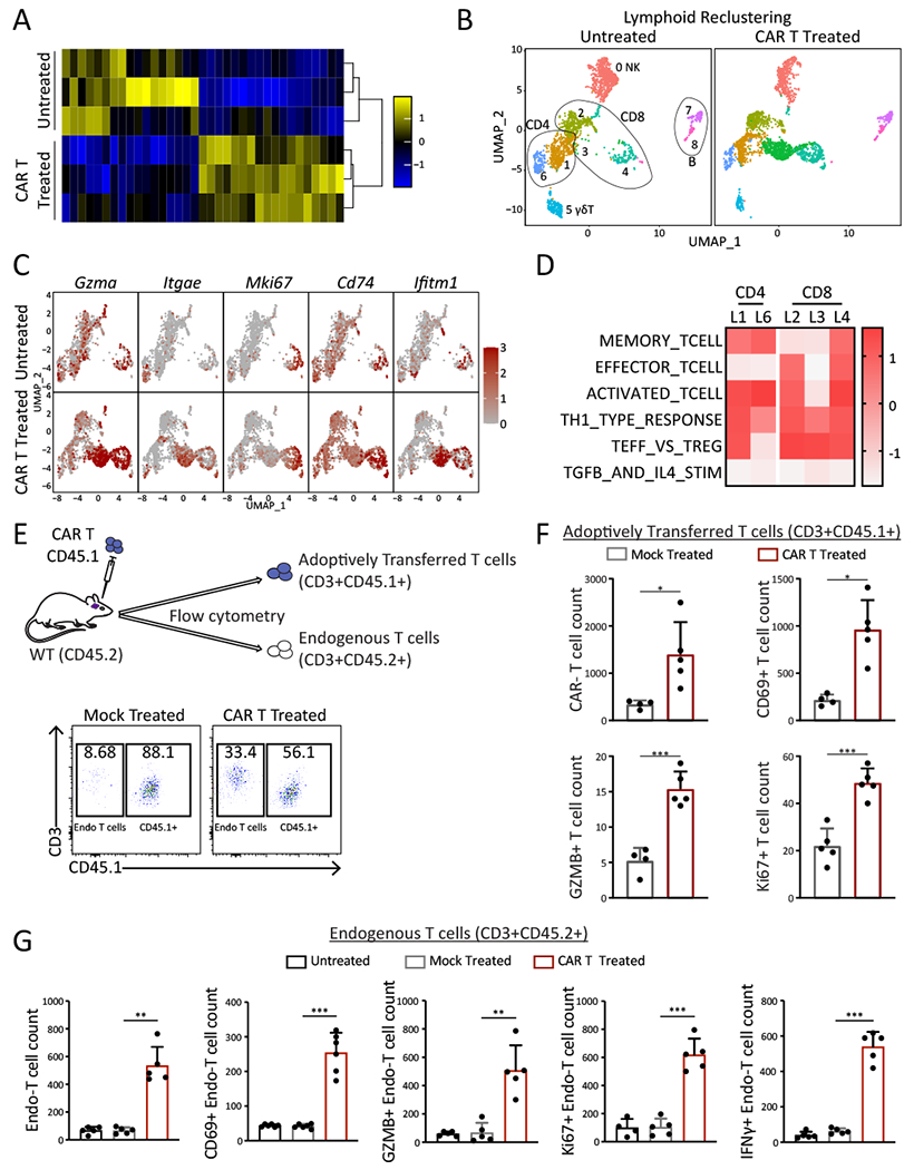
A, Nanostring analysis shows global changes in gene expression of intratumoral T cells (CD3+) isolated from untreated or CAR T-treated mice 3 days post-therapy. B, UMAP plots depict changes in lymphoid compartments in the glioma TME after CAR T therapy. C, Feature plots demonstrate phenotypic characterization of T cell subclusters and enriched pathways within CD8 and CD4 T cell subclusters post therapy. D, Heatmap of enrichment scores (GSEA analysis) shows enriched pathways in T cell subclusters. E, Experimental design demonstrating the adoptive transfer of CD45.1+ mock or CAR T cells (top) and a representative flow cytometry analysis show frequency of endogenous (CD3+CD45.2+) or adoptively transferred T cells (CD3+CD45.1+) in glioma TME (bottom). F, Bar graphs compare adoptively transferred mock (CD3+CD45.1+) or CAR T cell (CD3+CD45.1+CD19+) number and phenotypic characterization (CD69, Ki67 and GZMB). G, Bar graphs compare endogenous T cell (CD3+CD45.2+) numbers and phenotypic characterization (CD69, Ki67 GZMB, and IFNγ) in untreated, mock or CAR T treated mice (n=5 per group). Data are representative of at least two independent experiments. Data are presented as means ± s.e.m. (F and G) and were analyzed by two-tailed, unpaired Student’s t-test. *p < 0.05, **p < 0.01, and ***p < 0.001 for indicated comparison.
To further characterize T cell populations post-CAR T cell therapy at cellular level and differentiate changes in endogenous versus adoptively transferred T cells, isogenically mismatched CD45.1 CAR T cells were used to treat IL13Rα2+ K-Luc tumors engrafted in CD45.2 mice (Fig. 3E). Following intracranial delivery, CAR T cells (CD3+ CD45.1+CD19+), but not mock-transduced T cells (CD3+CD45.1+CD19−), displayed a significant increase in T cell count and expression in markers of activation (CD69+), cytotoxic function (GZMB+) and proliferation (Ki67+) (Fig. 3F), establishing that the observed effector activity was CAR-dependent. Importantly, only after CAR T therapy, a significant increase in the endogenous T cell (CD3+CD45.2+) count with activated (CD69+), proliferative (Ki67+), and cytotoxic (GZMB+) phenotypes was observed, which was not detected in untreated or mock treated controls (Fig. 3G). This was in line with qPCR analysis demonstrating upregulation of GZMA, GZMB, and PRF1 genes in intratumoral CD3+ T cells after CAR T therapy (Supplementary Fig. S13). These results establish that CAR T cells promote endogenous T cell activation and expansion, and are consistent with our previous finding that endogenous intratumoral T cells isolated post CAR T cell therapy have antitumor activity (Fig.2H–I).
We next evaluated changes in the innate myeloid cells, including microglia/macrophages, as they represent a dominant immune population in the glioma tumors and have decisive role in glioma pathogenesis(30). Analysis of intratumoral myeloid populations at single cell level identified 17 distinct myeloid subsets which underwent a striking remodeling following CAR T therapy (Fig. 4A and Supplementary Fig. S14).
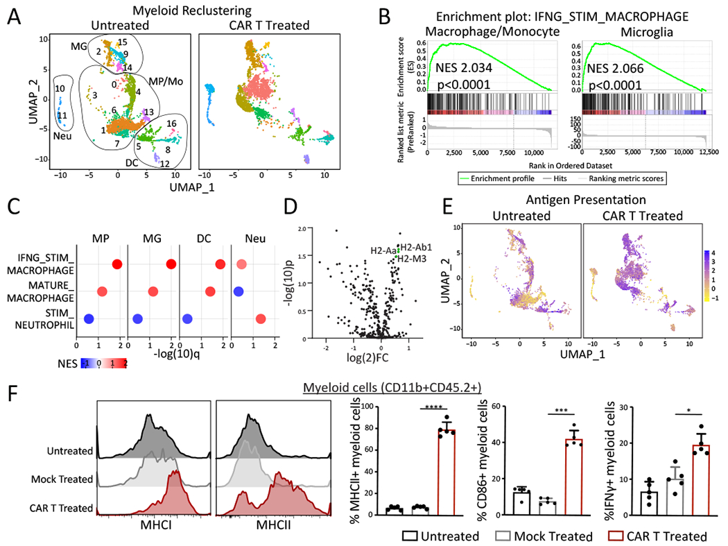
A, UMAP plots of scRNAseq depict changes in intratumoral myeloid cells from CAR T-treated or untreated mice (3-days post therapy). B, Enrichment plot of IFNγ signaling pathways in intratumoral macrophage and microglia cells in CAR T-treated compared with untreated, as identified by the GSEA computational method. C. GSEA analysis reveals upregulation of population specific pathways in myeloid subclusters following CAR T treatment (MP: macrophage; MG: microglia; DC: dendritic cells; Neu: neutrophils). D, Nanostring analysis show global changes in gene expression of myeloid cells (CD11b+) isolated from CAR T-treated vs untreated mice. E, UMAP projections indicate relative expression levels of antigen presentation gene signatures at a single-cell level within the myeloid compartment. F, Representative flow cytometry histograms (left) and summary bar graphs (right) show intratumoral CD11b+CD45.2+ cells expressing MHCII, MHCI, CD86, and IFNγ. Data are presented as means ± s.e.m and analyzed by two-tailed, unpaired Student’s t-test. *p < 0.05, **p < 0.01, ***p < 0.001, ****p<0.0001 for indicated comparison.
While some macrophage/monocyte subpopulations decreased in frequency, other populations expanded and re-shaped the TME. Seven major monocyte/macrophage (Itgam and Cd68), four microglia (Tmem119 and P2ry12), four DC (Ccr7 and Cd209a), and two neutrophil (S100A9) subpopulations were identified (Supplementary Fig. S14). Gene set enrichment analysis (GSEA) revealed enrichment of genes associated with IFNγ-stimulated macrophage and microglia in CAR-treated groups (Fig. 4B). Assessment of the main myeloid populations (macrophage, microglia, and neutrophils) identified higher expressions of genes associated with mature and IFNγ-activated macrophages as well as stimulated neutrophils (Fig. 4C), further confirming that resident innate immune cells have been exposed to IFNγ-mediated activation.
Nanostring analysis of intratumoral microglia/macrophages (CD11b+) from the TME 3 days post-CAR T therapy showed enrichment of genes that mediate antigen processing and presentation (e.g., H2-Ab1, H2-Aa, H2-Eb1) (Fig. 4D). Further analyses with scRNAseq confirmed that majority of macrophage/microglia subclusters may be involved in antigen processing and presentation (Fig. 4E). Assessing CAR T cell mediated changes in resident microglia/macrophage populations by flow cytometry, we found an increased frequency of activated brain-resident macrophage/microglia cells (CD86+, MHCIhigh, MHCIIhigh) in CAR T-treated mice (Fig. 4F) which also corresponded to increase number of activated myeloid cells (Supplementary Fig. S15A). Assessment of myeloid compartment also revealed a significant increase in the frequency and number of M1 type macrophages as measured by IFNγ+CD11b+ cells (Fig. 4F and Supplementary Fig. S15A). Conversely, expression of M2 type macrophages (CD163/CD206) decreased in the TME after CAR T therapy (Supplementary Fig. S15B). Collectively, these data show that CAR T therapy changes the GBM immune landscape and activates the host innate and adaptive immune cells. These results also further reveal a major role for IFNγ in inducing activation of local immune cells.
Lack of IFNγ in CAR T cells impairs in vivo antitumor activity and activation of host immune cells
Given that the myeloid cells constituted the largest population in the glioma TME (31) (Supplementary Fig. S15B) and our scRNAseq analysis identified gene-signatures related to IFNγ-stimulation within the macrophages and microglia subclusters (Fig. 4B), we reasoned that IFNγ produced by antigen-stimulated CAR T cells may play a role in modulating the activation of resident macrophage/microglia cells and subsequent priming and induction of adaptive immune response.
IFNγ is one of the key effector cytokines abundantly produced by CAR T cells upon activation and is a prototypic macrophage activator (14). To investigate whether IFNγ secreted by CAR T cells is responsible for changes observed in phenotype and function of resident macrophages/microglia cells, CAR T cells were generated from WT (CAR Twt) or IFNγ−/− (CAR TIFNγ−/−) mice (Fig. 5A) and characterized accordingly. CAR transduction efficiency, cell viability, expansion in both CAR T cell populations (CAR Twt and CAR TIFNγ−/−) showed comparable therapeutic products with some difference in ratio of CD4:CD8 T cells (p < 0.05) (Fig. 5B). We next verified the functionality of CAR T cells derived from IFNγ−/− mice by conducting an in vitro killing assay in comparison with CAR T cells from WT mice, which demonstrated comparable killing potency at a 1:1 effector to target ratio (Fig. 5C). Assessment of CAR T polyfunctionality demonstrated comparable production of TNFα and GZMB in both CAR Twt and CAR TIFNγ−/− cells with expected lack of IFNγ production in CAR TIFNγ−/− (Fig. 5D). In vivo, mice that received CAR TIFNγ−/− exhibited poor overall survival compared to mice treated with CAR Twt, indicating that IFNγ deficiency in CAR T cells dampens their antitumor activity in vivo (Fig. 5E and and5F).5F). Gene expression analysis of tumor and the associated TME 3 days post CAR therapy revealed enhanced expression in genes involved in activation and proinflammatory cytokines in mice that received CAR Twt and conversely reduced expression of genes involved in suppressive phenotype and function of intratumoral immune infiltrates (Fig. 5G) indicating that lack of IFNγ secretion by CAR T cells changes the glioma TME. IFNγ is a pleiotropic cytokine that induces activation of CD8 T cells (17), promotes polarization of Th1 CD4 cells (32) and reprograms or activates macrophage/microglia cells (14,15). Therefore, we then assessed if lack of IFNγ secreted by CAR T cells impacted the host immune cells. Flow cytometry analysis of TME 3 days post CAR T cell therapy revealed a significant decrease in T cell number, both endogenous and CAR T cells, that correlated with a reduction in activated (CD69+) T cells (Fig. 5H and and5I).5I). Furthermore, a significant increase in frequency of MHCI+/MHCII+ and CD86+ macrophage/microglia cell activation in tumor bearing mice that received CAR Twt compared with CAR TIFNγ−/− cells (Fig. 5J) was observed. Importantly, lack of IFNγ secretion by the CAR T cells resulted in higher M2 type intratumoral macrophages in mice that received CAR TIFNγ−/− cells compared with CAR Twt cells (Supplementary Fig. S16). Thus, IFNγ production as a consequence of CAR T antitumor activity, resulted in activation and reinvigoration of T cells, and reprogramming of macrophage/microglia cells to enhance their activation and antigen presentation potential.
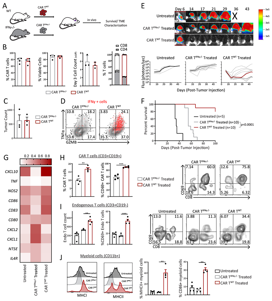
A, Schematic of the experimental design. B, Comparison of percent CAR positivity, viability, expansion, and CD4:CD8 ratio in CAR Twt and CAR TIFNγ−/− . C, In vitro killing of CAR Twt and CAR TIFNγ−/− against K-Luc-mIL13Rα2+ cells (E:T, 1:1). D, Representative flow cytometry plot depicts intracellular cytokine levels (TNFα, GZMB and IFNγ) in wt and IFNγ−/− CAR T cells after exposure to K-Luc-mIL13Rα2+ tumors. E, Representative bioluminescent (BLI) images (top) and flux values (bottom) show tumor growth in untreated, CAR Twt or CAR TIFNγ-/−. Individual mice are represented with dotted lines and median flux is shown in thick line. F, Survival curve of mice bearing K-Luc-mIL13Rα2+ tumors in untreated, CAR Twt treated and CAR TIFNγ−/− treated groups. G, Heatmap indicates normalized expression of genes associated with immune activation and suppression in the TME. H, Bar graphs (left) and representative flow cytometry plots (right) comparing CAR T cell (CD3+CD19+) number and activation phenotype (CD69). I. Bar graphs (left) and representative flow cytometry plots (right) comparing endogenous T cell (CD3+CD19−) number and activation phenotype (CD69). J, Representative histograms (left) and bar graphs (right) showing phenotype in myeloid (CD11b+) compartment. Data are representative of at least two independent experiments. Each symbol represents one individual (H, I, and J). Data are presented as means ± s.e.m and were analyzed by two-tailed, unpaired Student’s t-test. Differences between survival curves (F) were analyzed by log-rank (Mantel–Cox) test. *p < 0.05, **p < 0.01, and ***p < 0.001 for indicated comparison.
Lack of IFNγ-signaling in the host results in dampened CAR T antitumor activity in vivo
Previous studies have reported IFNγ signaling as a signature of response to immunotherapies such as anti-PD1 treatment (33). In order to investigate whether host IFNγ signaling plays a role in the CAR T-mediated immune response, CAR Twt cells were adoptively transferred into K-Luc-bearing WT or IFNγR−/− mice (Fig. 6A). IFNγR−/− mice that received CAR Twt demonstrated a survival disadvantage, suggesting that lack of IFNγ signaling in the host immune cells dampens the antitumor activity of CAR T cells and the overall survival (Fig. 6B and and6C6C).
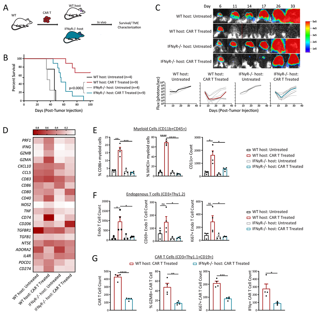
A, Schematic of the experimental design. B, Survival curve of mice bearing K-Luc-mIL13Rα2+ tumors in untreated or CAR T treated in wt host or IFNγR−/− host. C, Representative bioluminescent (BLI) images (top) and flux values (bottom) show tumor growth. Individual mice are represented with dotted lines, while median flux is represented by the thick line. D, Heatmap indicate normalized expression of genes associated with immune activation or suppression in the TME. E, Bar graphs of myeloid cells (CD11b+ CD45+) show phenotypic changes (CD86, MHCII, and CD11c). F, Bar graphs of endogenous T cells (CD3+Thy1.2) show changes in T cell count, CD69 and Ki67. G, Bar graphs of CAR T cells (CD3+Thy1.1+CD19+) demonstrate changes in number, GZMB, Ki67, and IFNγ. Data are representative of at least two independent experiments. Each symbol represents one individual mouse. Data are presented as means ± s.e.m. (E-G) and were analyzed by two-tailed, unpaired Student’s t-test. Differences between survival curves (B) were analyzed by log-rank (Mantel–Cox) test. *p < 0.05, **p < 0.01, ***p < 0.001and ****p < 0.0001 for indicated comparison.
Gene expression analysis of TME 3 days post CAR therapy, which includes tumor, tumor associated immune and stroma cells, revealed that lack of IFNγ responsiveness in the host immune cells resulted in reduced expression of genes involved in activation and proinflammatory responses (CD40, NOS2, TNFα, GZMA, GZMB, PRF1, and IFNγ) (Fig. 6D). To further investigate CAR T cell-mediated changes in the immune landscape, we adoptively transferred isogenically mismatched (Thy1.1+) CAR Twt cells into K-Luc-bearing WT or IFNγR−/− mice. Flow cytometry analysis of immune subsets in the TME revealed a significant increase in activated macrophage/microglia cells (CD86+ and MHCII+), M1 markers (IFNγ+TNFα+) and decrease in M2 markers (CD163+CD206+) in WT compared to IFNγR−/− mice 3 days post CAR T cell therapy (Fig. 6E and Supplementary Fig. S17A and S17B). Compared to WT mice, the number of endogenous T cells (Thy1.2+CD3+), activated with proliferative and effector-cytokine producing capacities was significantly lower in IFNγR−/− mice (Fig. 6F). Lower frequency of regulatory T cells (CD4+CD25+Foxp3+) and higher effector type T cells (CD8+GZMB+) were detected in WT mice that received CAR T cells as compared to IFNγR−/− CAR T treated groups (Supplementary Fig. S18A, S18B and S18C). Furthermore, a significant increase in exhausted T cells was observed in IFNγR−/− compared to WT host at later time point after CAR T therapy, (Supplementary Fig. S18D). Interestingly, higher levels of adoptively transferred CAR T cells (Thy1.1+CD3+) were detected in WT mice, and these T cells were more proliferative (Ki67+) and cytotoxic (IFNγ+ and GZMB+) relative to IFNγR−/− mice (Fig. 6G) suggesting IFNγ-dependent positive feedback from host immune cells that promote CAR T cell activity in the TME. Collectively, these results confirm that there is an interplay between host and adoptively transferred immune cells and host IFNγ signaling in the glioma TME is necessary for mounting a potent immune response during CAR T therapy.
Human CAR T cell therapy modulates patient host immune cells
To evaluate the clinical relevance of IFNγ-signaling for human CAR T cell therapy, we next investigated the impact of CAR T cell antitumor activity on human endogenous immune cells in GBM patients. To assess if CAR T cells are able to promote activation of GBM patient monocytes or macrophages, we developed an in vitro assay to phenotypically characterize patient myeloid populations in the presence of CAR T cell antitumor activity. Conditioned media (CM) from co-culture of human CAR T cells and patient-derived glioma tumor lines were collected and subsequently incubated with glioma patient derived-monocytes (Supplementary Fig. S19A), ex vivo differentiated patient macrophages or total CD3+ patient T cells (Fig. 7A). Phenotypic and morphological assessments revealed that CM from CAR T plus tumor coculture promoted differentiation and activation of patient derived-monocytes and macrophages, as demonstrated by increase in expression of activation markers (CD14+CD86+/CD80+ and CD14+HLADr+) (Fig. 7B, ,7C,7C, ,7D7D and Supplementary Fig. S19B and C), Gene analysis demonstrated increased expression of genes associated with classical M1 macrophages (Fig. 7E and Supplementary Fig. S19D). Accordingly, exposure to CM from CAR T plus tumor coculture resulted in induced activation of isolated T cells from GBM patient blood, as evidenced by increased expression of CD69 (Fig. 7F and and7G).7G). Importantly, blockade of IFNγ signaling in macrophages and T cells resulted in reduced activation (Fig. 7B–F), highlighting the impact of IFNγ in the CAR T-mediated activation of host immune cells in GBM patients. Taken together these findings suggest that IFNγ secreted by CAR T cell antitumor function promotes polarization of monocytes into activated macrophages, further induce activation of differentiated macrophages and promote T cell activation. These findings confirm our preclinical studies that an effective CAR T therapy has the potential to stimulate the host immune cells and highlights the important role for IFNγ signaling and the important role host immune cells play in a successful CAR T treatment.
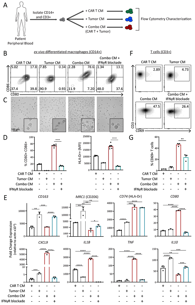
A, Schema of experimental design. B, Representative flow cytometry, C, microscopy images and D, bar graph summary of phenotypic changes of patient macrophages after incubation in conditioned media (CM) collected from unactivated T cells (CAR T CM), tumor only (Tumor CM) versus tumor-activated CAR T cells (Combo CM) or tumor-activated CAR T cells with IFNγR blockade. E, qPCR analysis shows genes associated with macrophage activation after incubation in condition media as described in B-D. F, Representative flow cytometry and G, bar graph summary of phenotypic changes in patient T cells after incubation in conditioned media as described in B-D. Each symbol represents one replicate. Data are presented as means ± s.e.m. (D, E, and G) and were analyzed by two-tailed, unpaired Student’s t-test. *p < 0.05, **p < 0.01, ***p < 0.001 and ****p < 0.0001 for indicated comparison.
DISCUSSION
Delineating the mechanisms that positively modulate host antitumor immunity is critical for development of cancer immunotherapy. Our studies indicate that, in addition to direct antitumor activity towards malignant cells, CAR T cells have immune-stimulatory effects which result in activation of host innate immune subsets and subsequent engagement of adaptive immunity. We find that IFNγ signaling both in CAR T cells and host tightly control adaptive and innate immunity. We employed well-characterized syngeneic glioma models to address fundamental questions about the role of resident immune cells in the glioma TME during the CAR T cell-mediated antitumor responses. The rationale for this study was based on our clinical observations, in which a patient with multifocal GBM had a complete response to IL13Rα2-CAR T therapy despite the heterogeneous expression of IL13Rα2 on the tumors. The current study provides insights into the immunological signatures required for an effective anti-glioma CAR T therapy, revealing that: 1) CAR T cells can activate host immune cells and induce tumor-specific T cells; 2) mice cured from CAR T therapy are immunized against antigen negative tumors; 3) effective CAR T cell antitumor activity is dependent on IFNγ-signaling, including both IFNγ secretion by CAR T and IFNγ-signaling in host immune cells; 4) clinically, IFNγ-dependent activation of patient host immune cells and generation of tumor-specific T cells can be induced by CAR T cell therapy.
The GBM microenvironment is characterized by high numbers of myeloid cells and lower T cell counts (31). Identifying mechanisms that regulate the activation of microglia/macrophages and/or increase the number of T cells in the TME are emerging strategies to overcome the immunosuppressive nature of GBM (34). We demonstrate that optimal CAR T therapy can activate host immune cells, resident microglia/macrophages and T cells. Accordingly, we also show that after CAR T therapy, the resident microglia/macrophage subpopulations displayed gene expression patterns with enhanced antigen presenting capacity. These findings are in line with recent studies demonstrating that lack of response in human solid tumors is not only due to presence of exhausted CD8 T cells or overexpression of PD-L1 in tumors, but also the failure of T cells to interact with antigen presenting myeloid populations in intratumoral niches (35). An important initiator of the immune response is antigen presentation with MHC proteins and accordingly our findings strongly suggest that the presence of myeloid subpopulations with antigen presenting phenotype is important in the CAR T-mediated anti-tumor immune response.
We also show evidence for an increase in number of endogenous T cells after CAR T therapy. This may be due to increased proliferation of existing T cells or increase in T cell infiltration. The elevated number of endogenous Ki67+ T cells and presence of Mki67 expressing subclusters may point towards potential proliferation of existing T cells in the TME. Detection of CD8+ lymphoid subclusters with higher CXCR3 expression post-CAR T therapy, however, also support increased infiltration of CD8 T cells. It should be noted that, in line with previous studies (35), the reduction in intratumoral CD4+ T regulatory cells (Treg) and reduction in the Treg subcluster after CAR T therapy indicate reduction in suppressive factors in the TME. Our finding that the survived mice post-CAR T cell therapy rejected the antigen-negative tumor cells validate the notion that CAR T cells mediate changes in immune landscape and subsequently induce endogenous memory response.
To investigate potential mechanisms of CAR T-mediated antitumor activity, we identified IFNγ as a major player in activation of the host immune cells in glioma TME. IFNγ, a key cytokine secreted by activated CAR T cells, is involved in inducing innate and adaptive immunity (14,16,33,36). IFNγ is critical in activation of macrophages (14,35) and microglia cells (15), and induction of memory alveolar macrophages post viral infection (37). IFNγ is also involved in recruitment and activation of cytotoxic T cells, polarization of CD4+ T cells into Th1 effector cells and reduction of tumor-promoting Treg development and function (16–18). These findings support our observation that IFNγ is a critical cytokine with important roles in driving CAR T cell associated effects on the endogenous TME within solid tumors, priming innate and adaptive immunity components against tumor cells and conferring long-lasting anti-tumor immunity.
While persistent IFNγ signaling paradoxically is linked to inhibitory feedback circuits through tumor cells and immune cells known as ‘adaptive resistance’, the IFNγ-mediated responses are still positively associated with patient’s survival in several cancers (16,38). Upregulation of cell surface MHC class I by IFNγ is crucial for host response to intracellular pathogens and tumor cells. IFNγ also upregulates cell surface MHC class II on antigen presenting cells. Thus, in the early phase of antigen recognition and the interaction between adaptive and innate immune cells, IFN responses are key to engaging the innate and adaptive arms for a potent CAR T therapy in GBM. While this study highlights the importance of IFNγ, our current results do not exclude other potential mechanisms of induction of host immune response, including tumor immune landscape and other inflammatory cytokines. These findings outline the adjuvant potential of CAR T cells that drives the generation of antitumor immune responses and endogenous memory responses.
Clinically, CAR T cell responses in GBM and other solid tumors have been inadequate for most patients. The heterogeneous nature of solid tumors and the suppressive TME create significant challenges for CAR T cell therapy. Further, long lasting antitumor immunity in patients is often times not observed likely due to evolution of the tumor under immune pressure leading to immune evasion (39). What will it take to improve CAR T cell response rates for solid tumors? This study highlights the importance of the immunomodulatory effects CAR T cell therapy, through production of inflammatory cytokines. Our findings are supported by clinical examples of productive CAR T cell responses in solid tumors. We have previously reported that IL13Rα2-CAR T-mediated complete response in a GBM patient was associated with increases in inflammatory cytokines and activated endogenous immune cells in the CSF (20). Likewise, a recent study reported evidence of endogenous immune reactivity after HER2-CAR T therapy which correlated with a positive patient response (21). These finding suggest that tumor-dependent characteristics (i.e., tumor mutational burden and IFNγ responsiveness) and features of the TME (i.e., CD3 infiltration) may impact CAR T cell responsiveness. Therefore, improving the effectiveness of CAR T cell therapy more broadly for solid tumors may require additional strategies to enhance the induction of host antitumor responses (8,13,40,41).
In conclusion, this report highlights the interplay between CAR T cells and the host immune cells in GBM. It is the first report that underlines the adjuvant potential of CAR T cells and the importance of host innate and adaptive immune cells in the context of CAR T therapy for solid tumors. Our findings provide mechanistic evidence that CAR T cell, a potent antitumor agent, functions as an immunomodulatory adjuvant therapy that engages resident innate immune population and generates tumor antigen–specific cytotoxic T cells. These insights strongly support the need to consider targeting/engaging both innate (e.g., macrophages) as well as adaptive (CD4+ and CD8+ T cells) immunity to improve the efficacy of CAR therapy for solid tumors. Our work advances the biological understanding of multifunctional aspect of CAR T therapy with tremendous potential to incorporate novel therapeutic strategies for patients with this devastating cancer. Such immune engagement and cytotoxic T cell initiation is desirable for current immunotherapies and would conceivably complement other modalities including immune checkpoint blockade or immune stimulatory adjuvant therapies.
METHODS
Mice and Cell lines
C57BL/6/J, CD45.1 (B6.SJL-PtprcaPepcb/BoyJ), Thy1.1 (B6.PL-Thy1a/CyJ), IFNγR−/− (B6.129S7-Ifngr1tm1Agt/J), and IFNγ−/− (B6.129S7-Ifngtm1Ts/J) mice were purchased from The Jackson Laboratory. NOD/Scid IL2RγCnull (NSG) mice were bred at City of Hope. All mouse experiments were approved by the City of Hope Institutional Animal Care and Use Committee (IACUC).
The luciferase-expressing murine GL261 (GL261-Luc) and KR158B (K-Luc) glioma cells were transduced with lentivirus to produce murine IL13Rα2 (mIL13Rα2) expressing sublines (GL261-Luc-mIL13Rα2 and K-Luc-mIL13Rα2). These tumor lines were maintained in DMEM (Gibco) supplemented with 10% fetal bovine serum (Hyclone Laboratories), 25mM HEPES (Irvine Scientific, Santa Ana, CA) and 2mM L-glutamine (Lonza). Cell surface expression of mIL13Rα2 was authenticated by flow cytometry and immunofluorescence imaging.
Patient-derived glioma cells (PBT030, UPN109, and UPN097) were isolated from GBM patient resections under protocols approved by the COH IRB and maintained as described previously (20). All tumor lines were authenticated for the desired antigen/marker expression by flow cytometry and cells were tested for mycoplasma and maintained in culture for less than 1-3 months.
CAR T cell Production
Human CAR T cells:
Naïve and memory T cells were isolated from healthy donors at City of Hope under protocols approved by the COH IRB (6,42). The construct of IL13Rα2-targeted CAR and CAR transduction was described in previous studies (6,20), (7). In brief, primary T cells were stimulated with Dynabeads Human T expander CD3/CD28 (Invitrogen) (T cells: beads = 1:3) for 24 hours and transduced with CAR lentivirus (multiplicity of infection [MOI] = 0.3). 7 days after CAR transduction, CD3/CD28 beads were removed and cells were resuspended and expanded in X-VIVO 15 media (Lonza) containing 10% FCS, 50 U/ml recombinant human IL-2 (Novartis), and 0.5 ng/ml recombinant human IL-15 (CellGenix) for additional 10-15 days before cryopreservation.
Murine CAR T cells:
The murine IL13Rα2 chimeric antigen receptor was constructed in a MSCV retroviral backbone (Addgene), containing the extracellular murine IL13 and murine CD8 hinge, murine CD8 transmembrane domain, and intracellular murine 4-1BB costimulatory and murine CD3ζ signals. Following a T2A ribosomal skip, a truncated murine CD19 was inserted as a transduction marker. The resulting plasmid was transfected into PlatE cells (a gift from Dr. Zuoming Sun lab) using Fugene (Promega). After 48 hours, the supernatant was collected and filtered using an 0.2 μm filter. The retroviral supernatant was aliquoted and frozen until the time of transduction.
Murine T cells were isolated from spleens of naïve C57BL/6J mice or appropriate strain (CD45.1, Thy1.1, or IFNγ−/−) with EasySep Mouse T cell Isolation Kit (STEMCELL Technologies) and stimulated with Dynabead Mouse T-Activator CD3/CD28 beads (Gibco) at a 1:1 ratio. T cells were transduced on RetroNectin-coated plates (Takara Bio USA) using retrovirus-containing supernatants (described above). Cells were then expanded for 4 days in RPMI-1640 (Lonza) supplemented with 10% FBS (Hyclone Laboratories), 55 mM 2-mercaptoethanol (Gibco), 50 U/mL recombinant human-IL-2 (Novartis), and 10 ng/mL recombinant murine IL-7 (Peprotech). Before in vitro and in vivo experiments, the beads were magnetically separated from the T cells and CAR expression was determined by flow cytometry.
In vivo studies
All mouse experiments were performed using protocols approved by the City of Hope IACUC. Orthotopic GBM models were generated as previously described (43). Orthotopic tumor model was established by stereotactically implanting 1×10⁵ tumor cells intracranially (i.c.) into the right forebrain of 8-10 week-old C57BL/6J, IFNγR−/−, or NSG mice. Engraftment was verified by bioluminescent imaging one day prior to CAR T cell injection, Mice were randomized into groups based on bioluminescent signal. Investigators were not blinded for randomization and treatment. Four or seven days after tumor injection, mice were treated intracranially with 1x10⁶ mIL13BBζ-CAR T cells. Tumor burden was monitored with SPECTRAL LagoX (Spectral Instruments Imaging) and analyzed using Aura software (v2.3.1, Spectral Instruments Imaging). Survival curves were generated by GraphPad Prism Software (v8).
For rechallenge experiments, clearance of tumor was verified by bioluminescent imaging prior to tumor rechallenge, where mice were injected with 104 K-Luc or 5x10⁴ GL261-Luc cells.
For subcutaneous studies, 1×10⁶ K-Luc-mIL13Rα2 in PBS was injected into the right and left flanks of 8-10 week-old C57BL/6J donor mice. Tumors were allowed to establish for 8 days, then 1x10⁶ CAR T cells were injected directly into the tumor. Three days later, the tumor mass were harvested, manually dissociated and sorted by flow cytometry into CD3+CD19− (endogenous T cells) or CD3+CD19+ (CAR T cells) using the BD AriaSORP (BD Biosciences). The purified T cell populations were either used as effector cells in in vitro coculture 10:1 (effector:target) ratio as described below or reinjected back into 5 day old subcutaneous K-Luc-mIL13Rα2 tumors, which tumor volume was measured over time using calipers.
Mice were also monitored by the Center for Comparative Medicine at City of Hope for survival and any symptoms related to tumor progression, with euthanasia applied according to the American Veterinary Medical Association Guidelines.
In vitro cytotoxicity:
For assessment of CAR T cell proliferation and cytotoxic activity, K-Luc-mIL13Rα2 or GL261-Luc-mIL13Rα2 tumor cells were cocultured with murine CAR T cells at 1:3 CAR+: tumor ratio for 48 hours. For coculture using effector T cells primed in vivo, T cells were plated at a 10:1 effector: tumor ratio for 72 hours. Cells were stained with anti-CD3, CD8, and CD19. Absolute number of viable tumor and CAR T cells was assessed by flow cytometry.
For the degranulation assay, CAR T cells and tumor cells were co-cultured at 1:1 effector: tumor ratio for 5 hours in the presence of GolgiStop Protein Transport Inhibitor (BD Biosciences). The cell mixture was stained with anti-CD3, CD8, and CD19 followed by intracellular staining with anti-IFNγ (BD Biosciences), GZMB and TNFα (eBiosciences) antibodies and analyzed by flow cytometry.
All samples were acquired on MACSQuant Analyzer (Miltenyi Biotec) and analyzed with FlowJo software (v10.7) and GraphPad Prism (v8).
Patient sample analysis
Conditioned media was generated by seeding patient-derived glioma cells, human CAR T cells, or the combination at a 1:1 ratio for 24 hours. The supernatant was collected and centrifuged to remove any cell debris. Peripheral blood from male and female GBM patients (obtained from scheduled blood draws under clinical protocols approved by the City of Hope) was lysed with PharmLyse buffer (BD Biosciences). CD3 and CD14 cells were isolated using selection kits (STEMCELL Technologies). CD14 and CD3 positive cells were incubated with conditioned media, in the presence or absence of IFNγR neutralizing antibody (10ng/ml) (R&D Systems). For macrophage differentiation, CD14 cells were incubated in the presence of M-CSF (20ng/ml) (BioLegend) for 7 days and then exposed to conditioned media, in the presence or absence of IFNγR neutralizing antibody (R&D Systems). After 48 hours, cells were visualized using Keyence microscope (Keyence), phenotyped by flow cytometry, and lysed for qPCR analysis.
Assessment of endogenous response in the unique responder (20) and a non-responder patient to CAR T therapy was conducted as previously reported (44). Briefly, T cells were isolated from total peripheral blood before and during therapy. Every two days, T cells were incubated with irradiated (40 Gy) autologous tumor cells in the presence of IL2 (50U/ml). After 14 days, T cells were purified and counted. T cells were cultured with fresh autologous tumor or irrelevant tumor line at a 10:1 (effector:target) ratio after 3 days, tumor counts were measured. IFNγ production was measure by stimulating the T cells with cell stimulation cocktail for additional 4 hours followed by flow cytometry for intracellular IFNγ.
Flow cytometry assays
Human tumor cells were stained with IL13Rα2 (BioLegend) and human monocyte/macrophages were stained with CD14, CD80, CD86 (BD Biosciences) HLA-Dr (eBioscience). All samples were run on MACSQuant (Miltenyi Biotec).
Mouse tumor cells expanded in vitro were stained with an unconjugated goat anti-mouse IL13Rα2 (R&D Systems) followed by secondary donkey anti-goat NL637 (R&D Systems). Live murine CAR T cells were stained with CD8 (BioLegend), CD3, CD4, CD62L (eBioscience) or CD45RA (BD Biosciences). CD19 (BD Biosciences) was used as a surrogate to detect the CAR.
Brains from euthanized mice were removed at the indicated time-points, and a rodent brain matrix was used to cut along the coronal and saggital planes to obtain a 4x4 mm section, centered around the injection site. These sections were minced manually, then passed through a 40 μm filter. Myelin was removed using Myelin Removal Beads II and LS magnetic columns (Miltenyi Biotec) according to the manufacturer’s instructions, then cells were counted. Cell were stained and analyzed using MACSQuant (Miltenyi Biotec). For flow sorting, cells were stained and sorted using BD AriaSORP (BD Biosciences). For gene expression analysis of TME, the remaining cells were lysed for RNA. Each experiment was repeated independently at least twice. List of all antibodies (mouse and human) are provided in Supplemental Table S1.
Immunofluorescence and Immunohistochemistry
For immunofluorescence, K-Luc and GL261-Luc parental or mIL13Rα2-transduced cells were cultured on coverslip, stained with unconjugated goat anti-mouse IL13Rα2 (R&D Systems) followed by secondary donkey anti-goat NL557 (R&D Systems), and actin. Slides were imaged using ZEISS LSM 700 laser scanning confocal microscope as previously described (45).
For immunohistochemistry, mice were euthanized 3 days after CAR T injection and were perfused with PBS followed by 4% PFA. Whole brains were dissected, and incubated in 4% PFA for 3 days, followed by 70% ethanol for 3 days before being embedded in paraffin. 10 μM transverse sections were cut and stained with hematoxylin and eosin or F4/80 (Abcam). Slides were digitized at 40x magnification using a NanoZoomer 2.0-HT digital slide scanner (Hamamatsu). The immunofluorescence of brain sections was performed on FFPE tissues as described previously (45). Myeloid cells in K-Luc tumors were evaluated using CD163 (ThermoFisher), CD68 and CD206 (Abcam). Images were acquired by ZEISS LSM 700 laser scanning confocal microscope.
Luminex Cytokine Analysis
To assess CAR T cell cytokine profile, mIL13Rα2-CAR+ T cells and tumor cells (GL261-Luc-mIL13Rα2 or K-Luc-mIL13Rα2) were incubated at 1:1 ratio for 1 day without exogenous cytokines. The cell-free supernatant was collected and assayed using the ProcartaPlex Mouse Th1/Th2 Cytokine Panel 11plex (ThermoFisher Scientific) according to the manufacturer’s instructions and acquired on the Bio-Plex 3D Suspension Array System (Bio-Rad Laboratories).
Quantitative PCR
All primers used for qPCR analysis are listed in Supplementary Table S2. RNA was isolated from myelin-removed brain tissue (either bulk tissue or flow sorted cells) using the RNeasy Mini Kit (Qiagen). cDNA was reverse transcribed using the SuperScript VILO Mastermix (Life Technologies) according to the manufacturer’s instructions. qPCR reactions were performed as previously described (42).
Nanostring gene expression analysis
RNA was purified from flow-sorted CD3+ or CD11b+ sorted cells using the RNEasyPlus micro kit, following the manufacturer’s instructions (Qiagen, Germantown, MD, USA). RNA samples were subsequently quantified and qualified using Nanodrop 1000 spectrophotometer (ThermoFisher, Waltham, MA, USA) and Bioanalyser Tape station (Agilent, Santa Clara, CA, USA) assays. The subsequent Nanostring analysis was performed at concentrations of 35ng/well and 25ng/well respectively for CD3+ cells and CD11b+ cells.
Samples were analyzed based on the nCounter® mouse PanCancer Immune profiling gene expression panel (NanoString Technologies, Seattle, WA, USA): Hybridation reaction was performed for 18h at 65°C. Fully automated nCounter FLEX analysis system; composed of an automated nCounter® Prep station and the nCounter® Digital Analyzer optical scanner (NanoString Technologies, Seattle, WA, USA) was used. Normalization was performed by using the geometric mean of the positive control counts as well as normalization genes present in the CodeSet Content: samples with normalization factors outside of the 0.3–3.0 range were excluded, samples with reference factors outside the 0.10–10.0 range were excluded as well. Gene expression analysis was performed using the nSolver v3.0 and Advanced analysis module softwares. (NanoString Technologies, Seattle, WA, USA).
Single cell RNA-sequencing
Seven days after K-Luc-mIL13Rα2 engraftment, CAR T cells were injected or not into the tumor as described above. Brains from CAR T treated or untreated mice (n=3 per group) were harvested and pooled three days after CAR T cell injection, manually minced, and myelin removed before flow sorting on the BD AriaSORP (BD Biosciences) for live (DAPI−) CD45 + (BD Biosciences) cells. Single cell suspensions were processed using the Chromium Single Cell 3′ v3 Reagent Kit (10x Genomics) and loaded onto a Chromium Single Cell Chip (10x Genomics) according to the manufacturer’s instructions. Raw sequencing data from each of two experiments were aligned back to mouse genome (mm10), respectively, using cellranger count command to produce expression data at a single-cell resolution according to 10x Genomics (https://support.10xgenomics.com/single-cell-gene expression/software/pipelines/latest/using/count). R package Seurat (46) was used for gene and cell filtration, normalization, principle component analysis, variable gene finding, clustering analysis, and Uniform Manifold Approximation and Projection (UMAP) dimension reduction. Briefly, matrix containing gene-by-cell expression data was imported to create a Seurat object individually for CAR T untreated and CAR T treated samples. Cells with <200 detectable genes and a percentage of mitochondrial genes >10% were further removed. Data were then merged and log-normalized for subsequent analysis. Principle component analysis (PCA) was performed for dimension reduction, and the first 20 principle components were used for clustering analysis with a resolution of 0.6. Clusters were visualized with UMAP embedding. In additional to the use of Immunologic Genome Project (ImmGen)(47,48), to facilitate cell type identification, the expression level of the following markers were plotted using VlnPlot(). They were Itagm, Cd3e, Cd19, Cd79a, Nkh7, Cd68 and Cd8a. Upon the identification of lymphoid and myeloid parental clusters, on each of them, we followed the above-mentioned strategy for subclustering to produce daughter clusters. In concert with ImmGen, key markers for distinguishing myeloid daughter clusters were Itgam, Cd68, S100a9, Itgax, Tmem119, and P2ry12, while for lymphoid Cd3e, Cd4, Cd8a, Cd79a, and Ncr1. To further visualize the average expression of a module of genes, CD74, H2-Aa, H2-Ab1, H2-Eb1, and MARCKS, across population in myeloid daughter clusters, AddModuleScore function was employed to generate a feature that could be rendered using FeaturePlot.
Gene set enrichment analysis
Differentially expressed (DE) genes between untreated and CAR T treated in each myeloid and lymphoid parental and daughter cluster were detected with function FindAllMarkers. The analysis on Gene ontology (GO), Kyoto encyclopedia of genes and genomes pathway, and Immunologic signatures collection (ImmuneSigDB) (49) was performed with the full list of DE genes of each cluster using GSEA function implemented in clusterProfiler package(50), then being plotted with ggplot2 (H. Wickham. ggplot2: Elegant Graphics for Data Analysis. Springer-Verlag New York, 2016).
Statistical analysis
Statistical significance was determined using Student t-test (two groups) or one-way ANOVA analysis with a Bonferroni (three or more groups). Survival was plotted using a Kaplan-Meier survival curve and statistical significance was determined by the Log-rank (Mantel-Cox) test. All analyses were carried out using GraphPad Prism software (v5). *, P < 0.05; **, P < 0.01; ***, P < 0.001; ****, P < 0.0001.
Supplementary Material
1
2
3
Acknowledgments
This work was supported in part by Mustang Bio. Inc., R01-CA236500 (D.A., R.A.W, C.E.B), CA234923 (D.W.), California Institute for Regenerative Medicine CLIN2-10248 (D.A., R.A.W, C.E.B.), Ben and Catherine Ivy Foundation (D.A., R.A.W, M.M, C.E.B.), Ligue Nationale contre le Cancer and SIRIC-BRIO (N.L.) and Cancer Center Support Grant P30 CA33572. S.G. is Parker Institute scholar. Patents associated with IL13Rα2-CAR T have been licensed by Mustang Bio., Inc., for which S.J.F. and C.E.B. receive royalty payments. All other authors declare no potential conflicts of interest. The authors would like to thank Leo D. Wang MD, PhD for critical paper review and feedback, and the technical assistance of Jinhui Wang, Ryan Urak, Supraja Saravanakumar, Charles Warden and Aniee Sarkissian.
Footnotes
Conflict of Interest
Patents associated with IL13Rα2-CAR-T have been licensed by Mustang Bio., Inc., for which S.J.F. and C.E.B. receive royalty payments. All other authors declare no potential conflicts of interest.
Genomic Data Reporting and Sharing
Single cell RNA sequencing datasets have been deposited to the Gene Expression Omnibus (GSE168115) and will be accessible to the public at the time of publication.
REFERENCES
Full text links
Read article at publisher's site: https://doi.org/10.1158/2159-8290.cd-20-1661
Read article for free, from open access legal sources, via Unpaywall:
https://cancerdiscovery.aacrjournals.org/content/candisc/11/9/2248.full.pdf
Citations & impact
Impact metrics
Citations of article over time
Alternative metrics

Discover the attention surrounding your research
https://www.altmetric.com/details/103579317
Article citations
Exploring the potential of the convergence between extracellular vesicles and CAR technology as a novel immunotherapy approach.
J Extracell Biol, 3(9):e70011, 26 Sep 2024
Cited by: 0 articles | PMID: 39328262 | PMCID: PMC11424882
Review Free full text in Europe PMC
Chimeric Antigen Receptor T Cells Targeting CD19 and GCC in Metastatic Colorectal Cancer: A Nonrandomized Clinical Trial.
JAMA Oncol, 19 Sep 2024
Cited by: 0 articles | PMID: 39298141 | PMCID: PMC11413756
Strategies to overcome tumor microenvironment immunosuppressive effect on the functioning of CAR-T cells in high-grade glioma.
Ther Adv Med Oncol, 16:17588359241266140, 15 Aug 2024
Cited by: 0 articles | PMID: 39156126 | PMCID: PMC11327996
Review Free full text in Europe PMC
Immunosuppressant therapy averts rejection of allogeneic FKBP1A-disrupted CAR-T cells.
Mol Ther, 32(10):3485-3503, 01 Sep 2024
Cited by: 1 article | PMID: 39222637
Reshaping the tumor immune microenvironment to improve CAR-T cell-based cancer immunotherapy.
Mol Cancer, 23(1):175, 26 Aug 2024
Cited by: 0 articles | PMID: 39187850 | PMCID: PMC11346058
Review Free full text in Europe PMC
Go to all (75) article citations
Data
Data behind the article
This data has been text mined from the article, or deposited into data resources.
BioStudies: supplemental material and supporting data
GEO - Gene Expression Omnibus
- (1 citation) GEO - GSE168115
Similar Articles
To arrive at the top five similar articles we use a word-weighted algorithm to compare words from the Title and Abstract of each citation.
Adoptive Transfer of IL13Rα2-Specific Chimeric Antigen Receptor T Cells Creates a Pro-inflammatory Environment in Glioblastoma.
Mol Ther, 26(4):986-995, 08 Feb 2018
Cited by: 51 articles | PMID: 29503195 | PMCID: PMC6079480
IL-13Rα2/TGF-β bispecific CAR-T cells counter TGF-β-mediated immune suppression and potentiate anti-tumor responses in glioblastoma.
Neuro Oncol, 26(10):1850-1866, 01 Oct 2024
Cited by: 1 article | PMID: 38982561
Off-the-shelf Vδ1 gamma delta T cells engineered with glypican-3 (GPC-3)-specific chimeric antigen receptor (CAR) and soluble IL-15 display robust antitumor efficacy against hepatocellular carcinoma.
J Immunother Cancer, 9(12):e003441, 01 Dec 2021
Cited by: 84 articles | PMID: 34916256 | PMCID: PMC8679077
Current progress in chimeric antigen receptor T cell therapy for glioblastoma multiforme.
Cancer Med, 10(15):5019-5030, 19 Jun 2021
Cited by: 12 articles | PMID: 34145792 | PMCID: PMC8335808
Review Free full text in Europe PMC
Funding
Funders who supported this work.
California Institute for Regenerative Medicine (1)
Grant ID: CLIN2-10248
Cancer Center Support (1)
Grant ID: P30 CA33572
Mustang Bio. Inc. (2)
Grant ID: CA234923
Grant ID: R01-CA236500
NCI NIH HHS (5)
Grant ID: K00 CA234923
Grant ID: P30 CA033572
Grant ID: P01 CA244118
Grant ID: F99 CA234923
Grant ID: R01 CA236500


