Abstract
Background
Colorectal cancer (CRC) is a highly prevalent malignancy worldwide, and new therapeutic targets urgently need to be found to prolong patient survival. 5-methoxytryptophan (5-MTP) is a tryptophan metabolite found in animals and humans. However, the effects of 5-MTP on proliferation and apoptosis of CRC cells are currently unknown.Aim
To investigate the effects of 5-MTP on the proliferation, migration, invasion, and apoptosis abilities of CRC cells. Additionally, we seek to explore whether 5-MTP has the potential to be utilized as a drug for the treatment of CRC.Methods
In order to evaluate the effect of 5-MTP on CRC cells, a series of experiments were conducted for evaluation. Colony formation assay and Cell Counting Kit 8 assays were used to investigate the impact of 5-MTP on the proliferation of CRC cell lines. Cell cycle assays were employed to examine the effect of 5-MTP on cellular growth. In addition, we investigated the effects of 5-MTP on apoptosis and reactive oxygen species in HCT-116 cells. To obtain a deeper understanding of how 5-MTP affects CRC, we conducted a study to examine its influence on the PI3K/Akt signaling pathway in CRC cells.Results
This article showed that 5-MTP promoted apoptosis and cell cycle arrest and inhibited cell proliferation in CRC cells. In many articles, it has been reported that PI3K/Akt/FoxO3a signaling pathway is one of the most important signaling pathways involved in internal regulating cell proliferation and differentiation. Nevertheless, 5-MTP combined with PI3K/Akt/FoxO3a signaling pathway inhibitors significantly promoted apoptosis and cell cycle arrest and inhibited cell proliferation in CRC cells compared with 5-MTP alone in our study.Conclusion
Therefore, there is strong evidence that 5-MTP can be used as an effective medicine for CRC treatment.Free full text

5-methoxytryptophan induced apoptosis and PI3K/Akt/FoxO3a phosphorylation in colorectal cancer
Abstract
BACKGROUND
Colorectal cancer (CRC) is a highly prevalent malignancy worldwide, and new therapeutic targets urgently need to be found to prolong patient survival. 5-methoxytryptophan (5-MTP) is a tryptophan metabolite found in animals and humans. However, the effects of 5-MTP on proliferation and apoptosis of CRC cells are currently unknown.
AIM
To investigate the effects of 5-MTP on the proliferation, migration, invasion, and apoptosis abilities of CRC cells. Additionally, we seek to explore whether 5-MTP has the potential to be utilized as a drug for the treatment of CRC.
METHODS
In order to evaluate the effect of 5-MTP on CRC cells, a series of experiments were conducted for evaluation. Colony formation assay and Cell Counting Kit 8 assays were used to investigate the impact of 5-MTP on the proliferation of CRC cell lines. Cell cycle assays were employed to examine the effect of 5-MTP on cellular growth. In addition, we investigated the effects of 5-MTP on apoptosis and reactive oxygen species in HCT-116 cells. To obtain a deeper understanding of how 5-MTP affects CRC, we conducted a study to examine its influence on the PI3K/Akt signaling pathway in CRC cells.
RESULTS
This article showed that 5-MTP promoted apoptosis and cell cycle arrest and inhibited cell proliferation in CRC cells. In many articles, it has been reported that PI3K/Akt/FoxO3a signaling pathway is one of the most important signaling pathways involved in internal regulating cell proliferation and differentiation. Nevertheless, 5-MTP combined with PI3K/Akt/FoxO3a signaling pathway inhibitors significantly promoted apoptosis and cell cycle arrest and inhibited cell proliferation in CRC cells compared with 5-MTP alone in our study.
CONCLUSION
Therefore, there is strong evidence that 5-MTP can be used as an effective medicine for CRC treatment.
Core Tip: Colorectal cancer (CRC) is insensitive to radiotherapy and has poor therapeutic efficacy, and there is an urgent need to find new therapeutic targets to prolong patient survival. 5-methoxytryptophan (5-MTP) is a tryptophan metabolite present in both animals and humans. 5-MTP has a wide range of physiological functions such as stabilizing endothelial function, anti-inflammation, and antioxidant to prevent cellular damage. Our study found that 5-MTP combined with an inhibitor of the PI3K/Akt/FoxO3a signaling pathway significantly promoted apoptosis and cell cycle arrest and inhibited cell proliferation in CRC cells compared with 5-MTP alone.
INTRODUCTION
The incidence and mortality of colorectal cancer (CRC) have declined over the past 30 years[1]. However, CRC morbidity and mortality are rising among young adults[2,3]. Patients with advanced CRC have a poor prognosis. Pathologic classification is used to assess prognosis and inform the treatment of CRC[4]. Great efforts have been made to develop noninvasive biomarkers to detect early cancers and/or reflect individual cancer risk, which is critical for reducing CRC mortality[5,6]. However, little progress has been made in improving disease-free survival in CRC patients. Because the pathomechanism of CRC progression is not fully understood, more studies are needed to discover and develop effective medicines for CRC treatment.
Traditional Chinese medicine (TCM) has a long history of treating malignant tumors and is of great significance in reducing the recurrence and metastasis rate, reducing adverse reactions to chemotherapy, prolonging survival, and improving quality of life[7,8]. Also, some previous study demonstrate the significant change of tryptophan after TCM treatment in cancer patients[9,10]. However, the potential anti-cancer role of tryptophan-related metabolites is still yet to be elucidated. 5-methoxytryptophan (5-MTP) is a tryptophan metabolite found in animals and humans[11]. Tryptophan is first hydroxylated by tryptophan type 1 or type 2 hydroxylases (TPH-1, TPH-2) to generate 5-hydroxytryptophan, then methylase hydroxyindole-O-methyltransferase further methylates 5-hydroxytryptophan to 5-MTP[12]. 5-MTP has anti-inflammatory and anti-fibrotic effects and has become an indispensable therapeutic factor in diseases such as myocardial infarction and renal fibrosis[11,12]. However, there have not been any reports of CRC, so more specific roles need to be further teased out.
In addition, 5-MTP was demonstrated for the first time to inhibit proliferation, invasion, and migration at the CRC cell level; promote apoptosis, reactive oxygen species (ROS) levels, and cell cycle arrest; and combined with PI3K/Akt/p-FoxO3a signaling pathway to play a stronger therapeutic role. Therefore, 5-MTP may be used as an adjuvant new strategy for treating CRC.
MATERIALS AND METHODS
Cell culture
The three major kinds of human colon cancer cell lines, including HCT-116, HCT15, and SW480, were purchased from American Type Culture Collection (https://www.atcc.org/) and cultured in McCoy’s 5A (Gibco, United States), RPMI-1640 (Gibco, United States) and DMEM medium (Gibco, United States), respectively, supplemented with 10% fetal bovine serum (Gibco, United States) and 1% penicillin/streptomycin (Sangon Biotech, China). The colon cancer cell lines were incubated in incubator (Thermo Scientific, United States) with 5% CO2 at 37 °C. We added 5-MTP (MedChemExpress, United States) in different concentrations during cell culture.
Cell cycle assays
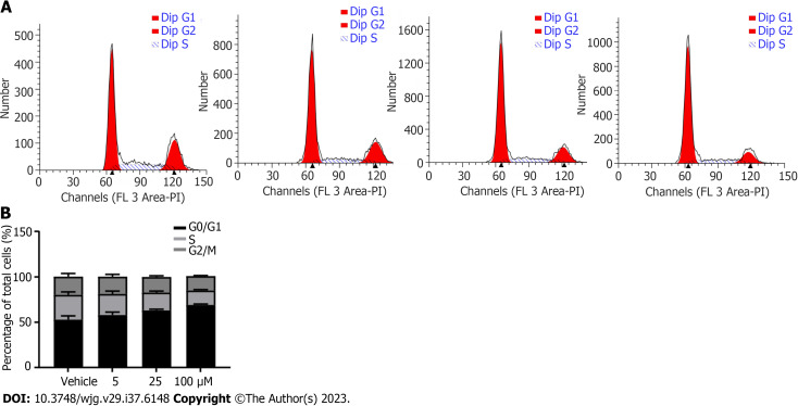
Cells were digested to make single-cell suspensions at logarithmic phase and uniformly seeded in 12-well plates at 1 × 10⁵ cells per well. After 48 h, cells were collected and washed with 1 mL of precooled phosphate buffered saline (PBS). The cell pellet was resuspended with 1 mL of precooled 70% ethanol and fixed overnight at 4 °C. On the second day, 70% ethanol was discarded by centrifugation at 1000 × g for 5 min at 4 °C, washed with 1 mL of precooled PBS. Each sample was added with 500 μL propyl iodide (PI) staining solution, gently mixed, incubated at 37 °C in the dark for 30 min, and the cells were filtered with a 400-mesh cell strainer to detect the cell cycle of each group by flow cytometry.
Apoptosis assays
Colon cancer cells in the logarithmic growth phase were seeded in 6-well plates, and 5-MTP-treated cells were added after the cells attached. Cells in each group were digested with ethylene diamine tetraacetic acid-free trypsin, washed with PBS, and collected at 1000 × g for 5 min. Binding buffer 100 μL was used to resuspend cells, and fluorescein isothiocyanate staining solution 5 μL and PI staining solution 10 μL were added. They were blown and mixed well by the pipette. The cells were allowed to stand at room temperature for 15 min. Before loading the machine, 400 μL of binding buffer solution was added to each tube and mixed well so that the final system was 500 μL. Apoptosis of cells in each group was detected by flow cytometry.
ROS assays
To test the ROS level in each group of cells, the colon cancer cells were inoculated into 6-well plates at logarithmic growth phase. The 2′,7′-dichlorofluorescin diacetate (Sangon Biotech, China) was added, incubated in a cell culture incubator at 37 °C for 20 minutes, and observed under a confocal microscope. Similarly, Hoechst 33342 Viable Cell Staining Solution (Sangon Biotech, China) was added and incubated in a 37 °C cell culture incubator for 10 min and observed under a confocal microscope.
JC-10 assays
Cells were treated with 5-MTP at various concentrations for 48 h at 12-well plate. Before incubation, 25 μL of 200X JC-10 concentrate (Sangon Biotech, China) was added to a 5 mL assay buffer to dilute JC-10. 500 μL of JC-1 solution was added to each well and incubated at 37 °C for 20 min. After incubated, washed twice with staining buffer, added 500 μL cell culture medium, and observed under an inverted fluorescence microscope.
Western blot assays
CRC cell lines were treated with 5-MTP for 48 h at logarithmic growth phase before protein extraction. Then, cells were washed with precooled PBS, 500 μL of RIPA buffer (Beyotime, China) was added to each dish, scraped by cell scraper, and transferred to a new centrifuge tube, lysed on ice for 30 min and centrifuged (12000 × g, 15 min, centrifugation radius 30 cm) to collect the supernatant. After adjusting the protein concentration, 4 × loading buffer was added and placed in a metal bath for heating denaturation (95 °C, 10 min). Sodium-dodecyl sulfate gel electrophoresis electrophoresis was performed to separate proteins (80 V), transferred by wet membranes (300 mA, 90 min), and blocked with 5% skimmed milk overnight (4 °C). Primary antibodies were used to incubate overnight (4 °C), including anti-AKT (1:1000, Cell Signaling Technology, #4685), anti-p-AKT (1:1000, Cell Signaling Technology, #13038), anti-FoxO3a (1:1000, Cell Signaling Technology, #12829), anti-p-FoxO3a (1:1000, Cell Signaling Technology, #9466), anti-β-actin (1:1000, Cell Signaling Technology, #4970), anti-Bax (1:1000, Cell Signaling Technology, #41162), anti-Bcl2 (1:1000, Cell Signaling Technology, #15071), anti-PARP (1:1000, Cell Signaling Technology, #9532), anti-Caspase3 (1:1000, Cell Signaling Technology, #9662), anti-Bim (1:1000, Cell Signaling Technology, #2933), anti-P27 (1:1000, Cell Signaling Technology, #3688) and anti-Cyclin D1 (1:1000, Cell Signaling Technology, #55506). Membranes were washed, incubated with secondary antibodies (1:10000) for 1 h at room temperature, and finally developed with ECL (Jiapeng Biotech, China), cassette luminescence, and band data analysis was performed by ImageJ software.
Colony formation assay
Cells were seeded in six-well plates at 800 cells/well in cell suspension, and 5-MTP was added when the cells were completely attached and cultured in an incubator for about 10 d, and > 50 cells/colony were considered as one clone, stained with crystal violet, photographed and counted.
Cell Counting Kit 8 assays
After adjusting the cell density of each group to 1 × 10⁴ cells/mL, 200 μL of cell suspension was added to a 96-well plate and cultured at 37 °C for 0, 24, 48, and 72 h. Following this, 10 μL of Cell Counting Kit 8 (CCK-8) solution was added to each well. After incubation at 37 °C for 2 h, absorbance values were measured using a microplate reader (Flash Biotech, China) at 450 nm.
Migration and invasion assays
Invasion assays were performed at 37 °C using transwell chambers coated with matrigel. The cell density was adjusted to 4 × 10⁵ cells/mL, 100 μL was added to the upper transwell chamber, and then 600 μL of medium containing 10% foetal bovine serum was added to the lower chamber. After incubation at 37 °C for 24 h, the cells on the lower surface of the membrane were fixed with 4% paraformaldehyde (Sangon Biotech, China) for 20 min and stained with 500 μL, 1% crystal violet (Sangon Biotech, China) for 30 min. Five fields were randomly selected to observe the number of cells in invasion using an inverted light microscope and photographed.
Statistical analysis
GraphPad Prism (v9.0.0) software was used for data processing. Measurement data were expressed as mean ± SD. An independent sample t-test was performed between two groups. A one-way analysis of variance was used to compare the means between multiple groups. P < 0.05 was considered with statistically significant.
RESULTS
5-MTP inhibited human CRC cell proliferation
ATL was selected for further analysis, and its structure is shown in Figure Figure1A.1A. To validate the role of 5-MTP in CRC, we selected HCT116, HCT15, and SW480 for subsequent experiments. In HCT116, HCT15, and SW480 cells, the results of CCK8 and colony formation assay showed that 5-MTP inhibited CRC cell proliferation gradually with increasing drug concentration (Figures (Figures1B1B--J).J). These data suggested that 5-MTP inhibited human CRC cell proliferation.
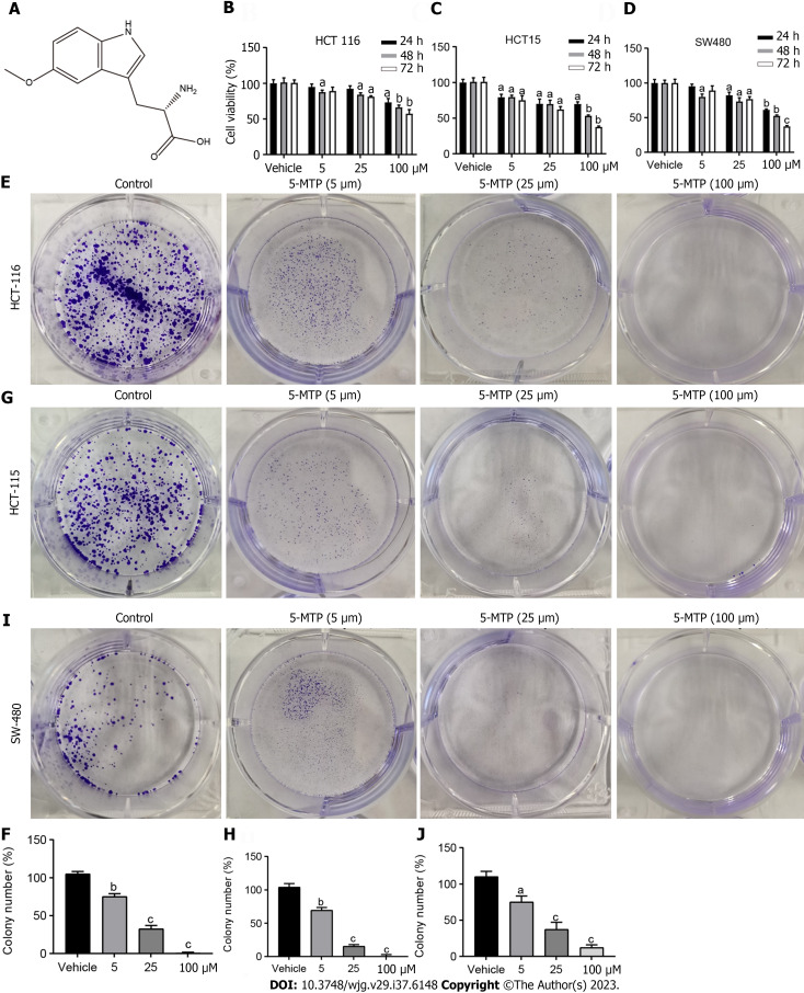
5-methoxytryptophan inhibited human colorectal cancer cell proliferation. A: The structure of 5-methoxytryptophan (5-MTP) is shown; B-D: The Cell Counting Kit 8 assays were used to measure the viability of the colorectal cancer cells treated with 5-MTP at the dose of 5, 25 and 100 μM; E-J: Colony formation assay were used to measure the viability of the HCT-116 (E and F), HCT-15 (G and H), SW480 (I and J) cells treated with 5-MTP at the dose of 5, 25 and 100 μM. aP < 0.05, bP < 0.01, c P < 0.001. 5-MTP: 5-Methoxytryptophan.
5-MTP induced apoptosis and ROS in HCT-116 cells
Next, we examined the effects of 5-MTP on CRC cell apoptosis by measuring levels of JC-1 and ROS, among others. In HCT-116 cells, Hoechst staining showed that 5-MTP inhibited CRC cell activity progressively (Figures (Figures2A2A and andB).B). Flow cytometry results showed that 5-MTP significantly inhibited JC-1 levels and promoted the apoptosis rate and ROS levels (Figures (Figures2C2C--HH).
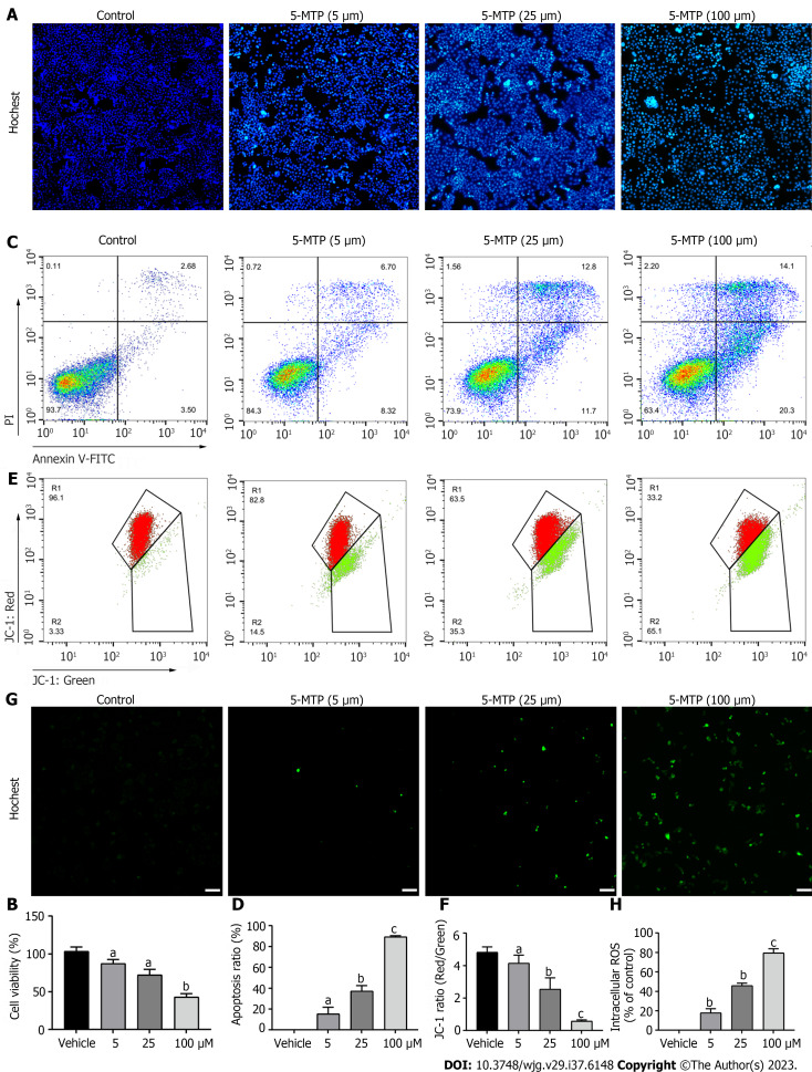
5-methoxytryptophan induced apoptosis and reactive oxygen species in HCT-116 cells. A and B: The Hoechst staining was used to measure the viability of HCT-116 cells treated with 5-methoxytryptophan (5-MTP) at the dose of 5, 25 and 100 μM; C and D: Apoptosis was detected by flow cytometry by double staining with PI and Annexin V of HCT-116 cells treated with 5-MTP at the dose of 5, 25 and 100 μM; E and F: Flow cytometry were used to analyses the JC-1 level of HCT-116 cells treated with 5-MTP at the dose of 5, 25 and 100 μM; G and H: 2’,7’-dichlorofluorescin diacetate assays were used to test the reactive oxygen species production of HCT-116 cells treated with 5-MTP at the dose of 5, 25 and 100 μM. aP < 0.05, bP < 0.01, cP < 0.001. 5-MTP: 5-Methoxytryptophan; ROS: Reactive oxygen species; PI: Propyl iodide.
5-MTP induced cell cycle arrest in HCT-116 cells
Flow cytometry results showed that the peak value became higher in the G2/M phase, that is, 5-MTP-induced HCT116 cell cycle arrest (Figures (Figures3A3A and andB).B). These results demonstrated that 5-MTP induced cell cycle arrest in HCT-116 cells.
5-MTP inhibited migration and invasion in HCT-116 cells
To further investigate the effect of 5-MTP on CRC cells, invasion and migration assays showed that 5-MTP inhibited CRC cell invasion and migration (Figures (Figures4A4A--C).C). Ly294002 (PI3K) inhibitors have been shown to play a role in various tumors, including proliferation, metastasis, and apoptosis[13-15]. However, the effects of 5-MTP combined with ly294002 on invasion and migration in CRC cells are further enhanced is unknown. It was found that 5-MTP combined with ly294002 resulted in significantly less cell invasion and migration than 5-MTP (Figures (Figures4D4D and andFF).
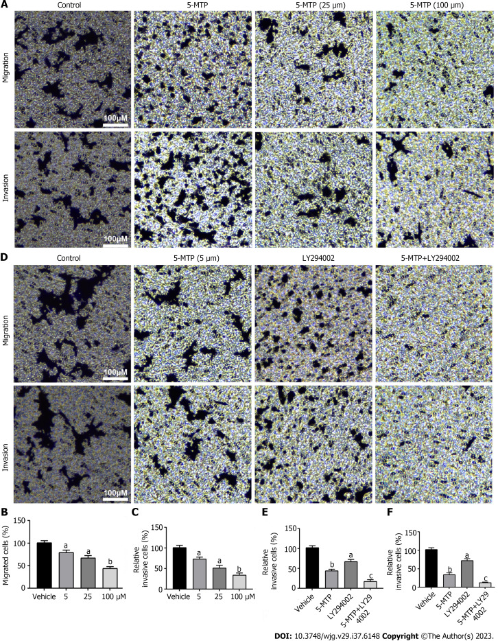
5-methoxytryptophan inhibited migration and invasion in HCT-116 cells. A-C: Detection of migration and invasion in HCT-116 cells were treated with 5-methoxytryptophan (5-MTP) at the dose of 5, 25 and 100 μM by transwell test; D-F: The HCT-116 cells were treated with 5-MTP and/or LY294002. Detection of migration and invasion in HCT-116 cells by transwell test. aP < 0.05, bP < 0.01, cP < 0.001. 5-MTP: 5-Methoxytryptophan.
5-MTP induced dose-response and time course inhibition of PI3K/Akt/FoxO3a signaling pathway in HCT-116 cells
The PI3K/Akt/FoxO3a signaling pathwa plays an important role in various pathological processes in various tumors, including proliferation, metastasis, and apoptosis[16-18]. However, Figure Figure44 confirmed that 5-MTP combined with ly294002 inhibited CRC cell invasion and migration. The next experimental results showed that 5-MTP inhibited the relative protein levels of p-Akt/t-AKT and p-FoxO3a/t-FoxO3a with increasing drug concentration in HCT116 cells (Figures (Figures5A5A--D).D). It was further found that the relative protein levels of p-Akt/t-AKT and p-FoxO3a/t-FoxO3a were also inhibited over time when the 5-MTP concentration was fixed, confirming the previous conclusions (Figures (Figures5E5E--H).H). Meanwhile, western blot results showed that Caspase3, PARP, and BAX protein levels were increased, while Bcl2 protein levels were significantly decreased (Figures (Figures6A6A--D).D). These results are the same as those in the Figure Figure1.1. Overall, our data indicated that 5-MTP induced dose-response and time course inhibition of PI3K/Akt/FoxO3a signaling pathway in HCT-116 cells.
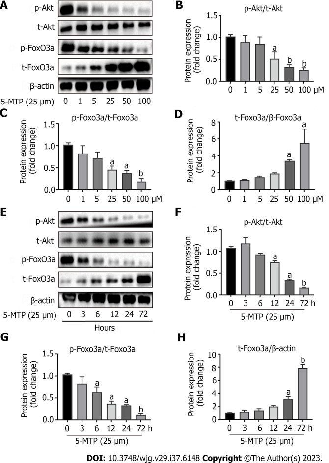
5-methoxytryptophan induced dose-response and time course inhibition of PI3K/Akt/FoxO3a signaling pathway in HCT-116 cells. A-D: The HCT-116 cells were treated with 5-methoxytryptophan (5-MTP) at the dose of 0, 1, 5, 25, 50 and 100 μM. The expression of PI3K/Akt/FoxO3a signaling pathway protein was detected by western blotting; E-H: The HCT-116 cells were treated with the dose of 25 μM of 5-MTP at the time of 0, 3, 6, 12, 24 and 72 h. The expression of p-Akt, t-AKT, p-FoxO3a and t-FoxO3a was detected by western blotting. aP < 0.05, bP < 0.01, cP < 0.001. 5-MTP: 5-Methoxytryptophan.
Apparent correlations between 5-MTP-induced cell cycle and apoptosis-associated proteins or inhibition of PI3K/Akt/FoxO3a signaling pathway in HCT-116 cells
To further verify that 5-MTP combined with PI3K/Akt/p-FoxO3a signaling pathway inhibitors played a better therapeutic effect, ly294002 (PI3K) inhibitor and AKT small interfering RNA (siRNA) were transfected into HCT116 cells. The results showed that the relative protein levels of p-Akt/AKT and p-FoxO3a/FoxO3a were significantly decreased in the 5-MTP combined with the ly294002 group compared with the 5-MTP group. At the same time, we also found that Cyclin D1 and P27 protein levels were significantly decreased while Bim was significantly increased in the 5-MTP combined ly294002 group compared with the 5-MTP group, suggesting that 5-MTP-induced cell cycle-related proteins are associated with the PI3K/Akt/p-FoxO3a signaling pathway (Figures (Figures7A7A--D).D). It was further found that the 5-MTP combined with the AKT siRNA group came to the same conclusion as the 5-MTP group (Figures (Figures7E7E--HH).
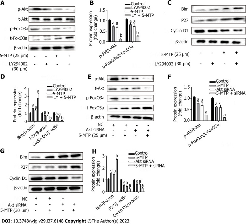
Apparent correlations between 5-methoxytryptophan-induced cell cycle and apoptosis-associated proteins or inhibition of PI3K/Akt/FoxO3a signaling pathway in HCT-116 cells. A-D: The HCT-116 cells were treated with 5-methoxytryptophan (5-MTP) and/or LY294002. The expression of PI3K/Akt/FoxO3a signaling pathway and cycle arrest protein was detected by Western blotting; E-H: The HCT-116 cells were treated with the dose of 25 μM of 5-MTP and/or AKT siRNA. The expression of p-Akt, t-AKT, p-FoxO3a, t-FoxO3a, Bim, Cyclin D1 and P27 was detected by western blotting. aP < 0.05, bP < 0.01, cP < 0.001. 5-MTP: 5-Methoxytryptophan; NC: Normal control; siRNA: Small interfering RNA.
DISCUSSION
CRC is a highly prevalent disease in countries around the world, and the incidence increases with age, accounting for nearly one-third of patients over 75 years of age[19,20]. The number of patients with early-onset CRC no more than 50 years of age also cannot be ignored[3,21]. The main treatments for CRC are surgery, radiotherapy, and chemotherapy[22]. However, 35% of patients are found to be in the advanced stage and lose the chance of radical surgery[23]. With the in-depth study of the formation, development, and treatment of CRC from the molecular and genetic levels, adjuvant therapy with TCM monomers optimizes the balance between tumor cell killing and non-targeted effects with the advantage of specific selection combined with oncogenic sites[24,25].
5-MTP, with a molecular formula of C12H14N2O3 and a molecular weight of 234.251, is a newly identified tryptophan metabolite produced by cells, such as fibroblasts, renal epithelial, smooth muscle and vascular endothelial cells[11,26,27]. Current studies have shown that 5-MTP has various physiological functions, such as stabilizing endothelial function, anti-inflammation, and anti-oxidation[11,28]. 5-MTP has been shown to be involved in regulating inflammatory responses and can maintain endothelial cell tight junctions to some extent[29]. Proinflammatory factors inhibit the expression of TPH-1, a key enzyme in 5-MTP synthesis in endothelial cells, reducing 5-MTP production, which leads to endothelial barrier disruption[30]. In addition, 5-MTP plays an important vaso-protective function by regulating vascular permeability, controlling systemic inflammation, and defending against systemic inflammation and multiple organ failure[31]. It has been found that 5-MTP may regulate cardiomyocyte growth-associated proteins, cytoskeleton, redox, and protein folding, thereby promoting wound healing, and preventing damaged cell death by maintaining redox balance and reducing endoplasmic reticulum stress[32].
PI3K/Akt signaling pathway is one of the most important signaling pathways involved in internal regulating cell proliferation and differentiation[33]. Akt is essential for the regulation of cell migration and growth[34]. P-Akt is the active form of Akt and is closely associated with CRC[35]. P-Akt plays an important role in CRC progression[36]. FOXO family members are major effector proteins of the PI3K/Akt signaling pathway[37]. The FOXO family is a subclass of the FOX family that contains four isoforms (FOXO1, FOXO3, FOXO4, and FOXO6), all of which structurally contain a Forkhead DNA-binding domain that binds to the same DAF-16-binding element binding site within the target gene promoter through a DNA domain, and thus regulates target gene expression[38]. FOXO3a has been found to be an important transcription factor for genes involved in cell cycle progression, apoptosis, metabolism, differentiation, and autophagy[39]. PI3K/Akt regulates the expression of a series of genes related to cell proliferation, apoptosis, and cycle arrest downstream of FOXO by working on phosphorylation sites on FOXO protein[40]. FOXO3A is involved in the biological effects of CRC, and this process may be related to cell proliferation and apoptosis[18]. For example, many FOXO factors are phosphorylated by Akt in the presence of growth factors, resulting in their translocation from the nucleus to the cytoplasm; PI3K/AKT pathway mediates hyperglycemia-induced apoptosis in ventricular myocytes of neonatal rats through translocation of FOXO3a to the nucleus[17,41].
In this study, the efficacy of 5-MTP in the treatment of CRC by in vitro cell experiments was investigated, and the effects of 5-MTP on CRC cells under different conditions were examined using CCK-8, colony formation, apoptosis rate detection, JC-1 level, ROS level and cell cycle detection. The results showed that 5-MTP promoted apoptosis and cell cycle arrest and inhibited cell proliferation in CRC cells. In subsequent experiments, the effect of 5-MTP combined with PI3K/Akt/p-FoxO3a signaling pathway inhibitors in the treatment of CRC was focused, confirming that combined PI3K/Akt/p-FoxO3a signaling pathway inhibitors played a more effective role in the treatment of CRC. There was a correlation between 5-MTP-induced cyclin-related proteins and PI3K/Akt/p-FoxO3a signaling pathway.
CONCLUSION
In summary, 5-MTP could inhibit proliferation and promote apoptosis of CRC cells, and combined ly294002 or AKT inhibitors played a more effective role in treating CRC. This study provided theoretical guidance for the clinical treatment of CRC with 5-MTP. However, there were some limitations, and in vivo experiments such as mice are still needed for further validation in the future.
ARTICLE HIGHLIGHTS
Research background
Colorectal cancer (CRC) is a highly prevalent malignant tumor. Research is needed to find and develop effective drugs for the treatment of CRC. 5-methoxytryptophan (5-MTP) is a tryptophan metabolite found in animals and humans. The effect of 5-MTP on the proliferation and apoptosis of CRC cells is still unclear.
Research motivation
Our work explored the effects of 5-MTP on the proliferation, migration, invasion and apoptosis of CRC cells. We tried to explore the potential of 5-MTP as a drug for the treatment of CRC.
Research objectives
Here, we studied that 5-MTP combined with PI3K/Akt/FOXO3a signaling pathway inhibitor can significantly promote apoptosis and cell cycle arrest of CRC cells, and inhibit cell proliferation. 5-MTP can be used as an effective drug in the treatment of CRC.
Research methods
In this study, a series of experiments were carried out. Colony forming assay and Cell Counting Kit were used to detect the effect of 5-MTP on the proliferation of CRC cell lines. The effect of 5-MTP on cell growth was detected by cell cycle analysis. In addition, the effects of 5-MTP on apoptosis and reactive oxygen species of HCT-116 cells were studied. In order to further explore the effect of 5-MTP on CRC, we studied the effect of 5-MTP on PI3K/Akt signaling pathway in CRC cells.
Research results
5-MTP can promote apoptosis and cell cycle arrest of CRC cells, and inhibit cell proliferation. However, compared with 5-MTP alone, 5-MTP combined with PI3K/Akt/FOXO3a signaling pathway inhibitors significantly promoted apoptosis and cell cycle arrest of CRC cells, and inhibited cell proliferation. It provides new insights into the mechanism of drug action.
Research conclusions
5-MTP combined with PI3K/Akt/FOXO3a signaling pathway inhibitor significantly promoted apoptosis and cell cycle arrest of CRC cells, and inhibited cell proliferation.
Research perspectives
This study confirmed that 5-MTP combined with PI3K/Akt/FOXO3a signaling pathway inhibitor could significantly promote the apoptosis and cell cycle arrest of CRC cells, and inhibit cell proliferation. These results provide a new direction for the drug treatment of CRC. However, the study of mechanism in this study is relatively limited. Therefore, further analysis, nude mouse experiments and more cell experiments are needed to explore its mechanism.
Footnotes
Institutional review board statement: The authors are accountable for all aspects of the work in ensuring that questions related to the accuracy or integrity of any part of the work are appropriately investigated and resolved. The study was approved by Institutional Review Board of Naval Medical Center of PLA.
Conflict-of-interest statement: All the authors report no relevant conflicts of interest for this article.
Provenance and peer review: Unsolicited article; Externally peer reviewed.
Peer-review model: Single blind
Peer-review started: September 6, 2023
First decision: November 1, 2023
Article in press: December 4, 2023
Specialty type: Gastroenterology and hepatology
Country/Territory of origin: China
Peer-review report’s scientific quality classification
Grade A (Excellent): 0
Grade B (Very good): 0
Grade C (Good): C, C
Grade D (Fair): 0
Grade E (Poor): 0
P-Reviewer: Cheng TH, Taiwan; Exbrayat JM, France S-Editor: Wang JJ L-Editor: A P-Editor: Cai YX
Contributor Information
Tian-Lei Zhao, Department of General Surgery, Naval Medical Center of PLA, Shanghai 200052, China.
Yue Qi, Department of General Surgery, Naval Medical Center of PLA, Shanghai 200052, China.
Yi-Fan Wang, Department of General Surgery, Naval Medical Center of PLA, Shanghai 200052, China.
Yi Wang, Department of General Surgery, Naval Medical Center of PLA, Shanghai 200052, China.
Hui Liang, Department of Gastroenterology, Naval Medical Center of PLA, Shanghai 200052, China.
Ya-Bin Pu, Department of General Surgery, Naval Medical Center of PLA, Shanghai 200052, China. moc.361@62upnibay.
Data sharing statement
The datasets generated during and/or analyzed during the current study are available from the corresponding author on reasonable request.
References
Articles from World Journal of Gastroenterology are provided here courtesy of Baishideng Publishing Group Inc
Citations & impact
Impact metrics
Article citations
The role of SIRT1 in autophagy and drug resistance: unveiling new targets and potential biomarkers in cancer therapy.
Front Pharmacol, 15:1469830, 30 Sep 2024
Cited by: 0 articles | PMID: 39403142 | PMCID: PMC11471651
Review Free full text in Europe PMC
Recommendations for articles and reviews in colorectal cancer-related research at the year-end of 2023.
World J Gastroenterol, 30(30):3548-3553, 01 Aug 2024
Cited by: 0 articles | PMID: 39193570 | PMCID: PMC11346154
Similar Articles
To arrive at the top five similar articles we use a word-weighted algorithm to compare words from the Title and Abstract of each citation.
Effects of Glut1 gene silencing on proliferation, differentiation, and apoptosis of colorectal cancer cells by targeting the TGF-β/PI3K-AKT-mTOR signaling pathway.
J Cell Biochem, 119(2):2356-2367, 18 Oct 2017
Cited by: 31 articles | PMID: 28884839
Endogenous tryptophan metabolite 5-Methoxytryptophan inhibits pulmonary fibrosis by downregulating the TGF-β/SMAD3 and PI3K/AKT signaling pathway.
Life Sci, 260:118399, 09 Sep 2020
Cited by: 16 articles | PMID: 32918977
Promethazine inhibits proliferation and promotes apoptosis in colorectal cancer cells by suppressing the PI3K/AKT pathway.
Biomed Pharmacother, 143:112174, 21 Sep 2021
Cited by: 11 articles | PMID: 34560542
A network pharmacology approach and experimental validation to investigate the anticancer mechanism and potential active targets of ethanol extract of Wei-Tong-Xin against colorectal cancer through induction of apoptosis via PI3K/AKT signaling pathway.
J Ethnopharmacol, 303:115933, 18 Nov 2022
Cited by: 21 articles | PMID: 36403742

