| PMC full text: | Published online 2023 Aug 23. doi: 10.3390/ijms241713116
|
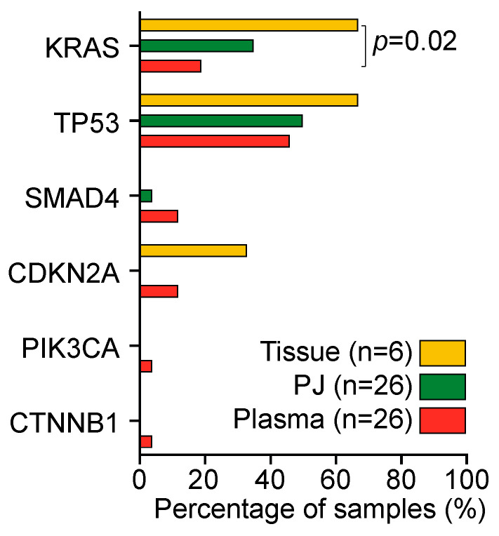
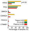
Figure 3

Detection rate of mutations in tissue (yellow), pancreatic juice (PJ; green), and plasma (orange). KRAS mutations were detected significantly more often in tissue as compared with plasma; KRAS detection rate between PJ and plasma was not different. Percentages were compared with a Kruskal–Wallis test.


