| PMC full text: |
|
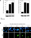
Figure 4.
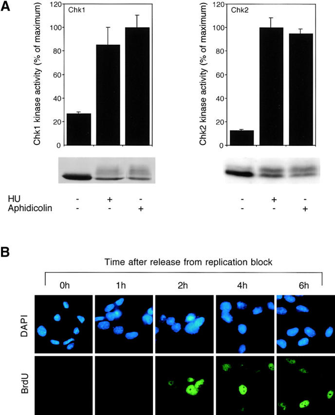
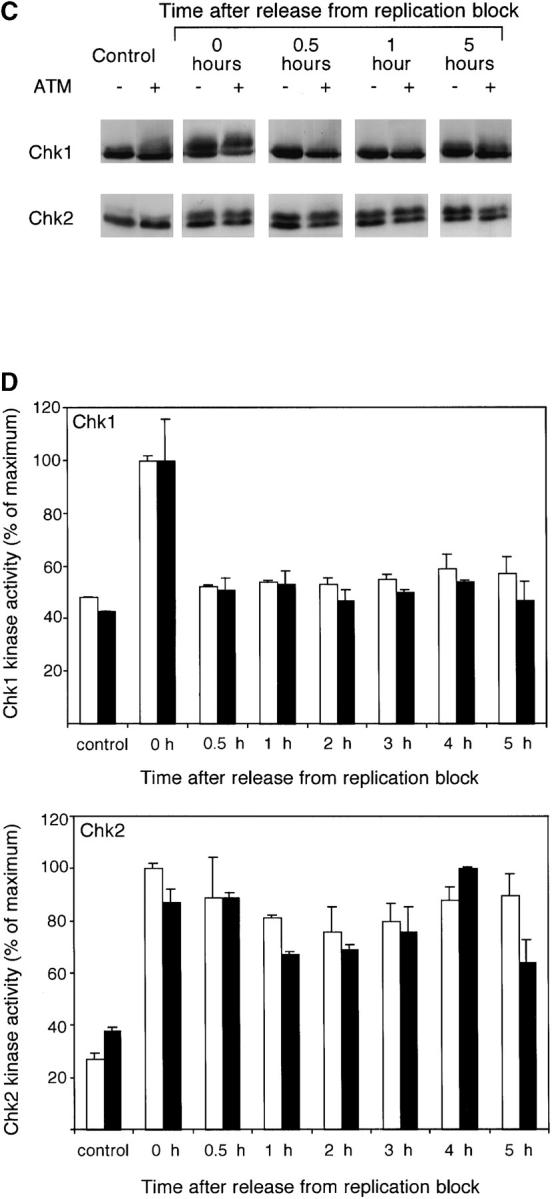
Differential inactivation of Chk1 and Chk2 following release from replication block. (A) Asynchronously growing HeLa cells were treated or not for 24 h with 2 mM HU or 5 μg/ml aphidicolin and lysates prepared. Kinase assays were performed on immunoprecipitated Chk1 and Chk2 (upper panels) as indicated. Lysates were subjected to SDS-PAGE and immunoblotted for Chk1 and Chk2 (lower panels). (B) Asynchronously growing AT221JE-T cells expressing ATM were treated with 2 mM HU for 24 h, released, and at times indicated pulse labeled with BrdU. Cells were fixed, stained with DAPI and anti-BrdU antibodies, and examined by indirect immunofluorescence microscopy. Identical results were observed with AT cells transfected with empty vector (not shown). (C and D) AT221JE-T cells containing either vector alone (−ATM) or vector expressing ATM (+ATM) were treated for 24 h with HU, and then released into fresh medium. (C) At the times indicated, lysates were prepared, subjected to SDS-PAGE, and immunoblotted for Chk1 and Chk2. (D) At times indicated, Chk1 (upper panels) and Chk2 (lower panels) IP kinase assays were performed on lysates from AT221JE-T cells containing vector alone (black bars) or vector expressing ATM (white bars) treated as above.
