| PMC full text: | Clin Cancer Res. Author manuscript; available in PMC 2007 Dec 26. Published in final edited form as: Clin Cancer Res. 2006 Oct 15; 12(20 Pt 1): 6106–6115. doi: 10.1158/1078-0432.CCR-06-1183 |
Fig. 2

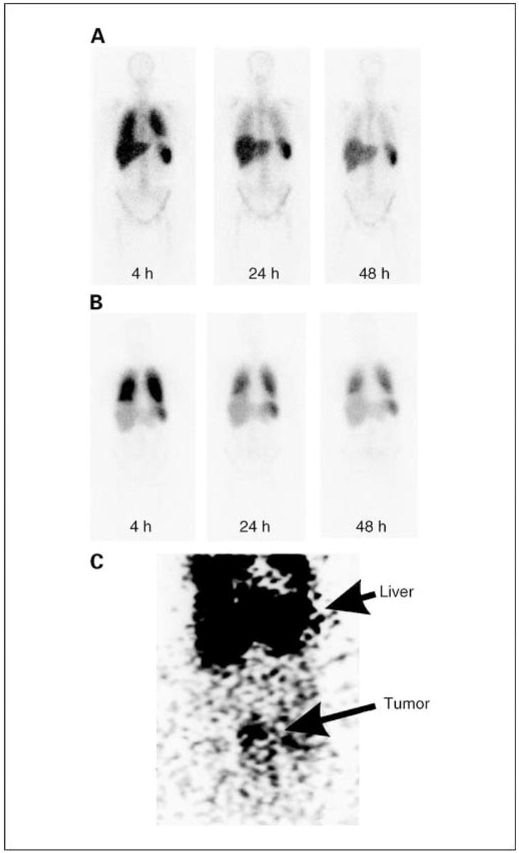
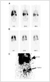
Biodistribution of radiolabeled T cells. Patients received up to 7.5 × 10⁹ 111In-labeledTcells, and imaging was done using a gamma camera at intervals followingTcell transfer. A, representative image of four transfers that were done withTcells that received a single stimulation with OKT3. B, representative image of three transfers that were done usingTcells that had received two stimulations with OKT3. Radioisotope signal was detected in lungs, liver, and spleen. Radiolabeled cells were preferentially retained in lungs of patients that received twice stimulated Tcells. C, anteroposterior image of the abdomen of patient 4 at 48 hours after receiving T cells in cycle 2 with evidence of T cell localization to a peritoneal tumor (bottom) in addition to localization to liver (top).


