| PMC full text: |
|
Figure 2

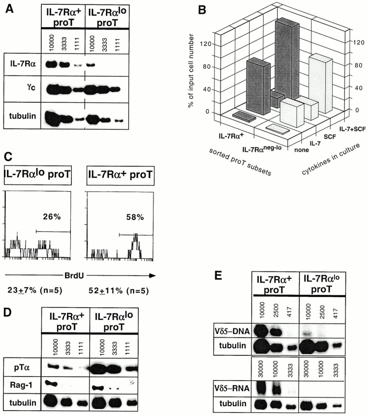
IL-7Rα+ and IL-7Rαneg-lo pro-T subsets exhibit distinct properties. (A) A representative radioactive RT-PCR analysis for IL-7Rα, γc, and tubulin transcripts in the sorted pro-T subsets of similar purity. Numbers indicate approximate cell equivalents used for IL-7Rα and γc-specific PCR. For tubulin PCR, the starting concentration was 3,333 cell equivalents, which was subject to two serial threefold dilutions. The IL-7Rα and γc autoradiographs were exposed for 3 d or overnight, respectively. No PCR products were detected when the RT step was omitted. (B) Proportion of input sorted cells surviving culturing the pro-T subsets for 4 d in the presence of IL-7 and/or SCF. (C) BrdU incorporation after a 3-h pulse with BrdU in vivo. Treated DN thymocytes were stained with mAbs for CD25-613, CD44-Cy5, IL-7Rα-biotin/strepavidin-PE, and BrdU-FITC. The levels of BrdU staining on gated CD25+CD44+IL-7Rα+ and CD25+CD44+IL-7Rαneg-lo TN thymocytes are presented. (D) RT-PCR analysis for pTα and Rag-1 transcripts. Results from one of four independent sorting experiments are shown for pTα expression analysis; two experiments showed a marginal difference (two- to threefold), whereas two others showed a larger (more than ninefold) difference. (E) Levels of Vδ5-Jδ1 rearrangements in genomic DNA (top) and Vδ5-Jδ1 transcripts in total RNA (bottom) from sorted pro-T cell subsets, determined by semiquantitative PCR or RT-PCR, respectively. Genomic DNA PCR was for 35 cycles; RT-PCR entailed 38 cycles for Vδ5-Jδ1 and 28 cycles for tubulin. No PCR products were detected when the RT step was omitted. The autoradiographs shown for Vδ5-Jδ1 and tubulin transcripts were exposed for 5 d and 4 h, respectively.


