| PMC full text: |
|
Figure 4.

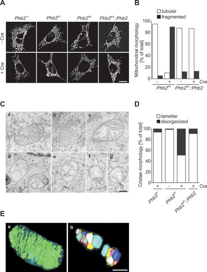
Defective cristae morphogenesis in Phb2−/− cells. (A) Fragmentation of mitochondria in prohibitin-deficient MEFs. Cell lines were transfected with mito-DsRed, treated with Cre-recombinase when indicated, and analyzed after 72 h by fluorescence microscopy. Bar, 10 μm. (B) Quantification of mitochondrial morphology in control (Phb2fl/fl Phb2) and prohibitin-deficient MEFs. Cells containing tubular (white bars) or fragmented (black bars) mitochondria were classified. More than 200 cells were scored per experiment. (C) Defective mitochondrial ultrastructure in Phb2−/− cells. Representative transmission electron micrographs of mitochondria in the following cell lines are shown: Phb2fl/fl (panel a), Phb2fl/+ +Cre (panel b), Phb2fl/fl
 Phb2 +Cre (panel c), and Phb2fl/fl +Cre (panels d–g). Bar, 500 nm. (D) Quantification of cristae morphology in Phb2fl/fl, Phb2fl/fl
 Phb2, and Phb2fl/fl MEFs transduced with Cre-recombinase when indicated. Approximately 50% of cells with disorganized cristae morphology contain vesicular cristae structures. Approximately 100 sections of individual cells were scored per experiment. (E) Three-dimensional reconstructions from serial transmission electron microscopy sections of Phb2fl/fl cells (panel a) transduced with Cre-recombinase (panel b). Bar, 500 nm.


