Abstract
Free full text

The histone methyltransferase SET8 is required for S-phase progression
Abstract
Chromatin structure and function is influenced by histone posttranslational modifications. SET8 (also known as PR-Set7 and SETD8) is a histone methyltransferase that monomethylates histonfe H4-K20. However, a function for SET8 in mammalian cell proliferation has not been determined. We show that small interfering RNA inhibition of SET8 expression leads to decreased cell proliferation and accumulation of cells in S phase. This is accompanied by DNA double-strand break (DSB) induction and recruitment of the DNA repair proteins replication protein A, Rad51, and 53BP1 to damaged regions. SET8 depletion causes DNA damage specifically during replication, which induces a Chk1-mediated S-phase checkpoint. Furthermore, we find that SET8 interacts with proliferating cell nuclear antigen through a conserved motif, and SET8 is required for DNA replication fork progression. Finally, codepletion of Rad51, an important homologous recombination repair protein, abrogates the DNA damage after SET8 depletion. Overall, we show that SET8 is essential for genomic stability in mammalian cells and that decreased expression of SET8 results in DNA damage and Chk1-dependent S-phase arrest.
Introduction
Genetic information in eukaryotes is organized in chromatin, a highly conserved structural polymer that supports and controls crucial functions of the genome. Chromatin undergoes dynamic changes, including massive structural reorganization, during genetic processes such as DNA replication and cell division, transcription, DNA repair, and recombination. Histones and particularly their N-terminal tails are modulated by a large number of posttranslational modifications, including lysine methylations that influence these fundamental biological processes (Kouzarides, 2007).
The contribution from the chromatin environment to DNA replication and DNA damage response processes is only starting to become evident. Recently, a link between histone lysine methylation and the DNA damage responses have been uncovered. The checkpoint mediator 53BP1 is directly recruited to chromatin regions flanking DNA double-strand breaks (DSBs). This occurs via interaction with histone H4 that is specifically mono- or dimethylated at Lys20 or with histone H3 dimethylated at Lys79 (Huyen et al., 2004; Botuyan et al., 2006). 53BP1 plays an important role in the cellular response to DNA damage by acting as an adaptor in the repair of DNA DSBs (Ward et al., 2006).
Histone H4 Lys20 (H4-K20) can be mono-, di-, or trimethylated, and SET8 (also known as PR-Set7 and SETD8) can catalyze the monomethylation (Fang et al., 2002; Nishioka et al., 2002; Couture et al., 2005; Xiao et al., 2005). Previously, the expression of SET8 in mammalian cells has been shown to increase during S phase until mitosis (Rice et al., 2002); however, the functional role of SET8 remains poorly understood. Key issues such as the consequences of SET8 depletion have not been reported. The fly SET8 homologue PR-Set7 has been deleted in Drosophila melanogaster larvae, in which tissues with higher rates of cell divisions were severely affected. In this organism, progression through early mitosis was delayed, and levels of the essential mitotic regulator cyclin B was reduced (Sakaguchi and Steward, 2007).
In this study, we have analyzed the functional role of human H4-K20 methyltransferase SET8. We establish that it is important for proper progression through the cell cycle. Inhibition of SET8 expression by siRNA results in the massive accumulation of DNA damage that subsequently activates a Chk1-dependent checkpoint. This leads to slower progression through S phase and decreased proliferation. We also show that SET8 interacts with proliferating cell nuclear antigen (PCNA) through a PCNA interaction motif and that there is a requirement for SET8 during replication fork progression. Collectively, our data suggest that SET8 plays an important role in securing the accurate completion of DNA replication and, for the first time, demonstrates such a role for a histone methyltransferase in protecting against genomic instability.
Results and discussion
Depletion of SET8 prevents cell proliferation and causes cell cycle delay in S phase
To investigate the role of SET8 depletion in cell cycle progression, we transfected U2OS cells with siRNA against SET8. U2OS cells are human osteosarcoma cells that are widely used in cell cycle studies. Cells were counted 48 and 96 h after siRNA treatment, and the SET8-depleted cells proliferated significantly slower than mock-treated cells (Fig. 1 A). We have not observed marked sub-G1 peaks or accumulating debris indicative of apoptosis/cell death at these time points. Depletion of SET8 also induced morphological alterations of the cells (Fig. 1 B), as depleted cells increased the size of their cytoplasm.
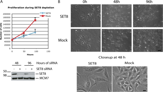
Removal of SET8 results in reduced cell proliferation. (A) U2OS cells were treated with either mock siRNA or siRNA against SET8. Cells were counted 48 and 96 h after siRNA treatment. Every time point represents three independent samples, and the experiment was repeated three times with similar results. (B) Pictures were taken of representative areas of the cell dishes before harvest. A fraction of the cells from A was collected and processed for Western blotting. MCM7 is a loading control. Molecular weights are indicated on the left side. Error bars represent SD. Bars: (top) 25 μm; (bottom) 4 μm.
To explore the nature of the cell cycle delay observed during SET8 depletion, cells were analyzed by flow cytometry (FACS). Addition of the mitotic spindle inhibitor nocodazole 16 h before harvesting resulted in the accumulation of cells in M phase in the mock-treated sample (Fig. 2 A). In contrast, inhibition of SET8 expression led to a significant accumulation of the cells in S phase, a defect that became more visible in the presence of nocodazole (Fig. 2 A). Western blotting of SET8-depleted cells supported the notion that SET8 is required for normal S-phase progression. These results were reproduced by two different individual siRNA as well as SMARTpool siRNA targeting SET8 (Fig. S1, A and B; available at http://www.jcb.org/cgi/content/full/jcb.200706150/DC1). As shown in Fig. 2 B, the levels of histone H3 Ser10 phosphorylation, a marker of mitotic cells, were low in SET8-depleted cells compared with mock cells. Consistently, the levels of cyclin A2, which is known to accumulate from the G1/S transition to G2 phase and is degraded in metaphase cells, were higher in SET8-depleted cells compared with mock cells.
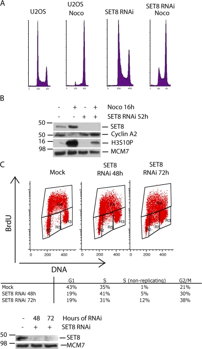
SET8 depletion leads to a delay in S-phase progression. (A) SET8 was depleted in U2OS cells using siRNA for 52 h. Nocodazole was added 16 h before harvest to the indicated samples. The samples were stained with PI and analyzed by flow cytometry analysis (FACS). (B) Cells were collected from a similar experiment as in A and were processed for immunoblotting. (C) U2OS cells were treated with siRNA against SET8 or mock for 48 and 72 h. Cells were pulsed with BrdU for 5 min, fixed, stained for BrdU and PI, and analyzed by FACS. The distribution of cells corresponding to the boxed areas is presented in the table beneath the graph. A fraction of the cells was collected and processed for immunoblotting.
Next, we wanted to determine whether decreasing SET8 levels would affect DNA replication. U2OS cells treated with SET8 or mock siRNA were pulse labeled with BrdU and analyzed by FACS. Remarkably, a significant fraction of cells in S phase were not incorporating BrdU (Fig. 2 C). Collectively, these data show that DNA replication is impaired in SET8-depleted cells, resulting in S-phase delay and, consequently, decreased cell proliferation.
Inhibition of SET8 expression results in DSBs
Next, we investigated whether the slower progression through S phase might be related to DNA replication–associated lesions. To address this, we stained U2OS cells using an antibody against phosphorylated H2AX (γ-H2AX), a well-established marker for DNA DSBs (Pilch et al., 2003). As shown in Fig. 3 A, inhibition of SET8 expression led to a dramatic increase in γ-H2AX–positive cells as early as 24 h after siRNA transfection, suggesting that SET8 depletion leads to massive DNA damage. To further corroborate these findings, we analyzed and quantified γ-H2AX levels in a FACS-based assay. Again, SET8 silencing lead to marked DNA damage (Fig. 3 B). Cells with DNA damage were negative for H4-K20 monomethylation, which is consistent with the concomitant loss of the monomethylase function of SET8 in these cells (Figs. 3 C and S1 C). γ-H2AX foci formation after SET8 depletion was also observed in HeLa cells (Fig. S1 D) as well as by direct analysis of DNA strand breaks using pulsed-field gel electrophoresis (Fig. 4 D). Collectively, our data demonstrate that SET8 has a critical role in maintaining correct genomic structure.
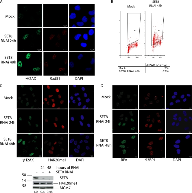
SET8 depletion leads to DSBs and checkpoint activation in S-phase cells. (A) SET8 was depleted in U2OS cells for 24 and 48 h. Cells were preextracted and fixed before staining with antibody toward γ-H2AX and Rad51. (B) U2OS cells were treated as in A and were fixed and stained for γ-H2AX and PI followed by FACS analysis. (C) Cells were treated as in A and stained with antibodies recognizing γ-H2AX and monomethylated H4 Lys20. Cells were also collected and processed for Western blotting with the indicated antibodies. Reduction of H4 Lys20 monomethylation upon SET8 depletion is quantified and indicated below the immunoblot. (D) Cells were treated as in A and were stained with 53BP1 and RPA antibodies. Bars, 10 μm.
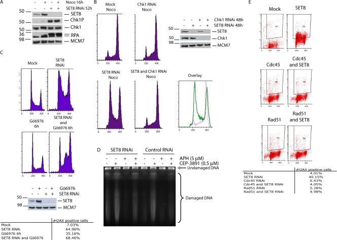
SET8 depletion leads to replication-dependent DNA damage that activates Chk1. (A) SET8 was depleted in U2OS cells using siRNA for 52 h. Nocodazole was added for the last 16 h as indicated. Cells were collected and processed for immunoblotting. (B) U2OS cells were treated with siRNA against mock, SET8, or Chk1 for 48 h. Nocodazole was added 13 h before harvest. Cells were fixed, stained with PI, and analyzed by FACS. A graph was generated by overlaying the FACS profiles from SET8-depleted cells and cells codepleted for SET8 and Chk1. (C) SET8 was depleted in U2OS cells using siRNA for 52 h. Nocodazole (16 h) and the Chk1 inhibitor Gö6976 (3 h) were added before harvest. Samples were fixed, stained with PI, and analyzed by FACS. (B and C) A fraction of the cells was collected and processed for immunoblotting. (D) 48 h after SET8 siRNA transfection, cells were treated with 5 μM aphidicolin and/or the Chk1 inhibitor CEP-3891 for an additional 24 h. Cells were then processed for pulsed-field gel electrophoresis. (E) U2OS cells were treated with siRNA against mock, SET8, Cdc45, or Rad51 for 48 h. Cells were then fixed and stained for γ-H2AX and PI followed by FACS analysis.
We reasoned that massive DNA damage observed after SET8 depletion could result from the inhibition of vital DNA repair processes because SET8 status could affect the recruitment of 53BP1 as well as other proteins to sites of DNA damage. 53BP1 is a checkpoint mediator involved in the initial sensing and signaling of DNA strand breaks (Ward et al., 2006). The protein has been suggested to be recruited to DNA DSBs via interaction with dimethylated histone H3-K79 and mono- or dimethylated H4-K20, and this interaction has been suggested to be dependent on SET8 (Botuyan et al., 2006). To understand whether the inhibition of SET8 expression affected the recruitment of key DNA repair proteins to sites of DNA damage, we investigated the nuclear accumulation of these proteins by confocal microscopy. As demonstrated in Fig. 3 (C and D), decreased SET8 expression lead to 53BP1 recruitment to sites of DNA damage and a marked increase in Rad51 and replication protein A (RPA) foci. Thus, inhibition of SET8 expression and reduction of H4-K20 monomethylation led to an increased recruitment of 53BP1. To test whether SET8 would be required for 53BP1 recruitment after exogenously induced DSB, we also treated SET8-depleted cells with ionizing irradiation. Yet again, 53BP1 readily relocated to radiation-induced DNA damage foci (Fig. S1 D). This strongly indicates that the presence of SET8 is not essential for the recruitment of 53BP1. 53BP1 was found primarily to bind dimethylated H4-K20, and we noticed that dimethylated H4-K20 persists after SET8 silencing (Fig. S2 A, available at http://www.jcb.org/cgi/content/full/jcb.200706150/DC1). This indicates that 53BP1 is recruited via persisting dimethylated H4-K20 or via interaction with accessible dimethylated H3-K79 or γ-H2AX (Huyen et al., 2004; Ward et al., 2006).
The S-phase delay in SET8-depleted cells is Chk1 mediated
DNA replication can cease for a variety of reasons before scheduled termination, including progression into areas with DNA damage lesions. Chk1 is a key regulator of the cellular response induced by stalled replication forks, a response that leads to the inhibition of DNA replication initiation at origins of replication (Feijoo et al., 2001). Therefore, we investigated whether Chk1 is activated in SET8-depleted cells. To do this, we transfected cells with SET8 siRNA. Nocodazole was added during the last 16 h, and cells were processed for immunoblotting analysis. The inhibition of SET8 expression led to a dramatic activation of Chk1 as measured by the phosphorylation of Ser317 on Chk1 (Fig. 4 A; Zhao and Piwnica-Worms, 2001). Phosphorylation of RPA was also markedly increased when SET8 was depleted. RPA is required for activation of the ataxia telangiectasia related kinase, which activates Chk1 in the presence of DNA damage (Zou et al., 2003). The activation of Chk1 suggested a role for the checkpoint kinase in mediating the cell cycle delay in SET8-depleted cells. To explore this, Chk1 was specifically long-term depleted using siRNA in combination with SET8 depletion. Inhibition of Chk1 prevented the delay in S phase (Fig. 4 B). Similar data were obtained with the Chk1 inhibitors Gö6976 (Fig. 4 C; Kohn et al., 2003), CEP-3891 (Sorensen et al., 2003), and UCN-01 (Sarkaria et al., 1999; and unpublished data). These cells progress through the cell cycle with markedly damaged DNA as judged by quantitative γ-H2AX FACS analysis (Figs. 4 C and S2 B) and pulsed-field gel electrophoresis (Figs. 4 D and S2 C). This is consistent with a critical role for Chk1 in restraining cell cycle progression after DNA damage.
DNA damage occurring after SET8 depletion requires replication and the functional homologous recombination repair pathway
We next wanted to address whether the lesions generated by SET8 silencing were dependent on DNA replication. Importantly, as shown in Fig. 4 D, the DNA replication inhibitor aphidicolin abrogated the DNA damage induced by SET8 depletion, suggesting that the lesions depend on ongoing DNA replication (Fig. 4 D). To corroborate this, we cosilenced several genes with an important role in DNA replication. Rapid accumulation of γ-H2AX foci was not observed by individual depletion of other replication-associated proteins such as Cdc45 (Syljuasen et al., 2005) and MCM4, which are both essential for the initiation of DNA replication (Fig. 4 E and not depicted; Bell and Dutta, 2002; Pacek and Walter, 2004). When codepleted with SET8, these proteins all reduced the DNA damage. This confirmed that DNA replication is necessary for SET8 silencing to cause DNA damage. The homologous recombination repair pathway plays an important role in the repair of DNA damage occurring during DNA replication (Saintigny et al., 2001; Helleday et al., 2007). Depletion of Rad51, a key component of this repair pathway, did not cause such massive DNA damage at the time points analyzed (Fig. 4 E). At such early time points, cells depleted for Rad51 were still viable and progressed through S phase like control depleted cells (Fig. S3 C, available at http://www.jcb.org/cgi/content/full/jcb.200706150/DC1). Notably, Rad51 depletion blocked DNA damage after SET8 depletion. We suggest that SET8 in unperturbed cells is involved downstream of Rad51 in resolving recombination structures forming spontaneously in cells during DNA replication. When SET8 is depleted, these structures are collapsed into DSBs.
Recent results have suggested that Drosophila PR-Set7 function is required for chromosome condensation and mitosis (Sakaguchi and Steward, 2007). Thus, to investigate whether the S-phase checkpoint observed in mammalian cells is dependent on progression through mitosis, we arrested cells at the G1/S transition by addition of the DNA replication inhibitor thymidine. At the same time, SET8 was depleted using siRNA, and cells were released 30 h after arrest and analyzed by FACS. As seen in Fig. 5 A, cells released from the thymidine arrest were also affected by the depletion of SET8, as these cells progressed slower through S phase compared with mock-treated cells. Therefore, we conclude that the S-phase delay in response to SET8 silencing can occur independently of progression through mitosis.
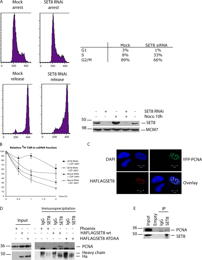
SET8 interacts with PCNA and is required for replication fork progression. (A) U2OS cells were transfected with siRNA against mock or SET8 and treated with thymidine for 30 h. Cells in the bottom panel were released from G1 arrest by the addition of new media. Nocodazole was added as the cells were released 10 h before harvest. (B) 48 h after SET8 siRNA transfection, cells were pulse labeled with [3H]thymidine for 30 min and were allowed to progress with and without Chk1 inhibitor CEP-3891 for the indicated times. Replication fork elongation from the incorporated [3H]thymidine was then measured as described in Fig. S3 D. 100% labeled single-stranded DNA (ssDNA) is equivalent to no fork progression. (C) Colocalization between HA-FLAG–tagged SET8 and YFP-PCNA transfected in U2OS cells. Cells were preextracted before fixation and processed for immunostaining with HA antibody. (D) Immunoprecipitation of HA-FLAG–tagged SET8 wild type and PIP-box mutant (LTDFY mutated to ATDAA) from HEK293 cells. Cells were transfected with the indicated constructs followed by lysis, immunoprecipitation, and immunoblotting. (E) Endogenous SET8 from HEK293 cells was immunoprecipitated and processed for immunoblotting. Bars, 10 μm.
Our results suggest that depletion of SET8 in the Drosophila and mammalian organisms may have different outcomes. This can partly be explained by the fact that Drosophila PR-Set7 and human SET8 only are moderately homologous. However, the different phenotypes could also be a consequence of the experimental approaches used. In the Drosophila study (Sakaguchi and Steward, 2007), the investigators used cells from PR-Set7 knockout flies. The cells originated from third instar larvae flies and, thus, had progressed through several cell cycles before analysis. In contrast, we used mammalian cells and investigated the defects of SET8 depletion after cells were released from a G1/S block. We cannot eliminate the possibility that cells depleted for SET8 experience defects in progression through M phase; however, such defects were not detected in our study.
SET8 is required for replication fork progression and interacts with PCNA
Having established that DNA damage after SET8 depletion is dependent on DNA replication, we further investigated whether SET8 plays a direct role in DNA replication. To achieve this, we monitored replication fork progression using a previously described method in which newly synthesized DNA is labeled with [3H]thymidine (Johansson et al., 2004). The assay is based on the principle that each replication fork consists of a pair of single-stranded ends. These ends become unwound in alkaline solution. If replication elongation is inhibited, the labeled DNA will be present in the single-stranded DNA fraction. On the contrary, replication elongation will lead to the presence of the labeled DNA in the double-stranded DNA fraction. Thus, 100% labeled single-stranded DNA is equivalent to no fork progression (Fig. S3 D). Using this assay, we found that replication progression was slowed significantly by SET8 depletion (Fig. 5 B), confirming that SET8 is required for efficient DNA replication. Fork progression was not increased by the coinhibition of Chk1; rather, this inhibition lead to further replication inhibition. This indicates that SET8 and Chk1 function on separate pathways to regulate replication fork progression.
Stimulated by this finding, we performed detailed sequence analysis of SET8 in Scan Prosite searching for conserved motifs that could link SET8 with DNA replication. Remarkably, we found a conserved PIP box in SET8, which is a short sequence that mediates binding to PCNA (Warbrick, 1998). The SET8 PIP-box sequence is N-X-X-L-X-X-F-Y, which is located from amino acid residues 178–185 in human SET8. To investigate the interaction between PCNA and SET8, we first performed immunofluorescence staining. This indeed revealed colocalization between SET8 and PCNA (Fig. 5 C). Next, we determined whether the two proteins could interact. As shown in Fig. 5 D, SET8 and PCNA interact in a manner dependent on a functional PIP box. We also detected an interaction at endogenous protein levels (Fig. 5 E), which, altogether, links SET8 directly with the replication machinery.
Collectively, we propose that SET8 supports the organization and maintenance of chromatin structures to facilitate DNA replication and efficient DNA repair. SET8 may also play a transcriptional role in regulating the expression of genes critical for S-phase progression; however, we have not observed abnormalities in the expression of the DNA replication–associated proteins analyzed so far. In conclusion, our results demonstrate that SET8 is required for normal S-phase progression. Inhibition of SET8 expression leads to a dramatic increase in Chk1 activity, resulting in Chk1-dependent inhibition of DNA replication.
Materials and methods
Cell culture and chemicals
The human U2-OS osteosarcoma cell line was grown in DME medium with 10% FBS. For siRNA-mediated ablation, cells were transfected using OligofectAMINE (Invitrogen) according to the manufacturer's protocol. The following oligonucleotide sequences were used: 5′-PACUUCAUGGCGCUCCGUACUU-3′ and 5′-PGAUUUGUCUCUCUAGUUGCUU-3′ (SET8 siRNA). Chk1 siRNA were purchased from Dharmacon (SMARTpool reagents M-003255-02; four siRNAs combined into a single pool). A control oligonucleotide targeting cyclophilin was also purchased from Dharmacon. Experiments with siRNA-transfected cells were performed as indicated in the figure legends. The PCNA construct was a gift from C. Lukas (Danish Cancer Society, Copenhagen, Denmark). Nocodazole (Sigma-Aldrich) was used at 100 ng/ml. Inhibition of Chk1 activity was achieved by the addition of 100 nM Gö6976 (Calbiochem). Thymidine and 2-deoxycytidine-HCl were purchased from Sigma-Aldrich and used at 2 mM and 24 μM, respectively.
FACS
Cells pulse labeled with 10 μM BrdU (Roche) for 5 min were analyzed according to standard protocols. In brief, cells were fixed in 70% ethanol and incubated in 2 M HCl for 30 min. Cells were stained with mouse antibody to BrdU (1:100) for 1 h followed by 1-h incubation with conjugated anti–mouse IgG (AlexaFluor488 at 1:1,000). DNA was then counterstained with 0.1 mg/ml propidium iodide (PI) containing RNase for 30 min at 37°C. γ-H2AX, Cdc45, and Rad51 were stained as described above for BrdU except for the omission of HCl treatment. Analysis was performed on a FACSCalibur (BD Biosciences) using CellQuest software (Becton Dickinson). Quantifications were performed using Modfit LT software (Verity Software House).
Microscopy and immunofluorescence
Cells were grown on coverslips and treated as indicated in the figure legends. The coverslips were then washed briefly in PBS followed by extraction for 10 min on ice with Triton buffer (0.5% Triton X-100 in 20 mM Hepes, pH 7.4, 50 mM NaCl, 3 mM MgCl, and 300 mM sucrose) and fixation for 10 min with 4% PFA. Samples were incubated with primary antibodies in PBS/1% FCS for 1 h at room temperature, washed in PBS/1% FCS three times for 10 min, and incubated with AlexaFluor488– or 594–conjugated secondary antibodies (1:1,000; Invitrogen) for 1 h at room temperature. Cells were washed in PBS three times for 10 min and mounted using Vectashield mounting medium containing DAPI (Vector Laboratories). Pictures were acquired on a microscope (Axiovert 200M LSM 510; Carl Zeiss, Inc.) using a 40× c-Apochromat objective with an NA of 1.2 in H2O. Pictures were taken at room temperature using LSM 510 META software and LSM image examiner software (Carl Zeiss, Inc.). Brightfield pictures were taken with a microscope (Axiovert 135; Carl Zeiss, Inc.) using a 10× Achroplan objective with an NA of 0.25. Pictures were acquired at room temperature with a camera (CoolSNAP cf2; Photometrics) using MetaMorph software (MDS Analytical Technologies). All pictures were exported in preparation for printing using Photoshop (Adobe).
Immunoprecipitation
Antibodies for immunoprecipitation were coupled to protein A beads for 1 h at 4°C. Extracts for immunoprecipitation were prepared using immunoprecipitation buffer (50 mM Hepes, pH 7.5, 150 mM NaCl, 1 mM EDTA, 2.5 mM EGTA, 10% glycerol, 0.1% Tween 20, and protease inhibitors leupeptin, aprotinin, and PMSF). The lysis mixture was incubated on ice for 15 min, sonicated three times with a digital sonifier for 3 s (20%; S250; Branson), and microfuged for 10 min at 4°C. Extracts were precleared with 20 μl of protein A–Sepharose and 2 μg of normal IgG. After microcentrifugation, 20 μl of protein A–Sepharose conjugated with 1 μg of antibody was added and allowed to precipitate for 1.5 h at 4°C with rotation. The immune complexes were pelleted by gentle centrifugation and washed four times with 1 ml of immunoprecipitation buffer. After a final wash with immunoprecipitation buffer, the buffer was aspirated completely, and beads were resuspended in laemmli buffer. Immunoblotting was performed as indicated in the next section.
Immunoblotting and antibodies
In brief, cells were lysed on ice in radioimmunoprecipitation assay buffer (50 mM Hepes, pH 7.5, 150 mM NaCl, 1 mM EDTA, 2.5 mM EGTA, 10% glycerol, 1% IgePal630, 1% deoxycholic acid [sodium salt], 0.1% SDS, 1 mM PMSF, 5 μg/ml leupeptin, 1% vol/vol aprotinin, 50 mM NaF, and 1 mM DTT). Proteins were separated on an SDS-PAGE gel and transferred to a nitrocellulose membrane. The membranes were incubated in primary antibody diluted in 5% milk as indicated in the figures followed by incubation with secondary antibody (peroxidase-labeled anti–mouse or –rabbit IgG; 1:10,000; Vector Laboratories). Films were developed using an x-ray machine (Valsoe; Ferrania). Phospho-Chk1 antibody (Chk1-pSer317) was purchased from Cell Signaling Technology. Antibodies to MCM7 (DCS-141) and Chk1 (DCS-310) have been described previously (Sorensen et al., 2005). Cdc25A (F-6), cyclin A1 (H-432), Rad51 (H-92), and 53BP1 (H-300) were obtained from Santa Cruz Biotechnology, Inc. Phosphorylated H2AX (JBW103), monomethylated histone H4 Lys20, and phosphorylated histone H3 Ser10 were purchased from Millipore. SET8 (ab3744) and PCNA (PC10) were purchased from Abcam, and RPA (Ab-3) was purchased from EMD.
Pulsed-field gel electrophoresis
48 h after SET8 siRNA transfection, cells were treated with 5 μM aphidicolin (Fluka) and/or 500 nM CEP-3891 (Cephalon) for an additional 24 h. 106 cells were inserted into 10 mg/ml InCert agarose (Cambrex) plugs and incubated for 48 h in 0.5 M EDTA, 1% N-laurylsarcosyl, and 2 mg/ml proteinase K at 20°C in darkness. After washing of plugs four times in Tris-EDTA buffer, separation was performed for 20 h as described previously (Lundin et al., 2005) on a 1% certified megabase agarose (Bio-Rad Laboratories) gel. The gel was then stained overnight with ethidium bromide. Three independent experiments were performed.
Replication fork elongation
Analysis of replication fork elongation was performed as described previously (Johansson et al., 2004). Basically, [3H]thymidine was incorporated in ongoing forks, and, after labeling, the fork progressed from the labeled area. In alkaline solution, unwinding was initiated from the single-stranded ends of the fork; thus, the amount of labeled single-stranded DNA is greater the slower the fork is moving. 24 h after SET8 siRNA transfection, cells were replated in 24-well plates (105 cells/well) and allowed to grow for an additional 24 h. Cells were then pulse labeled with 37 kBq/ml [3H]thymidine (GE Healthcare) in DME at 37°C with 5% CO2 for 30 min, washed, and incubated with prewarmed media ±500 nM CEP-3891 at 37°C with 5% CO2 for the indicated times. After incubation, replication was terminated by rinsing the cells with ice-cold 0.15 M NaCl before the addition of 0.5 ml of ice-cold 0.15 M NaCl and 0.03 M NaOH unwinding solution. After 30-min incubation on ice in darkness, unwinding was terminated by the addition of 1 ml of 0.02-M NaH2PO4. DNA was fragmented to ~3 kb by sonication (B-12 sonifier with micro-tip; Branson) for 15 s, and SDS was added to a final concentration of 0.25%. After overnight storage at −20°C, single- and double-stranded DNA was separated on hydroxy apatite columns at 60°C. Three independent experiments were performed.
Online supplemental material
Fig. S1 shows the specificity of SET8 removal by RNAi, the inverse relationship between γ-H2AX and H4-K20me1–positive cells, and the recruitment of 53BP1 in γ-irradiated HeLa cells during SET8 depletion. Fig. S2 shows the unchanged level of H4-K20me2 in U2OS cells after the depletion of SET8, a quantification of DNA damage during removal of SET8 combined with the inhibition of Chk1 using CEP-3891, and a quantification of replication-dependent DNA damage after SET8 depletion from Fig. 4 D. Fig. S3 shows the down-regulation of SET8, Rad51, and Cdc45 corresponding to Fig. 4 E, the progression of U2OS cells through the cell cycle after depletion of Rad51 for 48 h, and a schematic presentation of the replication fork progression assay. Online supplemental material is available at http://www.jcb.org/cgi/content/full/jcb.200706150/DC1.
Acknowledgments
We thank Randi G. Syljuåsen and Adrian P. Bracken for thorough and critical reading of the manuscript. We thank Claudia Lukas for the YFP-PCNA construct and Reidar Albrechtsen for helpful assistance during image acquisition.
We thank the Novo Nordisk Foundation, the Danish Cancer Society, the Danish Medical Research Council, the Danish National Research Foundation, the Swedish Cancer Society, the Swedish Research Council, the Swedish Pain Relief Foundation, and the Medical Research Council for financial support.
Notes
Abbreviations used in this paper: DSB, double-strand break; PCNA, proliferating cell nuclear antigen; PI, propidium iodide; RPA, replication protein A.
References
- Bell, S.P., and A. Dutta. 2002. DNA replication in eukaryotic cells. Annu. Rev. Biochem. 71:333–374. [Abstract] [Google Scholar]
- Botuyan, M.V., J. Lee, I.M. Ward, J.E. Kim, J.R. Thompson, J. Chen, and G. Mer. 2006. Structural basis for the methylation state-specific recognition of histone H4-K20 by 53BP1 and Crb2 in DNA repair. Cell. 127:1361–1373. [Europe PMC free article] [Abstract] [Google Scholar]
- Couture, J.F., E. Collazo, J.S. Brunzelle, and R.C. Trievel. 2005. Structural and functional analysis of SET8, a histone H4 Lys-20 methyltransferase. Genes Dev. 19:1455–1465. [Europe PMC free article] [Abstract] [Google Scholar]
- Fang, J., Q. Feng, C.S. Ketel, H. Wang, R. Cao, L. Xia, H. Erdjument-Bromage, P. Tempst, J.A. Simon, and Y. Zhang. 2002. Purification and functional characterization of SET8, a nucleosomal histone H4-lysine 20-specific methyltransferase. Curr. Biol. 12:1086–1099. [Abstract] [Google Scholar]
- Feijoo, C., C. Hall-Jackson, R. Wu, D. Jenkins, J. Leitch, D.M. Gilbert, and C. Smythe. 2001. Activation of mammalian Chk1 during DNA replication arrest: a role for Chk1 in the intra-S phase checkpoint monitoring replication origin firing. J. Cell Biol. 154:913–923. [Europe PMC free article] [Abstract] [Google Scholar]
- Helleday, T., J. Lo, D.C. van Gent, and B.P. Engelward. 2007. DNA double-strand break repair: from mechanistic understanding to cancer treatment. DNA Repair (Amst.). 6:923–935. [Abstract] [Google Scholar]
- Huyen, Y., O. Zgheib, R.A. Ditullio Jr., V.G. Gorgoulis, P. Zacharatos, T.J. Petty, E.A. Sheston, H.S. Mellert, E.S. Stavridi, and T.D. Halazonetis. 2004. Methylated lysine 79 of histone H3 targets 53BP1 to DNA double-strand breaks. Nature. 432:406–411. [Abstract] [Google Scholar]
- Johansson, F., A. Lagerqvist, K. Erixon, and D. Jenssen. 2004. A method to monitor replication fork progression in mammalian cells: nucleotide excision repair enhances and homologous recombination delays elongation along damaged DNA. Nucleic Acids Res. 32:e157. [Europe PMC free article] [Abstract] [Google Scholar]
- Kohn, E.A., C.J. Yoo, and A. Eastman. 2003. The protein kinase C inhibitor Go6976 is a potent inhibitor of DNA damage-induced S and G2 cell cycle checkpoints. Cancer Res. 63:31–35. [Abstract] [Google Scholar]
- Kouzarides, T. 2007. Chromatin modifications and their function. Cell. 128:693–705. [Abstract] [Google Scholar]
- Lundin, C., M. North, K. Erixon, K. Walters, D. Jenssen, A.S. Goldman, and T. Helleday. 2005. Methyl methanesulfonate (MMS) produces heat-labile DNA damage but no detectable in vivo DNA double-strand breaks. Nucleic Acids Res. 33:3799–3811. [Europe PMC free article] [Abstract] [Google Scholar]
- Nishioka, K., J.C. Rice, K. Sarma, H. Erdjument-Bromage, J. Werner, Y. Wang, S. Chuikov, P. Valenzuela, P. Tempst, R. Steward, et al. 2002. PR-Set7 is a nucleosome-specific methyltransferase that modifies lysine 20 of histone H4 and is associated with silent chromatin. Mol. Cell. 9:1201–1213. [Abstract] [Google Scholar]
- Pacek, M., and J.C. Walter. 2004. A requirement for MCM7 and Cdc45 in chromosome unwinding during eukaryotic DNA replication. EMBO J. 23:3667–3676. [Europe PMC free article] [Abstract] [Google Scholar]
- Pilch, D.R., O.A. Sedelnikova, C. Redon, A. Celeste, A. Nussenzweig, and W.M. Bonner. 2003. Characteristics of gamma-H2AX foci at DNA double-strand breaks sites. Biochem. Cell Biol. 81:123–129. [Abstract] [Google Scholar]
- Rice, J.C., K. Nishioka, K. Sarma, R. Steward, D. Reinberg, and C.D. Allis. 2002. Mitotic-specific methylation of histone H4 Lys 20 follows increased PR-Set7 expression and its localization to mitotic chromosomes. Genes Dev. 16:2225–2230. [Europe PMC free article] [Abstract] [Google Scholar]
- Saintigny, Y., F. Delacote, G. Vares, F. Petitot, S. Lambert, D. Averbeck, and B.S. Lopez. 2001. Characterization of homologous recombination induced by replication inhibition in mammalian cells. EMBO J. 20:3861–3870. [Europe PMC free article] [Abstract] [Google Scholar]
- Sakaguchi, A., and R. Steward. 2007. Aberrant monomethylation of histone H4 lysine 20 activates the DNA damage checkpoint in Drosophila melanogaster. J. Cell Biol. 176:155–162. [Europe PMC free article] [Abstract] [Google Scholar]
- Sarkaria, J.N., E.C. Busby, R.S. Tibbetts, P. Roos, Y. Taya, L.M. Karnitz, and R.T. Abraham. 1999. Inhibition of ATM and ATR kinase activities by the radiosensitizing agent, caffeine. Cancer Res. 59:4375–4382. [Abstract] [Google Scholar]
- Sorensen, C.S., R.G. Syljuasen, J. Falck, T. Schroeder, L. Ronnstrand, K.K. Khanna, B.B. Zhou, J. Bartek, and J. Lukas. 2003. Chk1 regulates the S phase checkpoint by coupling the physiological turnover and ionizing radiation-induced accelerated proteolysis of Cdc25A. Cancer Cell. 3:247–258. [Abstract] [Google Scholar]
- Sorensen, C.S., L.T. Hansen, J. Dziegielewski, R.G. Syljuasen, C. Lundin, J. Bartek, and T. Helleday. 2005. The cell-cycle checkpoint kinase Chk1 is required for mammalian homologous recombination repair. Nat. Cell Biol. 7:195–201. [Abstract] [Google Scholar]
- Syljuasen, R.G., C.S. Sorensen, L.T. Hansen, K. Fugger, C. Lundin, F. Johansson, T. Helleday, M. Sehested, J. Lukas, and J. Bartek. 2005. Inhibition of human Chk1 causes increased initiation of DNA replication, phosphorylation of ATR targets, and DNA breakage. Mol. Cell. Biol. 25:3553–3562. [Europe PMC free article] [Abstract] [Google Scholar]
- Warbrick, E. 1998. PCNA binding through a conserved motif. Bioessays. 20:195–199. [Abstract] [Google Scholar]
- Ward, I., J.E. Kim, K. Minn, C.C. Chini, G. Mer, and J. Chen. 2006. The tandem BRCT domain of 53BP1 is not required for its repair function. J. Biol. Chem. 281:38472–38477. [Europe PMC free article] [Abstract] [Google Scholar]
- Xiao, B., C. Jing, G. Kelly, P.A. Walker, F.W. Muskett, T.A. Frenkiel, S.R. Martin, K. Sarma, D. Reinberg, S.J. Gamblin, and J.R. Wilson. 2005. Specificity and mechanism of the histone methyltransferase Pr-Set7. Genes Dev. 19:1444–1454. [Europe PMC free article] [Abstract] [Google Scholar]
- Zhao, H., and H. Piwnica-Worms. 2001. ATR-mediated checkpoint pathways regulate phosphorylation and activation of human Chk1. Mol. Cell. Biol. 21:4129–4139. [Europe PMC free article] [Abstract] [Google Scholar]
- Zou, L., D. Liu, and S.J. Elledge. 2003. Replication protein A-mediated recruitment and activation of Rad17 complexes. Proc. Natl. Acad. Sci. USA. 100:13827–13832. [Europe PMC free article] [Abstract] [Google Scholar]
Articles from The Journal of Cell Biology are provided here courtesy of The Rockefeller University Press
Full text links
Read article at publisher's site: https://doi.org/10.1083/jcb.200706150
Read article for free, from open access legal sources, via Unpaywall:
https://rupress.org/jcb/article-pdf/179/7/1337/1401780/jcb_200706150.pdf
Free to read at intl.jcb.org
http://intl.jcb.org/cgi/content/abstract/179/7/1337
Free after 6 months at intl.jcb.org
http://intl.jcb.org/cgi/content/full/179/7/1337
Free after 6 months at intl.jcb.org
http://intl.jcb.org/cgi/reprint/179/7/1337.pdf
Citations & impact
Impact metrics
Citations of article over time
Alternative metrics
Smart citations by scite.ai
Explore citation contexts and check if this article has been
supported or disputed.
https://scite.ai/reports/10.1083/jcb.200706150
Article citations
Endonucleosis mediates internalization of cytoplasm into the nucleus.
Nat Commun, 15(1):5843, 11 Jul 2024
Cited by: 0 articles | PMID: 38992049 | PMCID: PMC11239883
RAD18 directs DNA double-strand break repair by homologous recombination to post-replicative chromatin.
Nucleic Acids Res, 52(13):7687-7703, 01 Jul 2024
Cited by: 1 article | PMID: 38884202 | PMCID: PMC11260465
Inhibition of FBP1 expression by KMT5A through TWIST1 methylation is one of the mechanisms leading to chemoresistance in breast cancer.
Oncol Rep, 52(2):110, 04 Jul 2024
Cited by: 0 articles | PMID: 38963044 | PMCID: PMC11258601
<i>Drosophila melanogaster</i> Set8 and L(3)mbt function in gene expression independently of histone H4 lysine 20 methylation.
Genes Dev, 38(9-10):455-472, 25 Jun 2024
Cited by: 3 articles | PMID: 38866557 | PMCID: PMC11216177
miR-17 ~ 92 suppresses proliferation and invasion of cervical cancer cells by inhibiting cell cycle regulator Cdt2.
Discov Oncol, 14(1):172, 14 Sep 2023
Cited by: 1 article | PMID: 37707654 | PMCID: PMC10501107
Go to all (152) article citations
Data
Data behind the article
This data has been text mined from the article, or deposited into data resources.
BioStudies: supplemental material and supporting data
Similar Articles
To arrive at the top five similar articles we use a word-weighted algorithm to compare words from the Title and Abstract of each citation.
PR-Set7-dependent lysine methylation ensures genome replication and stability through S phase.
J Cell Biol, 179(7):1413-1426, 24 Dec 2007
Cited by: 110 articles | PMID: 18158331 | PMCID: PMC2373513
CRL4(Cdt2)-mediated destruction of the histone methyltransferase Set8 prevents premature chromatin compaction in S phase.
Mol Cell, 40(1):22-33, 01 Oct 2010
Cited by: 159 articles | PMID: 20932472 | PMCID: PMC2957874
Direct interaction between SET8 and proliferating cell nuclear antigen couples H4-K20 methylation with DNA replication.
J Biol Chem, 283(17):11073-11077, 03 Mar 2008
Cited by: 91 articles | PMID: 18319261 | PMCID: PMC2431066
Roles for the methyltransferase SETD8 in DNA damage repair.
Clin Epigenetics, 14(1):34, 04 Mar 2022
Cited by: 7 articles | PMID: 35246238 | PMCID: PMC8897848
Review Free full text in Europe PMC
Funding
Funders who supported this work.
Medical Research Council (1)
MRC/CR-UK Gray Institute for Radiation Oncology and Biology, Oxford - MRC Core Grant
Professor William Gillies McKenna, University of Oxford
Grant ID: G0700730


