| PMC full text: | Cancer Res. Author manuscript; available in PMC 2010 Oct 1. Published in final edited form as: Cancer Res. 2009 Oct 1; 69(19): 7569–7576. Published online 2009 Sep 22. doi: 10.1158/0008-5472.CAN-09-0529 |
Figure 2

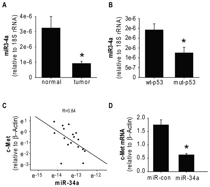
A) miR-34a levels were measured by qRT-PCR in 11 glioblastoma surgical specimens and 6 normal brain samples and normalized to 18S rRNA measured in the same samples (arbitrary units). The results show that average levels of miR-34a in gliomas are lower than in normal brain. B) The p53 status of the glioma specimens described in (A) were determined. The blots show that average expression of miR-34a in wild-type p53 glioblastoma tumors (n=7) is significantly higher than miR-34a expression in mutant p53 tumors. C) c-Met expression in the same tissues described in (A) was measured by RT-PCR and normalized to β-Actin. Plotting of miR-34a vs. c-Met expressions shows an inverse correlation between them. D) Glioma cells were transfected with miR-34a for 48 hrs prior to measurement of c-Met mRNA levels by RT-PCR. The results show that miR-34a reduces c-Met mRNA levels. * = p<0.05.


