| PMC full text: | Cell. Author manuscript; available in PMC 2010 Jan 9. Published in final edited form as: Cell. 2009 Jan 9; 136(1): 163–174. doi: 10.1016/j.cell.2008.11.046 |
Figure 5

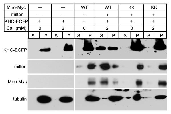
Ca++, via Miro, Releases KHC from Microtubules
Lysates of HEK cells, transfected as indicated, were mixed with Taxol-stabilized microtubules in either 0 or 2 mM Ca++ buffer prior to sedimentation of the microtubules and microtubule-bound proteins by centrifugation (pellet, P), leaving unbound proteins in the supernatant fraction (S). Equivalent fractions of the supernatant and pellet were assayed for KHC-ECFP, milton, Miro-Myc, and tubulin.


