Abstract
Free full text

Imaging with total internal reflection fluorescence microscopy for the cell biologist
Abstract
Total internal reflection fluorescence (TIRF) microscopy can be used in a wide range of cell biological applications, and is particularly well suited to analysis of the localization and dynamics of molecules and events near the plasma membrane. The TIRF excitation field decreases exponentially with distance from the cover slip on which cells are grown. This means that fluorophores close to the cover slip (e.g. within ~100 nm) are selectively illuminated, highlighting events that occur within this region. The advantages of using TIRF include the ability to obtain high-contrast images of fluorophores near the plasma membrane, very low background from the bulk of the cell, reduced cellular photodamage and rapid exposure times. In this Commentary, we discuss the applications of TIRF to the study of cell biology, the physical basis of TIRF, experimental setup and troubleshooting.
Introduction
The plasma membrane is the barrier that all molecules must cross to enter or exit the cell, and a large number of biological processes occur at or near the plasma membrane. These processes are difficult to image with traditional epifluorescence or confocal microscopy techniques, because details near the cell surface are easily obscured by fluorescence that originates from the bulk of the cell. Total internal reflection fluorescence (TIRF) microscopy – also known as evanescent wave or evanescent field microscopy – provides a means to selectively excite fluorophores near the adherent cell surface while minimizing fluorescence from intracellular regions. This serves to reduce cellular photodamage and increase the signal-to-noise ratio. TIRF primarily illuminates only fluorophores very close (e.g. within 100 nm) to the cover-slip–sample interface. The background fluorescence is minimized because the excitation of fluorophores further away from the cover slip is reduced. The plasma membrane of an adherent cell lies well within the region of excitation, allowing imaging of processes occurring at or near the membrane. On the basis of these unique features, TIRF has been employed to address numerous questions in cell biology.
This Commentary details key issues for researchers who are using, or are considering using, TIRF for live cell imaging. We begin with a brief selection of specific areas of cell biological research in which the use of TIRF imaging has made a major impact. Subsequently, we describe the physical basis of TIRF, and discuss key issues to consider when setting up and employing TIRF. Finally, we identify several potential pitfalls and provide helpful suggestions. A basic knowledge of fluorescence microscopy is assumed. For general background on fluorescence microscopy, we refer readers to North (North, 2006) and Waters (Waters, 2009) For reviews containing an extensive treatment of TIRF theory and advanced applications, we refer readers to Axelrod (Axelrod, 2003; Axelrod, 2008).
Cellular processes visualized with TIRF
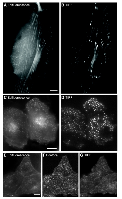
TIRF microscopy has been used in many different types of studies for the visualization of the spatial-temporal dynamics of molecules at or near the cell surface, particularly in cases in which the signal would otherwise be obscured by cytosolic fluorescence. Some of the advantages of TIRF for imaging near the cell surface are illustrated in Fig. 1. Actin (LifeAct–GFP in Fig. 1A,B), clathrin (GFP–clathrin light chain in Fig. 1C,D) and caveolin (caveolin-1–EGFP in Fig. 1E–G) have been imaged by both conventional epifluorescence (Fig. 1A,C,E) and TIRF (Fig. 1B,D,G). In each case, it is apparent that TIRF minimizes the out-of-focus intracellular fluorescence, resulting in images with a much higher signal-to-noise ratio. Similarly, although confocal microscopy (Fig. 1F) shows a reduced cytosolic signal relative to epifluorescence (Fig. 1E), the corresponding TIRF image (Fig. 1G) provides the greatest amount of information for fluorophores associated with the plasma membrane. The suppression of background fluorescence is crucial for studying each of the areas of cell biology on which TIRF has had a major impact.

Comparison of images obtained using epifluorescence and TIRF. In both cases, the microscope was focused at the adherent plasma membrane and images were acquired with one of three modes of excitation: epifluorescence, TIRF or confocal. (A,B) Actin (LifeAct–GFP) in a migrating MDCK cell. (C,D) Clathrin (clathrin light chain–GFP) in a HeLa cell. (E–G) Caveolin-1 (caveolin-1–EGFP) in MDCK cells. In each case, TIRF clearly eliminates of out-of-focus fluorescence and reveals details at or near the cell surface. Scale bars: 10 μm.
TIRF has had an impact on many varied areas of cell biology, including HIV-1 virion assembly (Jouvenet et al., 2006) and intraflagellar transport in the Chlamydomonas flagella (Engel et al., 2009), and in single-molecule experiments. Below, we highlight several areas of cell biology – the cytoskeleton, endocytosis, exocytosis, cell–substrate contact regions and intracellular signaling – that have particularly benefited from investigation by TIRF.
Cytoskeleton
The dynamics of the cytoskeleton near the plasma membrane have been studied with TIRF (Fig. 1A,B), leading to new insights. Before TIRF was used to study vesicle trafficking, it was not known that a cortical microtubule network extended immediately adjacent to the plasma membrane, and that secretory vesicles remained attached to these microtubules until the moment of vesicle fusion (Schmoranzer and Simon, 2003). This finding was made possible, in part, by the spatial restriction and high signal-to-noise ratio of the excitation field, as well as by the rapid acquisition rates that are possible when using wide-field acquisition. Furthermore, the increased excitation of fluorophores near the cover slip permitted the quantification of microtubule motility in the axial direction, revealing how microtubules are targeted to focal adhesions (Krylyshkina et al., 2003). TIRF also provided the spatial and temporal resolution to study the dynamics of actin and actin-associated proteins near the plasma membrane in several endocytosis studies (Kaksonen et al., 2005; Merrifield et al., 2002). Finally, fluorescence speckle microscopy, in which a limiting amount of cytoskeleton monomers are fluorescently labeled, combined with TIRF was able to deliver important information about the dynamics and flow of cytoskeleton filaments (Danuser and Waterman-Storer, 2006).
Endocytosis
The formation of endocytic vesicles involves the recruitment of cytosolic proteins to the adherent plasma membrane. When viewed with standard epifluorescence, the surface patches of the vesicle coat protein clathrin are difficult to discern from background fluorescence and intracellular clathrin structures (Fig. 1C). The initial report to image clathrin during endocytosis in live cells employed epifluorescence and was thus restricted to analyzing only those events that occurred in the cell periphery, because of out-of-focus signals (Gaidarov et al., 1999). By contrast, when TIRF is used, the clathrin patches on or near the membrane appear as distinct features (Fig. 1D). Imaging the dynamics of endocytosis is aided by rapid image acquisition, background elimination and the exponential decrease in excitation intensity with distance from the cover slip. TIRF has made it possible to gain insight into the components that are necessary for vesicle formation and the dynamics of this process (Rappoport, 2008). A main focus of studies that have applied TIRF to analyze endocytosis has been to determine the ‘life history’ of the formation of individual clathrin-coated vesicles. These studies have demonstrated that some proteins are present throughout the endocytosis process (e.g. clathrin and epsin) (Rappoport et al., 2006; Rappoport and Simon, 2003), whereas the localization of other proteins to the coated vesicle either increased over time (e.g. dynamin) (Merrifield et al., 2002; Rappoport et al., 2008; Rappoport and Simon, 2003) or decreased (e.g. AP-2) (Rappoport et al., 2003; Rappoport et al., 2005).
Exocytosis
The thin TIRF excitation field allows identification of secretory carriers near the membrane (Lang et al., 1997). Specific criteria, which involve quantifying the total fluorescence, as well as its peak and spread, have been established to quantify and characterize the fusion of vesicles with the plasma membrane (Schmoranzer et al., 2000). The exponential decay of the TIRF excitation field allows the small motions of individual fluorescence-marked secretory carriers in the direction normal to the substrate and plasma membrane (the axial or z-direction) to be manifested as intensity changes. The precision of tracking such axial movements can be as small as 2 nm, which is considerably smaller than the resolution limit of light microscopy (Allersma et al., 2006). Because of the high contrast and low background, the position of the centers of vesicles can be measured with an accuracy of about 10 nm and motions before fusion that are smaller than the granule diameter can be followed. By observing single vesicles as they approached and fused with a membrane, it was found that many of the vesicles did not deliver all of their cargo in a single fusion step, but required two or more fusions to fully discharge their cargo (Jaiswal et al., 2009; Schmoranzer et al., 2000).
Cell–substrate contact regions
TIRF has also been used to investigate cell–substrate contact regions using several different approaches. Similar to the examples above, the restricted excitation field was shown to be crucial for studying focal adhesions with regard to their location, composition, motion and specific biochemistry (Axelrod, 1981; Choi et al., 2008). One technique for identifying cell–substrate contacts involves adding a fluorophore to the surrounding solution (Todd et al., 1988). The intensity of fluorescence can be used to calculate the distance of the membrane from the surface and to ‘map’ the bottom surface of the cell (Gingell et al., 1987). This technique requires the thin excitation field of TIRF, as other imaging methods would result in overwhelming background fluorescence. Recent work has analyzed focal adhesion disassembly in real time with TIRF and has demonstrated an unexpected role for clathrin in this process (Ezratty et al., 2009). This was made possible by the ability to rapidly acquire images of two spectrally distinct fluorophores, with axial information, at the adherent plasma membrane.
Intracellular signaling
TIRF has also been used to visualize different modes of intracellular signaling. For instance, TIRF has been instrumental in studies addressing plasma membrane recruitment and spatial distributions of signaling molecules such as phosphoinositide lipids (Haugh et al., 2000). Furthermore, a plasma-membrane-targeted biosensor revealed temporal oscillations of cAMP signaling, indicating a previously unidentified basis for regulation of upstream targets (Dyachok et al., 2006). Single plasma membrane Ca2+ channels have been imaged with good spatial and temporal resolution, providing information inaccessible to electrophysiological means, and revealing an uneven spatial distribution and diversity in kinetics (Demuro and Parker, 2004). TIRF and patch-clamp methods have been successfully combined to demonstrate the localization and coordination of open calcium channels and ER calcium-sensing molecules, revealing the spatial dynamics of intracellular Ca2+ signaling (Luik et al., 2006).
Physical basis of TIRF
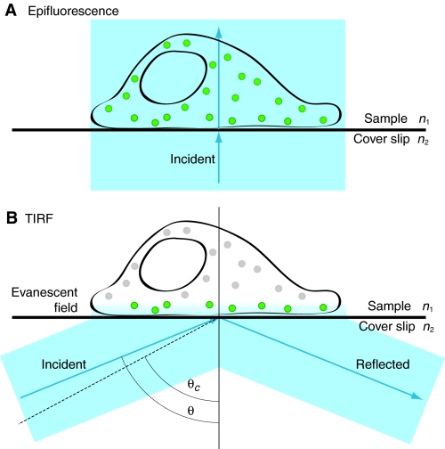
To understand the setup, optimization and common pitfalls of TIRF, it is important to understand the physical basis of the technique. In the following section, we provide a brief description of some key parameters in the context of cellular imaging. We begin by considering the excitation light encountering the cover-slip–sample interface (Fig. 2). It is this interaction that produces the thin excitation field used in TIRF microscopy.

The physical basis of epifluorescence and TIRF illumination. Schematic illustrating the cover-slip–sample interface. (A) Epifluorescence. The excitation beam travels directly through the cover-slip–sample interface. All of the fluorophores in the entire sample are excited. (B) TIRF. The excitation beam enters from the left at incidence angle θ, which is greater than the critical angle, θc (indicated by the dashed line). Angles are measured from the normal. The excitation beam is reflected off the cover-slip–sample interface and an evanescent field is generated on the opposite side of the interface, in the sample. Only fluorophores in the evanescent field are excited, as indicated by the green color. The refractive index of the sample (n1) must be less than the index of refraction of the cover slip (n2) to achieve TIR.
In the configuration most commonly used for cellular imaging, through-the-objective TIRF, the laser beam is focused off-axis at the back focal plane (BFP) of the objective lens. When the light exits the objective lens, it passes through the immersion oil and into the cover slip, which are matched in refractive index. When the excitation light beam propagating through the glass cover slip encounters the interface with the aqueous sample, the direction of travel of the light beam is altered (Fig. 2B). If the angle of incidence of the excitation light on the interface, θ, is greater than the ‘critical angle’, the light beam undergoes total internal reflection (TIR) and does not propagate into the sample. The critical angle, θc, is given by Snell's law:
where n1 and n2 are the refractive indices of the sample and the cover slip, respectively. To achieve TIR, the refractive index of the sample must be less than that of the cover slip.
If the angle of incidence is less than θc, most of the excitation light propagates through the sample; this is what occurs in epifluorescence (Fig. 2). However, if the angle of incidence is greater than θc, the excitation light is reflected off the cover-slip–sample interface back into the cover slip. In this case, some of the incident energy penetrates through the interface, creating a standing wave called the evanescent field. This is the excitation field employed in TIRF microscopy.
The intensity (I) of the evanescent field decays exponentially with distance from the interface (z). Therefore, a fluorophore that is closer to the interface will be excited more strongly than a fluorophore that is further from the interface. The intensity of the evanescent field at any position z is described by:
where I0 is the intensity of the evanescent field at z=0. I0 is related to the intensity of the incident beam by a complex function of θ and polarization (Axelrod, 1989). The depth of the evanescent field, d, refers to the distance from the cover slip at which the excitation intensity decays to 1/e, or 37%, of I0. Depth d is defined by:
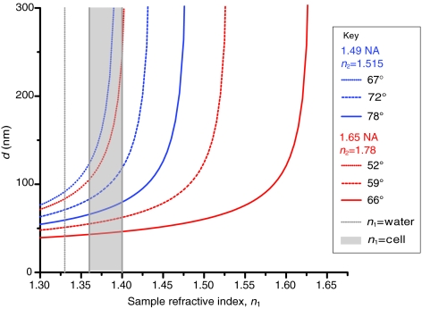
where λ0 is the wavelength of the excitation light in a vacuum. Typical values for d are in the range 60–100 nm. The depth of the evanescent field is affected by several parameters, including incidence angle, wavelength, and the refractive indices of the sample and the cover slip. The depth decreases (i.e. the excitation field becomes more narrow) as the incidence angle increases. The longer the wavelength, the greater the depth (i.e. the thicker the excitation volume). In addition, for a given incidence angle, the depth depends on the refractive index of the sample. As the refractive index of the sample increases, the depth increases (Fig. 3).

The depth of the evanescent field depends on the refractive index of the sample. The depth of the evanescent field (d) is a function of the index of refraction of the sample (n1). Incidence angles were chosen as 1.5° above the critical angle (dotted lines), the midpoint of all possible TIRF angles (dashed lines) and 1.5° less than the maximum incidence angle (solid lines). As the angle of incidence is increased, the sensitivity of d to the sample refractive index decreases in the range for cells. The 1.65 NA objective has a larger range with lower sensitivity to sample refractive index than the 1.49 NA objective.
Practically speaking, the excitation wavelength and refractive index of the cover slip are set, and the user controls the angle of incidence. Generally, this angle should be increased until it surpasses the critical angle. It is not advisable to ‘set’ a predefined angle of incidence without evaluating the evanescent field, because the critical angle will change depending on the refractive index of the sample. Biological samples have unknown and probably variable refractive indices. Moreover, the refractive index can vary within one sample as well as between samples. Thus, careful and repeated adjustment of the angle of incidence is strongly recommended because variations in sample refractive indices are the cause of many problems in TIRF microscopy, as discussed in detail below.
TIRF setup
Commercial TIRF systems are available; however, many researchers use ‘homemade’ TIRF setups. Some of these were constructed before major microscope manufacturers began selling off-the-shelf TIRF systems, whereas other setups are used by researchers who require flexible and easily modified platforms. Instructions on how to construct a homemade TIRF system are available (Axelrod, 2003). For the purpose of this article, we consider commercial TIRF systems, which are available from major microscopy companies (Leica, Nikon, Olympus and Zeiss) and third-party suppliers. These systems have the same basic ability to deliver through-the-objective TIRF illumination, but the individual system details vary. When evaluating different systems, we recommend viewing an experimental sample of interest, as well as each of the two test samples described in Box 1, to assess performance differences. In the best-case scenario, identical or very similar samples should be evaluated to exclude sample variation as a confounding variable.
When assembling a TIRF system, many of the same rules of thumb apply as when assembling any other high-resolution fluorescence microscopy system. Some important considerations include selection of an appropriate camera, dichroic mirror, emission filters, excitation light source, acquisition software and sample environment. Hardware considerations of particular importance in TIRF microscopy are described below.
TIRF excitation
In the majority of commercial systems, the excitation light is introduced to the sample through the same objective lens as the fluorescence is collected (Stout and Axelrod, 1989). However, there are various configurations for delivering the excitation beam to the sample, some of which use the objective and others a prism. The latter can be beneficial because it is generally inexpensive to set up and produces a ‘cleaner’ evanescent field with less scattered light, lower background and a greater range of incidence angles. Prism-based TIRF is often employed for in vitro studies, but is not the system of choice for most cell biologists because it can restrict sample accessibility and the choice of objectives. By contrast, through-the-objective TIRF allows rapid and easy access to the sample for changes to the media or drug addition, and exchange of samples is similarly quick and simple. Through-the-objective TIRF systems also tend to be more user friendly, requiring minimal maintenance and alignment. Thus, it is no surprise that through-the-objective TIRF is the configuration most commonly used by cell biologists.
Through-the-objective TIRF
To satisfy the physical requirements for TIRF, the excitation laser must be directed to the cover-slip–sample interface at an angle greater than the critical angle, as discussed above. In addition, all of the excitation light should be incident on the interface at the same angle. To achieve this uniform angle of illumination, the laser beam is focused on the BFP of the objective and therefore emerges in a collimated form, with all its ‘rays’ in parallel at a single angle. There is a one-to-one correspondence between the angle at which the light emerges from the objective and the position of the focused light on the BFP. The further off-axis the focused light is, the larger the angle of incidence. The angle of incidence is generally controlled either with a micrometer or through software.
High-numerical-aperture objective
One important requirement in through-the-objective TIRF is the numerical aperture (NA) of the objective. The NA of an objective describes its light-gathering power – specifically, the angle through which it is able to collect light. The NA also describes the maximum angle at which the excitation light can emerge from the objective. For an oil-immersion objective, this angle is also the maximum incidence angle of the excitation beam on the cover-slip–sample interface. The NA of the objective must be greater than the refractive index of the sample, preferably by a substantial margin. For example, if the sample is water (n2=1.33), then a 1.4 NA objective is sufficient for TIR. However, the refractive index of a cell is greater than that of water and is variable. The refractive index of the cell interior is typically 1.38, although it varies among cell types, thus making TIRF imaging of cells challenging with a 1.4 NA objective (Fig. 3).
Fortunately, there are several commercial objectives available with a NA greater than 1.4; these are often marketed exclusively as TIRF objectives. The most common objectives are 1.45 NA and 1.49 NA, and are both available in 60×, 100× and 150× magnification. All these objectives should be used with standard glass cover slips (n=1.515) and standard immersion oil. In addition, a 100× 1.65 NA objective is also available from Olympus and has a number of advantages. First, the higher NA allows larger incidence angles, which can be crucial for imaging certain cell types (i.e. chromaffin cells) that have unusually high refractive indices. Second, there is a range of incidence angles where the depth of the evanescent field produced by the 1.65 NA objective is not very sensitive to the sample refractive index within the range of typical cellular refractive indices (Fig. 3). This is important because if there are any local variations in cellular refractive index (perhaps due to vesicles or organelles), the 1.65 NA objective can create a uniform evanescent field. The major drawbacks of the 1.65 NA objective are that it requires a special and volatile immersion oil, and expensive non-standard cover slips (n=1.78).
Laser light source
It is possible to use either a laser or an arc lamp such as xenon or mercury for TIRF excitation. Some benefits of using an arc lamp are that it enables easy selection of excitation wavelengths with a filter wheel and the illumination field contains no interference fringes (see below). In this configuration, any light from the arc lamp that would arrive at the sample at less than the critical angle must be discarded. Therefore, a significant percent of the excitation power is lost, resulting in lower excitation intensity and dimmer images. Arc lamp sources for TIRF are commercially available and work well for samples that are intrinsically bright.
Lasers are the most common source for TIRF excitation and the TIRF system should have one laser line optimized for each fluorophore. The lasers can be mounted together and combined so that multiple fluorophores can be imaged either simultaneously or alternately. An acousto-optic tunable filter (AOTF) or filter wheel can be used for rapid switching between excitation wavelengths.
Camera
TIRF is a technique that captures the full field of an image, rather than scanning a single point. For the collection of images, a cooled charge-coupled device (CCD) camera is used. There is a wide range of CCD cameras to choose from, including electron multiplying (EM) CCDs. When rapid imaging in very low light situations is required, EMCCDs offer benefits over conventional CCDs. However, TIRF does not require an EMCCD and camera selection should be determined on the basis of the same considerations as for wide-field fluorescence imaging (Moomaw, 2007; Spring, 2007). When intensity changes are to be quantified, the linearity of the camera in response to incoming photons is an important consideration.
Image splitter
TIRF illumination is restricted within a single focal plane and relatively short exposure times (e.g. many frames per second) are routinely employed, making the technique especially useful for imaging dynamic processes. One of the developments that have made TIRF particularly powerful is the ability to image multiple fluorophores, either simultaneously or in very rapid succession. Image splitters, such as those sold by Cairn (Cairn Research Limited, UK) and Photometrics (Tucson, AZ, USA), allow simultaneous acquisition of emission from two to four spectrally distinct fluorophores on different regions of a CCD camera. This is an optimal method for imaging very rapid events; however, it reduces the size of the field that can be imaged. Alternatively, splitters that project two spectrally distinct images on two separate cameras are also available. Both methods can generate potential problems when aligning and overlaying the spectrally distinct channels, also known as image registration.
Sample environment
Live cell imaging often requires stable maintenance of the sample at 37°C, as temperature gradients can be a major source of focal drift. The thin optical section imaged with TIRF makes it particularly sensitive to small changes in focus, which degrade image quality. A number of solutions to this problem involve controlling temperature, and possibly also humidity and pCO2 (partial pressure of CO2). Stage-top incubators combined with objective heaters are one strategy for maintaining a stable temperature, although whole-scope incubators allow the entire system to equilibrate with fewer problems. Several companies are now offering focus-maintaining solutions, which reduce or eliminate focal drift resulting from temperature gradients.
Selection, preparation and analysis of TIRF samples
TIRF is ideal for imaging events occurring at a surface. However, there are several variables that must be considered before embarking on experiments.
Selection of cell type
The cells must be adherent, because TIRF illuminates only the region near the cover slip and cannot be used to image non-adherent cells. For some cell lines, it can be necessary to coat cover slips with extracellular matrix molecules or substances such as polylysine or collagen to ensure cell adherence. On the other hand, confluent monolayers of cells can generate areas of high refractive index, which can make imaging with TIRF difficult.
As outlined above, the refractive index of the cells should be below the NA of the objective. For example, chromaffin cells have a very high refractive index, which can make it difficult to obtain TIRF images using standard 1.45 NA or 1.49 NA objectives; the 1.65 NA objective was shown to yield good TIRF images (Allersma et al., 2004). Moreover, apoptotic cells generally have a higher index of refraction than non-apoptotic cells. Also, it is important to keep in mind that intracellular organelles have different refractive indices. Attempting TIRF imaging of a cell with a high refractive index can result in scattering of propagated light through the sample. To address this problem, it might be possible to increase the angle of incidence until the propagated light disappears. If this is not effective, the sample might require a 1.65 NA objective or the use of a prism-based system.
Sample preparation
TIRF is ideal for imaging live cells. Because of the thin excitation field, most of the imaged cell is not exposed to the excitation light, leading to fewer phototoxic effects. If cells are fixed, they should be mounted in a low refractive index media, such as PBS. Mounting medium that hardens or contains glycerol is useful for long-term sample storage, but it usually has a higher refractive index, rendering imaging of these samples with TIRF impossible. Finally, it should be noted that some dyes commonly used in cell biology (i.e. FM4-64 and DiI) adhere to the cover-slip surface and can obscure imaging unless the sample is properly washed after staining.
Data interpretation
When interpreting TIRF data, it is important to remember that the excitation field is not discrete, but exponentially decaying. The penetration depth, which is usually between 60 and 100 nm, describes the distance from the cover slip at which the excitation intensity is 1/e, or ~37%, of its value at the cover slip. The evanescent field exponentially decays, and objects located further than 100 nm from the surface of the cover slip can still be excited and imaged.
Particular care must be taken when interpreting the intensity of objects in an image obtained by TIRF. The intensity is affected by more than just the number of fluorophores; other factors include the axial (z) position and the orientation of the excitation dipole of the fluorophore relative to the polarization of the evanescent field. For example, 100 fluorophores positioned at z=0 will have the same intensity as 370 fluorophores at z=d (I=1/e×370=100). Thus, intensity alone cannot be used to infer relative number of fluorophores or z positions between objects.
It follows that changes in intensity from a single object can be due to changes in multiple parameters, including the number of associated fluorophores, movements in z, the orientation of the object or the occurrence of photobleaching. The intensity of an object will increase if it gains fluorophores or moves closer to the cover slip, or if the excitation dipole rotates to align with the polarization of the evanescent field. In some cases, the specific biological context of the experiment can help to clarify the source of the intensity change; for example, the number of fluorophores on the inside of a secretory vesicle typically remains constant and, therefore, alteration in intensity can generally be interpreted as movement in z (Allersma et al., 2006). Also, in endocytosis, a loss of fluorescence can be interpreted as due to internalization only when it can be clearly distinguished from photobleaching. Alternatively, an epifluorescence image can be used for normalization and movement in z can be interpreted regardless of fluctuations in the number of fluorophores (Saffarian and Kirchhausen, 2008).
Troubleshooting and practical advice
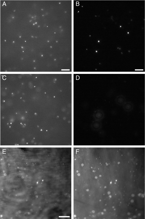
In this section, we discuss commonly encountered problems when using TIRF microscopy. One of the most common is contamination of the image with propagating light, which can arise from a subcritical incidence angle or scattering from the sample. Both these scenarios significantly degrade the quality of TIRF images. Propagating light skimming at an angle through the sample will have similar characteristics to epifluorescence. Fig. 4A–D demonstrates the differences between TIRF and epifluorescence. The most notable characteristics are that, in epifluorescence, there is out-of-focus fluorescence and the sample can be focused in more than one plane. In TIRF, the background fluorescence is eliminated and the sample can only be focused in one plane. If a system is misaligned, it is possible for the image to appear to be half in TIR and half in epifluorescence. If a sample has areas of higher refractive index, ‘comets’ of propagating light can be seen. As a simple rule of thumb, if the image can be focused in more than one z plane, then propagating light is a problem (Fig. 4A–D; supplementary material Movie 1). The first step to overcome this problem is to vary the incidence angle. It is likely that the source of the problem is that the excitation angle is very close to the critical angle, or that dense organelles with a high refractive index found deeper in the cell are causing propagated light. This can be eliminated by increasing the incidence angle. However, if this is not the source of the problem, several other potential causes of propagating light can originate in the physical set up and/or the sample. It is simplest to begin by checking the parameters of the set up before exploring the sample itself as the cause of the propagated light.

TIRF test samples. The differences between epifluorescence and TIRF and the quality of the evanescent field can be examined using a test sample of fluorescent beads (A–D). In epifluorescence (incidence angle θ=0), there is a large fluorescent background and beads appear in focus at both the cover slip z=0 (A) and deeper into the sample z>0 (C). In TIRF (θ>θc), the background is significantly less than in epifluorescence (B) and there are no beads in focus deeper into the sample (z>0) because the excitation field does not extend deep into the sample (D). The region imaged was constant, and identical exposure times and scalings were applied to all four images. DiI deposited on a cover slip reveals (E) interference fringes. For contrast, the same region is shown with elimination of the interference fringes (F). Scale bars: 10 μm.
Laser alignment
The most basic reason for contamination with propagated light is an improperly aligned excitation source. This will often manifest itself as a field that is half in and half out of TIRF, or the complete inability to obtain TIRF. As discussed above, the excitation laser beam must be focused on the BFP of the objective. If this is not the case, light will emerge from the objective at a variety of angles. It is important to follow manufacturer's instructions to check and correct the focus on the BFP.
Similarly, proper setting of the incidence angle is essential. The onset of TIRF should be obvious as a sudden darkening of the background and a flat two-dimensional look to the features near the surface (Fig. 1; supplementary material Movie 1). When the sample is viewed in TIRF, no new features should become apparent if the microscope is focused up into the sample, because there is only one plane of focus. Specific details of what should be observed for each of the two test samples are described in Box 1 and Fig. 4.
Interference fringes
Laser illumination can produce interference fringes, which are caused by optical imperfections in the beam path. The interference fringes manifest themselves as an alternating light-dark pattern of excitation intensity at the sample plane (Fig. 4E). Although it is possible to obtain a correction image with which the image can be normalized, fringes are often specific to the sample and position in the xy plane, and are unlikely to be the same in the sample and the correction image. There are several alterations to the TIRF set up that will eliminate fringes and create a uniform excitation field (Fig. 4F) (Fiolka et al., 2008; Inoue et al., 2001; Kuhn and Pollard, 2005; Mattheyses et al., 2006), although none are currently commercially available. When ordering filters and dichroics, it is recommended to specify that they will be used for TIRF imaging to reduce problems with interference fringes. Cleaning dust off optical surfaces, ensuring that the dichroic mirror is strain free and selecting the best of several objectives can reduce the number and severity of interference fringes. It is important to keep in mind that the excitation field might not be uniform; therefore, changes in intensity between objects located in different xy positions might be due to differences in excitation and not differences in the objects. The xy uniformity of the field can be examined with test samples, as detailed in Box 1 and Fig. 4.
Photobleaching
Photobleaching is the photon-induced decomposition of a fluorophore. It generally causes a permanent loss of fluorescence and dimming of the observed sample over time. In TIRF, it is important to keep in mind the unusual geometry of the excitation field. Fluorophores must be close to the origin of the evanescent field to be photobleached. Depending on the residence time of the fluorescent protein in the evanescent field, this will produce different effects. A fluorescently tagged membrane protein will be photobleached with normal kinetics because it resides in the excitation field. However, soluble fluorescently tagged proteins that diffuse in and out of the excitation field will not photobleach as quickly, because of the constant exchange between molecules in the evanescent field and those in the bulk of the cytoplasm. The intensity of the evanescent field is strongest closer to the cover slip and it is important to keep in mind the potential for photobleaching or photolysis when labeled molecules are present close to the cover slip (Jaiswal et al., 2007). Heat and free radicals generated by excitation light in any form of fluorescence microscopy can damage cellular proteins and structures, causing, for example, vesicle rupture or even cell death.
Conclusions
TIRF is a useful and accessible imaging technique used in cell biology for selective excitation of fluorophores at or near the cell membrane, while eliminating background fluorescence. TIRF facilitates the collection of information regarding processes in living cells that occur at the membrane, and enables the analysis of individual cellular and molecular events. This Commentary has provided a brief technical review of the physical basis of TIRF, highlighted some common issues that can arise when setting up and employing TIRF microscopy, suggested solutions to some of these potential problems, and provided examples of different types of cellular processes that can be effectively analyzed by TIRF imaging. The past few years have seen a great upswing in the application of TIRF microscopy in cell biology; of the nearly 1000 papers concerning TIRF published since approximately 1980, about 70% were written in the past five years! In the future, we can anticipate dissemination of this powerful technique to all areas of cell biology. Combining the ability to selectively probe dynamics at or near the cell membrane with different techniques, and the development of novel methods to make use of the properties of fluorophores and the geometry of TIRF will lead to great advances in our understanding of cell biology. Spectroscopic properties of fluorophores such as polarization (Anantharam et al., 2010; Sund et al., 1999) and anisotropic emission of fluorescence (Hellen and Axelrod, 1987) offer additional avenues for the development of novel techniques that will explore the environment and orientation of molecules. Furthermore, combining TIRF with other techniques, such as fluorescence recovery after photobleaching (FRAP) (Thompson et al., 1981), fluorescence correlation spectroscopy (FCS) (Lieto et al., 2003; Ohsugi et al., 2006), fluorescence resonance energy transfer (FRET) (Riven et al., 2006; Wang et al., 2008) or atomic force microscopy (AFM) (Brown et al., 2009; Kellermayer et al., 2006), will provide a wide variety of data on molecular dynamics in living cells. The superior background reduction provided by TIRF has allowed the development of several super-resolution techniques (Patterson et al., 2010). In the future, TIRF will continue to provide a unique view of cell biology.
Acknowledgments
J.Z.R. is funded through BBSRC grant BB/H002308/1. The authors thank Alexandre Benmerah for GFP-tagged clathrin, Ari Helenius for GFP-tagged caveolin-1 and Roland Wedlich-Soldner for GFP-tagged LifeAct. The authors also thank Natalie Poulter for assistance in the generation of Fig. 1. The TIRF microscope used in this research to generate Fig. 1 was obtained through the Birmingham Science City Translational Medicine Clinical Research and Infrastructure Trials Platform, with support from Advantage West Midlands (AWM). S.S.M. is funded through NIH grant R01 GM087977. Deposited in PMC for release after 12 months.
Footnotes
Supplementary material available online at http://jcs.biologists.org/cgi/content/full/123/21/3621/DC1
References
- Allersma M. W., Wang L., Axelrod D., Holz R. W. (2004). Visualization of regulated exocytosis with a granule membrane probe using total internal reflection microscopy. Mol. Biol. Cell 15, 4658-4668 [Europe PMC free article] [Abstract] [Google Scholar]
- Allersma M. W., Bittner M. A., Axelrod D., Holz R. W. (2006). Motion matters: secretory granule motion adjacent to the plasma membrane and exocytosis. Mol. Biol. Cell 17, 2424-2438 [Europe PMC free article] [Abstract] [Google Scholar]
- Anantharam A., Onoa B., Edwards R. H., Holz R. W., Axelrod D. (2010). Localized topological changes of the plasma membrane upon exocytosis visualized by polarized TIRFM. J. Cell Biol. 188, 415-428 [Europe PMC free article] [Abstract] [Google Scholar]
- Axelrod D. (1981). Cell-substrate contacts illuminated by total internal reflection fluorescence. J. Cell Biol. 89, 141-145 [Europe PMC free article] [Abstract] [Google Scholar]
- Axelrod D. (1989). Total internal-reflection fluorescence microscopy. Methods Cell Biol. 30, 245-270 [Abstract] [Google Scholar]
- Axelrod D. (2003). Total internal reflection fluorescence microscopy in cell biology. Biophotonics B 361, 1-33 [Abstract] [Google Scholar]
- Axelrod D. (2008). Total internal reflection fluorescence microscopy. In Biophysical Tools for Biologists, Vol. 2, In Vivo Techniques (ed. Correia J. J., Detrich H. W.), pp. 169-221 San Diego, CA: Academic Press; [Google Scholar]
- Brown A. E. X., Hategan A., Safer D., Goldman Y. E., Discher D. E. (2009). Cross-correlated TIRF/AFM reveals asymmetric distribution of force-generating heads along self-assembled, “synthetic” myosin filaments. Biophys. J. 96, 1952-1960 [Europe PMC free article] [Abstract] [Google Scholar]
- Choi C. K., Vicente-Manzanares M., Zareno J., Whitmore L. A., Mogilner A., Horwitz A. R. (2008). Actin and alpha-actinin orchestrate the assembly and maturation of nascent adhesions in a myosin II motor-independent manner. Nat. Cell Biol. 10, 1039-1050 [Europe PMC free article] [Abstract] [Google Scholar]
- Danuser G., Waterman-Storer C. M. (2006). Quantitative fluorescent speckle microscopy of cytoskeleton dynamics. Annu. Rev. Biophys. Biomol. Struct. 35, 361-387 [Abstract] [Google Scholar]
- Demuro A., Parker I. (2004). Imaging the activity and localization of single voltagegated Ca2+ channels by total internal reflection fluorescence microscopy. Biophys. J. 86, 3250-3259 [Europe PMC free article] [Abstract] [Google Scholar]
- Dyachok O., Isakov Y., Sagetorp J., Tengholm A. (2006). Oscillations of cyclic AMP in hormone-stimulated insulin-secreting beta-cells. Nature 439, 349-352 [Abstract] [Google Scholar]
- Engel B. D., Lechtreck K. F., Sakai T., Ikebe M., Witman G. B., Marshall W. F. (2009). Total internal reflection fluorescence (TIRF) microscopy of chlamydomonas flagella. Methods Cell Biol. 93, 157-177 [Europe PMC free article] [Abstract] [Google Scholar]
- Ezratty E. J., Bertaux C., Marcantonio E. E., Gundersen G. G. (2009). Clathrin mediates integrin endocytosis for focal adhesion disassembly in migrating cells. J. Cell Biol. 187, 733-747 [Europe PMC free article] [Abstract] [Google Scholar]
- Fiolka R., Belyaev Y., Ewers H., Stemmer A. (2008). Even illumination in total internal reflection fluorescence microscopy using laser light. Microsc. Res. Tech. 71, 45-50 [Abstract] [Google Scholar]
- Gaidarov I., Santini F., Warren R. A., Keen J. H. (1999). Spatial control of coated-pit dynamics in living cells. Nat. Cell Biol. 1, 1-7 [Abstract] [Google Scholar]
- Gingell D., Heavens O. S., Mellor J. S. (1987). General electromagnetic theory of total internal-reflection fluorescence-the quantitative basis for mapping cell substratum topography. J. Cell Sci. 87, 677-693 [Abstract] [Google Scholar]
- Haugh J. M., Codazzi F., Teruel M., Meyer T. (2000). Spatial sensing in fibroblasts mediated by 3′ phosphoinositides. J. Cell Biol. 151, 1269-1279 [Europe PMC free article] [Abstract] [Google Scholar]
- Hellen E. H., Axelrod D. (1987). Fluorescence emission at dielectric and metal-film interfaces. J. Opt. Soc. Am. B 4, 337-350 [Google Scholar]
- Inoue S., Knudson R. A., Goda M., Suzuki K., Nagano C., Okada N., Takahashi H., Ichie K., Iida M., Yamanaka K. (2001). Centrifuge polarizing microscope. I. Rationale, design and instrument performance. J. Microsc. 201, 341-356 [Abstract] [Google Scholar]
- Jaiswal J. K., Fix M., Takano T., Nedergaard M., Simon S. M. (2007). Resolving vesicle fusion from lysis to monitor calcium-triggered lysosomal exocytosis in astrocytes. Proc. Natl. Acad. Sci. USA 104, 14151-14156 [Europe PMC free article] [Abstract] [Google Scholar]
- Jaiswal J. K., Rivera V. M., Simon S. M. (2009). Exocytosis of post-Golgi vesicles is regulated by components of the endocytic machinery. Cell 137, 1308-1319 [Europe PMC free article] [Abstract] [Google Scholar]
- Jouvenet N., Neil S. J. D., Bess C., Johnson M. C., Virgen C. A., Simon S. M., Bieniasz P. D. (2006). Plasma membrane is the site of productive HIV-1 particle assembly. PLoS Biol. 4, 2296-2310 [Europe PMC free article] [Abstract] [Google Scholar]
- Kaksonen M., Toret C. P., Drubin D. G. (2005). A modular design for the clathrin- and actin-mediated endocytosis machinery. Cell 123, 305-320 [Abstract] [Google Scholar]
- Kellermayer M. S. Z., Karsai A., Kengyel A., Nagy A., Bianco P., Huber T., Kulcsar A., Niedetzky C., Proksch R., Grama L. (2006). Spatially and temporally synchronized atomic force and total internal reflection fluorescence microscopy for imaging and manipulating cells and biomolecules. Biophys. J. 91, 2665-2677 [Europe PMC free article] [Abstract] [Google Scholar]
- Krylyshkina O., Anderson K. I., Kaverina I., Upmann I., Manstein D. J., Small J. V., Toomre D. K. (2003). Nanometer targeting of microtubules to focal adhesions. J. Cell Biol. 161, 853-859 [Europe PMC free article] [Abstract] [Google Scholar]
- Kuhn J. R., Pollard T. D. (2005). Real-time measurements of actin filament polymerization by total internal reflection fluorescence microscopy. Biophys. J. 88, 1387-1402 [Europe PMC free article] [Abstract] [Google Scholar]
- Lang T., Wacker I., Steyer J., Kaether C., Wunderlich I., Soldati T., Gerdes H. H., Almers W. (1997). Ca2+-triggered peptide secretion neurotechnique in single cells imaged with green fluorescent protein and evanescent-wave microscopy. Neuron 18, 857-863 [Abstract] [Google Scholar]
- Lieto A. M., Cush R. C., Thompson N. L. (2003). Ligand-receptor kinetics measured by total internal reflection with fluorescence correlation spectroscopy. Biophys. J. 85, 3294-3302 [Europe PMC free article] [Abstract] [Google Scholar]
- Luik R. M., Wu M. M., Buchanan J., Lewis R. S. (2006). The elementary unit of store-operated Ca2+ entry: local activation of CRAC channels by STIM1 at ER-plasma membrane junctions. J. Cell Biol. 174, 815-825 [Europe PMC free article] [Abstract] [Google Scholar]
- Mattheyses A. L., Shaw K., Axelrod D. (2006). Effective elimination of laser interference fringing in fluorescence microscopy by spinning azimuthal incidence angle. Microsc. Res. Tech. 69, 642-647 [Abstract] [Google Scholar]
- Merrifield C. J., Feldman M. E., Wan L., Almers W. (2002). Imaging actin and dynamin recruitment during invagination of single clathrin-coated pits. Nat. Cell Biol. 4, 691-698 [Abstract] [Google Scholar]
- Moomaw B. (2007). Camera technologies for low light imaging: overview and relative advantages. In Digital Microscopy, 3rd edn (ed. Sluder G., Wolf D. E.), pp. 251-283 San Diego, CA: Elsevier Academic Press; [Abstract] [Google Scholar]
- North A. J. (2006). Seeing is believing? A beginners' guide to practical pitfalls in image acquisition. J. Cell Biol. 172, 9-18 [Europe PMC free article] [Abstract] [Google Scholar]
- Ohsugi Y., Saito K., Tamura M., Kinjo M. (2006). Lateral mobility of membrane-binding proteins in living cells measured by total internal reflection fluorescence correlation spectroscopy. Biophys. J. 91, 3456-3464 [Europe PMC free article] [Abstract] [Google Scholar]
- Patterson G., Davidson M., Manley S., Lippincott-Schwartz J. (2010). Superresolution imaging using single-molecule localization. Annu. Rev. Phys. Chem. 61, 345-367 [Europe PMC free article] [Abstract] [Google Scholar]
- Rappoport J. Z. (2008). Focusing on clathrin-mediated endocytosis. Biochem. J. 412, 415-423 [Abstract] [Google Scholar]
- Rappoport J. Z., Simon S. M. (2003). Real-time analysis of clathrin-mediated endocytosis during cell migration. J. Cell Sci. 116, 847-855 [Abstract] [Google Scholar]
- Rappoport J. Z., Taha B. W., Lemeer S., Benmerah A., Simon S. M. (2003). The AP-2 complex is excluded from the dynamic population of plasma membrane-associated clathrin. J. Biol. Chem. 278, 47357-47360 [Abstract] [Google Scholar]
- Rappoport J. Z., Benmerah A., Simon S. M. (2005). Analysis of the AP-2 adaptor complex and cargo during clathrin-mediated endocytosis. Traffic 6, 539-547 [Europe PMC free article] [Abstract] [Google Scholar]
- Rappoport J. Z., Kemal S., Benmerah A., Simon S. M. (2006). Dynamics of clathrin and adaptor proteins during endocytosis. Am. J. Physiol. Cell Physiol. 291, C1072-C1081 [Abstract] [Google Scholar]
- Rappoport J. Z., Heyman K. P., Kemal S., Simon S. M. (2008). Dynamics of dynamin during clathrin mediated endocytosis in PC12 cells. PLoS ONE 3, e2416 [Europe PMC free article] [Abstract] [Google Scholar]
- Riven I., Iwanir S., Reuveny E. (2006). GIRK channel activation involves a local rearrangement of a preformed G protein channel complex. Neuron 51, 561-573 [Abstract] [Google Scholar]
- Saffarian S., Kirchhausen T. (2008). Differential evanescence nanometry: live-cell fluorescence measurements with 10-nm axial resolution on the plasma membrane. Biophys. J. 94, 2333-2342 [Europe PMC free article] [Abstract] [Google Scholar]
- Schmoranzer J., Simon S. M. (2003). Role of microtubules in fusion of post-Golgi vesicles to the plasma membrane. Mol. Biol. Cell 14, 1558-1569 [Europe PMC free article] [Abstract] [Google Scholar]
- Schmoranzer J., Goulian M., Axelrod D., Simon S. M. (2000). Imaging constitutive exocytosis with total internal reflection fluorescence microscopy. J. Cell Biol. 149, 23-31 [Europe PMC free article] [Abstract] [Google Scholar]
- Spring K. R. (2007). Cameras for digital microscopy. In Digital Microscopy, 3rd edn (ed. Sluder G., Wolf D. E.), pp. 171-187 San Diego, CA: Elsevier Academic Press; [Google Scholar]
- Stout A. L., Axelrod D. (1989). Evanescent field excitation of fluorescence by epi-illumination microscopy. Appl. Opt. 28, 5237-5242 [Abstract] [Google Scholar]
- Sund S. E., Swanson J. A., Axelrod D. (1999). Cell membrane orientation visualized by polarized total internal reflection fluorescence. Biophys. J. 77, 2266-2283 [Europe PMC free article] [Abstract] [Google Scholar]
- Thompson N. L., Burghardt T. P., Axelrod D. (1981). Measuring surface dynamics of biomolecules by total internal-reflection fluorescence with photobleaching recovery or correlation spectroscopy. Biophys. J. 33, 435-454 [Europe PMC free article] [Abstract] [Google Scholar]
- Todd I., Mellor J. S., Gingell D. (1988). Mapping cell glass contacts of dictyostelium amebas by total internal-reflection aqueous fluorescence overcomes a basic ambiguity of interference reflection microscopy. J. Cell Sci. 89, 107-114 [Abstract] [Google Scholar]
- Wang L., Bittner M. A., Axelrod D., Holz R. W. (2008). The structural and functional implications of linked SNARE motifs in SNAP25. Mol. Biol. Cell 19, 3944-3955 [Europe PMC free article] [Abstract] [Google Scholar]
- Waters J. C. (2009). Accuracy and precision in quantitative fluorescence microscopy. J. Cell Biol. 185, 1135-1148 [Europe PMC free article] [Abstract] [Google Scholar]
Articles from Journal of Cell Science are provided here courtesy of Company of Biologists
Full text links
Read article at publisher's site: https://doi.org/10.1242/jcs.056218
Read article for free, from open access legal sources, via Unpaywall:
http://jcs.biologists.org/content/123/21/3621.full.pdf
Free after 6 months at jcs.biologists.org
http://jcs.biologists.org/cgi/content/full/123/21/3621
Free to read at jcs.biologists.org
http://jcs.biologists.org/cgi/content/abstract/123/21/3621
Citations & impact
Impact metrics
Article citations
The effects of light emitting diodes on mitochondrial function and cellular viability of M-1 cell and mouse CD1 brain cortex neurons.
PLoS One, 19(8):e0306656, 30 Aug 2024
Cited by: 0 articles | PMID: 39213294 | PMCID: PMC11364243
Recent Advances in Fluorescent Nanoparticles for Stimulated Emission Depletion Imaging.
Biosensors (Basel), 14(7):314, 21 Jun 2024
Cited by: 0 articles | PMID: 39056590 | PMCID: PMC11274644
Review Free full text in Europe PMC
Cargo-specific effects of hypoxia on clathrin-mediated trafficking.
Pflugers Arch, 476(9):1399-1410, 31 Jan 2024
Cited by: 1 article | PMID: 38294517 | PMCID: PMC11310247
Sialylation of EGFR by ST6GAL1 induces receptor activation and modulates trafficking dynamics.
J Biol Chem, 299(10):105217, 01 Sep 2023
Cited by: 7 articles | PMID: 37660914 | PMCID: PMC10520885
Study liquid-liquid phase separation with optical microscopy: A methodology review.
APL Bioeng, 7(2):021502, 09 May 2023
Cited by: 5 articles | PMID: 37180732 | PMCID: PMC10171890
Review Free full text in Europe PMC
Go to all (161) article citations
Data
Data behind the article
This data has been text mined from the article, or deposited into data resources.
BioStudies: supplemental material and supporting data
Similar Articles
To arrive at the top five similar articles we use a word-weighted algorithm to compare words from the Title and Abstract of each citation.
Live-Cell Total Internal Reflection Fluorescence (TIRF) Microscopy to Investigate Protein Internalization Dynamics.
Methods Mol Biol, 2438:45-58, 01 Jan 2022
Cited by: 5 articles | PMID: 35147934
The physical basis of total internal reflection fluorescence (TIRF) microscopy and its cellular applications.
Methods Mol Biol, 1251:1-23, 01 Jan 2015
Cited by: 16 articles | PMID: 25391791
Review
Total internal reflection fluorescence (TIRF) microscopy.
Curr Protoc Microbiol, Chapter 2:Unit 2A.2.1-2A.2.22, 01 Aug 2008
Cited by: 7 articles | PMID: 18729056
Total internal reflection fluorescence (TIRF) microscopy illuminator for improved imaging of cell surface events.
Curr Protoc Cytom, Chapter 12:Unit 12.29, 01 Jul 2012
Cited by: 12 articles | PMID: 22752951
Funding
Funders who supported this work.
Biotechnology and Biological Sciences Research Council (1)
Systematic analysis of polarised trafficking during cell motility
Dr Rappoport, University of Birmingham
Grant ID: BB/H002308/1
NIGMS NIH HHS (2)
Grant ID: R01 GM087977-02
Grant ID: R01 GM087977


