| PMC full text: | Published online 2011 May 23. doi: 10.1172/JCI44771
|
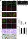
Figure 8
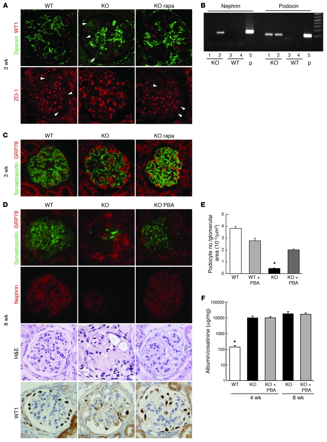
(A) EMT-like phenotypic changes were induced in PcTSC1KO podocytes. Double staining with desmin and WT1 antibodies was performed in renal tissues from the indicated animals and analyzed by confocal microscopy (top row). Arrowheads indicate desmin expression in the podocytes. Rapamycin treatment was performed for a week. ZO-1 expression was analyzed by confocal microscopy (bottom row). Arrowheads indicate the liner expression pattern of ZO-1. (B) Detection of nephrin and podocin mRNA in PcTSC1KO urinary sediments. Twenty-four-hour urine samples were collected from the indicated animals. RT-PCR was performed using purified mRNA from the sediments. Glomerular mRNA was used as a positive control (p). (C) Accumulation of GRP78 in PcKOTsc1 podocytes (3 weeks of age). Double staining using anti-GRP78 and anti-synaptopodin antibodies in the indicated glomeruli is shown. Rapamycin treatment was performed for 1 week. (D) PBA treatment protects podocyte loss in PcKOTsc1 mice. Staining with GRP78 plus synaptopodin, nephrin, H&E, and WT1 is shown in the indicated animals (8 weeks of age). PBA treatment (400 mg/kg/d, i.p.) was performed for 6 weeks. (E) Podocyte number in the indicated glomerular sections from 8-week-old animals was measured. The ratio (number of WT1-positive cells/glomerular tuft area [μm2]) was determined in 15~30 glomeruli from the indicated animals as in Figure Figure3E.3E. *P < 0.001 versus other groups, mean ± SEM, n = 4~8. (F) Urinary albumin concentrations were measured in the indicated animals. *P < 0.001 versus other groups; no statistical significance was observed between KO and KO with PBA treatment; mean ± SEM, n = 4~8. Original magnification, ×400 (A, C, and D).

