| PMC full text: | Mol Microbiol. Author manuscript; available in PMC 2012 Aug 19. Published in final edited form as: Mol Microbiol. 2009 Mar; 71(6): 1574–1590. Published online 2009 Feb 10. doi: 10.1111/j.1365-2958.2009.06623.x |
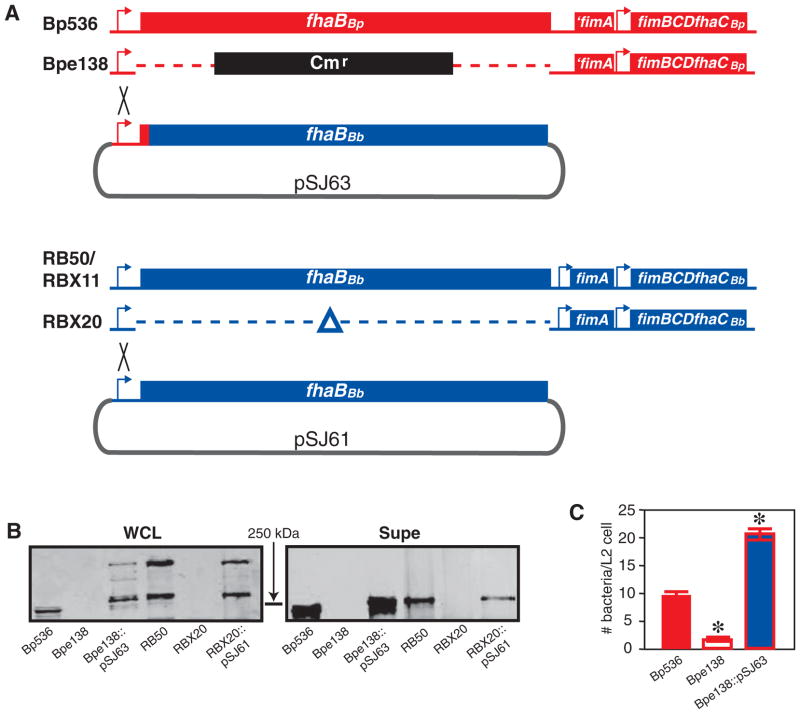
Fig. 1

A. Schematic of strains used. The B. pertussis fhaB locus is shown in red and the B. bronchiseptica fhaB locus is shown in blue. The regions of integration of pSJ63 and pSJ61 into the chromosomes of Bpe138 and RBX20 to form Bpe138::pSJ63 and RBX20::pSJ61, respectively, are indicated by the black crosses.
B. Western blot showing FhaB (~370 kDa) and FHA (~250 kDa) in whole cell lysates (WCLs) and concentrated supernatants (Supe) of the various B. pertussis and B. bronchiseptica strains as indicated below each lane. Blots were probed with the anti-CRD antibody. The position of the 250 kDa molecular mass marker is shown.
C. Adherence of wild-type and mutant B. pertussis strains to L2 cells (moi = 200). Asterisks indicate a statistically significant (P < 0.05).


